

What are the quadriceps muscles?
The quadriceps femoris is a group of four muscles that forms the bulk of the front of the thigh: rectus femoris and three vastus muscles. TeachMeAnatomy calls it one of the most powerful muscles in the body, and Kenhub calls it the most powerful extensor of the knee. (TeachMeAnatomy: Muscles of the Anterior Compartment of the Thigh.)
OpenStax describes the quadriceps femoris group as four muscles that make up the anterior compartment of the thigh and extend and stabilise the knee. The muscles of this compartment flex the thigh at the hip and extend the leg at the knee. (OpenStax Anatomy and Physiology 2e, 11.6: Appendicular Muscles of the Pelvic Girdle and Lower Limbs.)
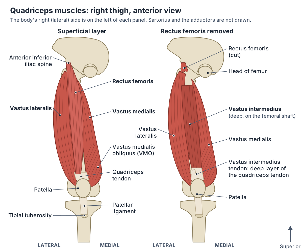
The four parts are arranged around the femur. Rectus femoris lies on the front of the thigh, vastus lateralis on the outer (lateral) side, vastus medialis on the inner (medial) side, and vastus intermedius between the two vasti, deep to rectus femoris. (OpenStax Anatomy and Physiology 2e, 11.6: Appendicular Muscles of the Pelvic Girdle and Lower Limbs.)
Radiopaedia sums up the layout simply: rectus femoris is superficial, vastus medialis medial, vastus lateralis lateral and vastus intermedius deep. All four are supplied by the femoral nerve. (Radiopaedia: Quadriceps muscles.)
All three thigh compartments: the quadriceps and sartorius in front, the adductors and the hamstrings. Thigh muscles.
Origins and insertion of the four quadriceps muscles
Rectus femoris is the only part of the quadriceps that starts on the pelvis. Its straight head arises from the anterior inferior iliac spine (AIIS) and its reflected head from a groove just above the acetabulum, the hip socket. It forms the central part of the quadriceps tendon on the base of the patella. (University of Washington Musculoskeletal Atlas: Rectus femoris.)
Vastus lateralis arises from a long line on the upper femur: the upper intertrochanteric line, the front and lower borders of the greater trochanter, the lateral part of the gluteal tuberosity and the upper lateral lip of the linea aspera. It inserts on the lateral base and border of the patella and forms the lateral patellar retinaculum. (University of Washington Musculoskeletal Atlas: Vastus lateralis.)
Vastus medialis arises from the lower intertrochanteric line, the spiral line, the medial lip of the linea aspera, the upper medial supracondylar ridge and the medial intermuscular septum. It inserts on the medial base and border of the patella and forms the medial patellar retinaculum. (University of Washington Musculoskeletal Atlas: Vastus medialis.)
Vastus intermedius arises from the upper two-thirds of the front and lateral surfaces of the femoral shaft and forms the deep part of the quadriceps tendon. TeachMeAnatomy gives the same origin, the anterior and lateral surfaces of the femoral shaft. (University of Washington Musculoskeletal Atlas: Vastus intermedius.)
All four end in the same place. Kenhub lists the insertions as the patella and, through the patellar ligament, the tibial tuberosity, the bump on the front of the upper tibia. So the vasti act only on the knee, while rectus femoris, which also crosses the front of the hip, acts on both joints. (Kenhub: Quadriceps femoris muscle.)
The largest part of the quadriceps: its origin line, iliotibial tract relations, injection and biopsy use. Vastus lateralis.
Quadriceps muscles: origin, insertion, nerve and roots, action
| Muscle | Origin | Insertion | Nerve (roots) | Action |
|---|---|---|---|---|
| Rectus femoris | Straight head: anterior inferior iliac spine; reflected head: groove above the acetabulum | Base of patella (central quadriceps tendon), then tibial tuberosity via the patellar ligament | Femoral nerve (L2–L4) | Extends knee; flexes hip |
| Vastus lateralis | Upper intertrochanteric line, greater trochanter, gluteal tuberosity, lateral lip of linea aspera | Lateral base and border of patella; lateral patellar retinaculum; then tibial tuberosity | Femoral nerve (L2–L4) | Extends knee; steadies patella |
| Vastus medialis (including VMO) | Lower intertrochanteric line, spiral line, medial lip of linea aspera, medial supracondylar ridge | Medial base and border of patella; medial patellar retinaculum; then tibial tuberosity | Femoral nerve (L2–L4) | Extends knee; VMO stabilises patella medially |
| Vastus intermedius | Upper two-thirds of anterior and lateral femoral shaft | Deep layer of quadriceps tendon to patella; then tibial tuberosity | Femoral nerve (L2–L4) | Extends knee |
Compiled from the University of Washington Musculoskeletal Atlas, TeachMeAnatomy, Kenhub and Radiopaedia. Root values L2–L4 are given for the femoral nerve by TeachMeAnatomy, Kenhub and Radiopaedia.
Quadriceps tendon, patella and patellar ligament
The quadriceps tendon is the tendon common to all four muscles. It inserts into the patella (kneecap) and continues below it as the patellar ligament, which attaches to the tibial tuberosity. OpenStax also calls the quadriceps tendon the patellar tendon, while Radiopaedia and OrthoInfo use "patellar tendon" for the part below the patella (the patellar ligament). (OpenStax Anatomy and Physiology 2e, 11.6: Appendicular Muscles of the Pelvic Girdle and Lower Limbs.)
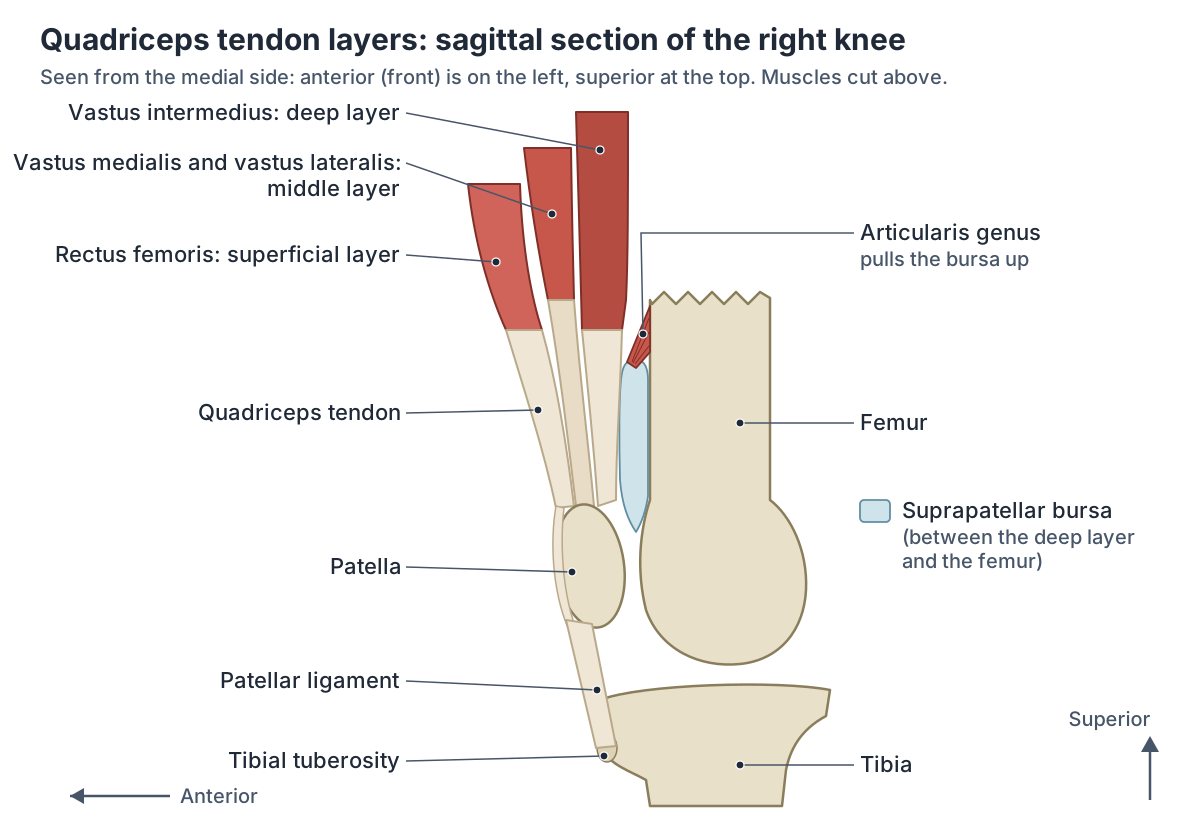
Radiopaedia describes the quadriceps tendon as classically trilaminar: rectus femoris forms the superficial layer, vastus medialis and vastus lateralis the middle layer, and vastus intermedius the deep layer. Below the patella the tendon continues as the patellar tendon. (Radiopaedia: Quadriceps tendon.)
A dissection study of ten cadaver legs by Grob and colleagues confirmed three layers but found them built from six muscle elements, with the lateral part of the vastus intermedius aponeurosis forming the deepest layer. (Grob K et al. New insight in the architecture of the quadriceps tendon. J Exp Orthop. 2016.)
The patella is a sesamoid bone, a bone that develops inside a tendon, and the largest in the body. It increases the leverage that the quadriceps tendon can exert, so knee extension is more efficient, and it protects the front of the knee joint. (TeachMeAnatomy: The Patella.)
The patella is triangular, with its base above and its apex below. The quadriceps tendon attaches to the base and the patellar tendon leaves the apex for the tibial tubercle. At its sides, the medial and lateral patellar retinacula, condensations of fascia rather than true ligaments, blend with the fascia of vastus medialis and of vastus lateralis. (Radiopaedia: Patella.)
OrthoInfo describes the whole unit, the four muscles, the quadriceps tendon, the patella and the patellar tendon, as a pulley mechanism that straightens the knee. (OrthoInfo (AAOS): Quadriceps Tendon Tear.)
The kneecap in 3D: its surfaces, the patellofemoral joint and patellar fractures. Patella.
Vastus medialis obliquus (VMO) and patellar stability
The vastus medialis obliquus (VMO) is the lowest part of vastus medialis, whose fibres run obliquely towards the inner edge of the patella. Radiopaedia describes VMO fibres inserting on the upper third of the medial patella, where they act as a medial stabiliser of the patella. (Radiopaedia: Vastus medialis muscle.)
In ten dissected limbs, El Sawy and colleagues found the upper fibres of vastus medialis (vastus medialis longus) more vertical and the lower fibres (VMO) more oblique. The VMO inserted into the upper medial margin of the patella and the knee capsule, and the authors describe its main job as medial stabilisation of the patella. Sources differ on where the VMO fibres arise: El Sawy and colleagues traced them largely to the attachment of adductor magnus, the medial lip of the linea aspera and the medial supracondylar line, while Radiopaedia gives the adductor tubercle and the intermuscular septum. (El Sawy MM et al. Morphometric analysis of vastus medialis oblique muscle and its influence on anterior knee pain. Anat Cell Biol. 2021.)
Why the patella needs this help: Radiopaedia explains that the quadriceps pull the patella obliquely and laterally. The lateral femoral condyle, the tension of the medial retinaculum and the direction of the vastus medialis fibres all resist that lateral pull. (Radiopaedia: Patella.)
Kenhub likewise notes that some authors call this part of the muscle the vastus obliquus, and that its nearly horizontal fibres counter the pull of the other vasti, helping to keep the patella from dislocating during extension. (Kenhub: Quadriceps femoris muscle.)
The patellofemoral and tibiofemoral joints, the ligaments and the menisci in 3D. Knee anatomy.
Vastus intermedius, articularis genus and the 'fifth head'
Vastus intermedius is the hidden quadriceps muscle. It lies on the front and side of the femur, deep to rectus femoris and between vastus lateralis and vastus medialis, and Kenhub describes it as completely surrounded by the other three. That is why most diagrams, including the right panel above, remove rectus femoris to show it. (Kenhub: Quadriceps femoris muscle.)
Articularis genus (articularis genu) is a small muscle under vastus intermedius. It arises from the front of the lower femoral shaft, deep to vastus intermedius, and attaches to the knee joint's synovial membrane and the upper border of the suprapatellar bursa. It pulls the capsule up during extension so that folds of synovium are not pinched, it is supplied by the femoral nerve, and it develops from vastus intermedius. (Radiopaedia: Articularis genu muscle.)
A study of 18 cadaver limbs from 12 donors found that articularis genus is linked so closely to vastus intermedius that it does not act alone. Articularis genus, vastus medialis and the medial part of vastus intermedius were always supplied by the same division of the femoral nerve, and the authors suggest that, with the support of those two vasti, articularis genus can retract the suprapatellar bursa. (Grob K et al. The anatomy of the articularis genus muscle and its relation to the extensor apparatus of the knee. JBJS Open Access. 2017.)
Grob and colleagues also describe a fifth muscle component, which they name tensor vastus intermedius, lying between vastus lateralis and vastus intermedius. Its aponeurosis joins the middle layer of the quadriceps tendon; in their ten dissected legs it was independent and strong in four. It is a research finding rather than standard textbook anatomy, so the quadriceps is still taught as four muscles. (Grob K et al. New insight in the architecture of the quadriceps tendon. J Exp Orthop. 2016.)
The thigh bone that the three vasti arise from: trochanters, linea aspera and shaft. Femur.
Nerve and blood supply of the quadriceps
The femoral nerve supplies all four parts of the quadriceps. It comes from the anterior rami of spinal nerves L2, L3 and L4 in the lumbar plexus, runs through psoas major, passes under the inguinal ligament and enters the thigh in the femoral triangle. Besides the quadriceps it supplies sartorius, pectineus and iliacus, and its saphenous branch carries sensation from the inner side of the leg and foot. (TeachMeAnatomy: The Femoral Nerve.)
TeachMeAnatomy gives the anterior compartment's nerve as the femoral nerve (L2 to L4), and Kenhub and Radiopaedia give the same roots for the whole quadriceps. (TeachMeAnatomy: Muscles of the Anterior Compartment of the Thigh.)
The arterial supply comes from the femoral artery and its deep (profunda femoris) branch. The University of Washington atlas lists the lateral circumflex femoral artery for rectus femoris, vastus lateralis and vastus intermedius, and Radiopaedia names branches of the lateral circumflex femoral and deep femoral arteries for vastus lateralis. (University of Washington Musculoskeletal Atlas: Rectus femoris.)
The lateral circumflex femoral artery branches from the profunda femoris and wraps round the front and outer side of the femur to supply the muscles on the lateral side of the thigh. (TeachMeAnatomy: Arteries of the Lower Limb.)
Vastus medialis has a separate supply from the femoral artery, the profunda femoris artery and the superior medial genicular branch of the popliteal artery. (Radiopaedia: Vastus medialis muscle.)
In the middle third of the thigh, vastus medialis forms the lateral wall of the adductor canal (Hunter's canal), with sartorius covering it and adductor longus and adductor magnus behind it. The canal carries the femoral artery and vein, the saphenous nerve and the nerve to vastus medialis. (Kenhub: Quadriceps femoris muscle.)
Higher up, in the femoral triangle, the femoral nerve lies lateral to the femoral vessels; about 4 cm below the inguinal ligament it divides into anterior and posterior divisions, and the posterior division gives the branches to the quadriceps. (TeachMeAnatomy: The Femoral Nerve.)
The lumbar and sacral plexuses, the femoral nerve's branches and the sciatic nerve. Nerves of the leg.
What do the quadriceps do?
The main job of the quadriceps is to extend (straighten) the knee. All four parts do this through the quadriceps tendon, patella and patellar ligament, and OpenStax adds that the four muscles also stabilise the knee. Each vastus also helps to steady the patella. (TeachMeAnatomy: Muscles of the Anterior Compartment of the Thigh.)
Rectus femoris has a second action. Because it crosses the front of the hip as well as the knee, it also flexes the thigh at the hip. OpenStax describes it moving the lower leg out in front of the body, as when kicking, and helping to raise the knee. (OpenStax Anatomy and Physiology 2e, 11.6: Appendicular Muscles of the Pelvic Girdle and Lower Limbs.)
The hamstrings at the back of the thigh are the antagonists of the quadriceps: the hamstrings flex the leg at the knee, whereas the quadriceps extend it. An antagonist is a muscle with the opposite action of the prime mover. (OpenStax Anatomy and Physiology 2e, 11.1: Interactions of Skeletal Muscles, Their Fascicle Arrangement, and Their Lever Systems.)
In everyday life the quadriceps are essential for climbing stairs and getting up from a chair, and StatPearls notes that their work also affects walking and posture. (Bordoni B, Varacallo MA. Anatomy, Bony Pelvis and Lower Limb: Thigh Quadriceps Muscle. StatPearls, 2023.)
That is why femoral nerve injury, which weakens knee extension, makes walking forward and climbing stairs difficult, as a neurosurgical examination guide lists. (Hamzah NA et al. An Approach to the Examination of the Lumbar Plexus for Neurosurgical Residents: A Video Manuscript. Malays J Med Sci. 2024.)
The two-joint quadriceps muscle: its straight and reflected heads, hip flexion and kicking injuries. Rectus femoris.
How the quadriceps and femoral nerve are tested
Quadriceps strength is a quick test of the femoral nerve. The person lies on their back with the knee slightly bent and straightens the leg against the examiner's resistance. Damage to the femoral nerve shows up as weak knee extension, and with a complete injury the quadriceps does not contract at all. (TeachMeAnatomy: Muscles of the Anterior Compartment of the Thigh.)
The knee-jerk reflex of the quadriceps is one of the standard leg reflexes: the tendon at the muscle's insertion is struck with a rubber mallet and the quadriceps contracts. For the knee jerk, Kenhub notes, the tap falls on the patellar ligament just below the kneecap. (OpenStax Anatomy and Physiology 2e, 16.4: The Sensory and Motor Exams.)
Sources give slightly different spinal segments for the knee jerk. A neurosurgical examination guide lists the patellar reflex as femoral nerve, L3 to L4, while Kenhub says it tests segments L2, L3 and L4, the same roots as the femoral nerve. The same guide lists wasting of the quadriceps and loss of the patellar reflex as signs of femoral nerve injury. (Hamzah NA et al. An Approach to the Examination of the Lumbar Plexus for Neurosurgical Residents: A Video Manuscript. Malays J Med Sci. 2024.)
Quadriceps tendon tears, Osgood-Schlatter disease and other problems
Most quadriceps injuries in sport are muscle strains and bruises (contusions); a review in Current Reviews in Musculoskeletal Medicine says these two make up the majority. (Kary JM. Diagnosis and management of quadriceps strains and contusions. Curr Rev Musculoskelet Med. 2010.)
Radiopaedia describes a spectrum of strains, tears, avulsions and contusions, up to rupture of the quadriceps tendon. Rectus femoris is the muscle most often involved: it crosses two joints and contracts forcefully while lengthening (eccentric contraction) in sprinting, jumping and kicking. A contusion comes from a direct blow to the front of the thigh, and Radiopaedia notes that vastus intermedius seems to be the muscle most often bruised. (Radiopaedia: Quadriceps injury.)
Kenhub says contusions are seen especially in high-impact sports and that most heal on their own over time. (Kenhub: Quadriceps femoris muscle.)
A contusion can be followed by myositis ossificans, bone forming inside the injured muscle, which the review calls a potentially disabling complication. (Kary JM. Diagnosis and management of quadriceps strains and contusions. Curr Rev Musculoskelet Med. 2010.)
A quadriceps tendon tear is uncommon. OrthoInfo says it most often affects middle-aged people who play running or jumping sports, often when a heavy load lands on a planted foot with the knee partly bent. With a complete tear the knee cannot be straightened and a gap may be felt above the kneecap; most complete tears need surgery, so a suspected tear needs prompt assessment by a clinician. (OrthoInfo (AAOS): Quadriceps Tendon Tear.)
Radiopaedia notes that most ruptures occur at or near the junction of tendon and bone at the upper pole of the patella. Conditions that weaken the tendon raise the risk, including chronic kidney disease, gout, diabetes, rheumatoid arthritis, lupus and hyperparathyroidism, as do corticosteroids and fluoroquinolone antibiotics. (Radiopaedia: Quadriceps tendon rupture.)
Osgood-Schlatter disease is a common cause of knee pain in growing adolescents. The quadriceps attach through the patellar tendon to the tibial tubercle, which covers a growth plate; during growth spurts, especially in running and jumping sports, the tubercle becomes painful, tender and swollen. (OrthoInfo (AAOS): Osgood-Schlatter Disease.)
Radiopaedia describes Osgood-Schlatter disease as a chronic fatigue injury from repeated small injuries where the patellar tendon inserts on the tibial tuberosity, seen in active adolescents who jump and kick. (Radiopaedia: Osgood-Schlatter disease.)
When the patella dislocates, it usually moves laterally. TeachMeAnatomy gives the causes as a high-force impact or a forceful, sudden twist of the knee, which is why the medial stabilisers described above matter. (TeachMeAnatomy: The Patella.)
The bones, muscle groups, nerves and vessels of the whole lower limb. Leg anatomy.
The quadriceps in the 3D model
Seen from the front, the quadriceps shows its layout clearly: rectus femoris is the superficial central strap, vastus lateralis and vastus medialis form the outer and inner bulges, and vastus intermedius lies deep, on the femur. (Radiopaedia: Quadriceps muscles.)
The 3D view highlights rectus femoris, vastus lateralis, vastus medialis and vastus intermedius on both thighs, seen from the front over a ghosted skeleton, with each part labelled on the subject's left thigh. Vastus intermedius lies mostly deep to rectus femoris; turn the model to follow it down the femur. Click any part to open its own page. The model is BodyParts3D, built from one adult male; it does not show the femoral nerve, the articularis genus or the layers of the quadriceps tendon, so the diagrams above show those.
Quadriceps in the 3D model
- Rectus femoris
- The only part of quadriceps femoris that crosses both the hip and knee.
- Vastus intermedius
- The deep quadriceps muscle lying beneath rectus femoris.
- Vastus lateralis
- The largest quadriceps muscle on the lateral thigh.
- Vastus medialis
- The medial quadriceps muscle with oblique fibers near the patella.
Frequently asked questions
What are the 4 quadriceps muscles?
The four quadriceps muscles are rectus femoris, vastus lateralis, vastus medialis and vastus intermedius. Rectus femoris runs down the front of the thigh from the pelvis, vastus lateralis lies on the outer side, vastus medialis on the inner side, and vastus intermedius lies deep, on the femur. All four join the quadriceps tendon on the patella and extend the knee.
Which quadriceps muscle is the largest?
Vastus lateralis, on the outer side of the thigh, is usually described as the largest of the four quadriceps muscles; Kenhub and Radiopaedia both call it the largest of the four. It arises from a long line on the upper femur, from the greater trochanter down the linea aspera, and inserts on the lateral side of the patella.
What is the VMO muscle?
The VMO (vastus medialis obliquus) is the lowest part of vastus medialis, just above and inside the kneecap. Its fibres run more obliquely than the rest of the muscle and insert on the upper part of the medial patella. Because the quadriceps tend to pull the patella outwards, the VMO helps to hold it in its groove as a medial stabiliser.
What nerve supplies the quadriceps?
The femoral nerve supplies all four parts of the quadriceps. It comes from spinal nerves L2, L3 and L4 in the lumbar plexus and enters the thigh under the inguinal ligament. Doctors test it by asking the person to straighten the knee against resistance and by tapping below the kneecap for the knee-jerk reflex.
Which quadriceps muscle crosses the hip?
Only rectus femoris. It starts on the pelvis, at the anterior inferior iliac spine and just above the hip socket, so it crosses the front of the hip as well as the knee. That lets it flex the hip as well as extend the knee, as in kicking. The three vasti start on the femur and act on the knee alone.
Where do the quadriceps attach at the knee?
The four quadriceps muscles join one tendon, the quadriceps tendon, which attaches to the top (base) of the patella. Below the kneecap the pull continues through the patellar ligament, also called the patellar tendon, to the tibial tuberosity on the front of the shinbone. The vastus lateralis and vastus medialis also send fibres to the sides of the patella.
What happens if the quadriceps tendon tears?
A complete quadriceps tendon tear stops the knee from straightening, and a dent or gap may be felt just above the kneecap, often with bruising and tenderness. It is uncommon and most often affects middle-aged people who play running or jumping sports; conditions and medicines that weaken tendons raise the risk. Most complete tears are repaired surgically, so anyone with these signs should see a clinician promptly.
References
- OpenStax. Anatomy and Physiology 2e, §11.6 Appendicular Muscles of the Pelvic Girdle and Lower Limbs. Rice University, 2022.
- OpenStax. Anatomy and Physiology 2e, §11.1 Interactions of Skeletal Muscles, Their Fascicle Arrangement, and Their Lever Systems. Rice University, 2022.
- OpenStax. Anatomy and Physiology 2e, §16.4 The Sensory and Motor Exams. Rice University, 2022.
- TeachMeAnatomy. Muscles of the Anterior Compartment of the Thigh.
- TeachMeAnatomy. The Femoral Nerve.
- TeachMeAnatomy. Arteries of the Lower Limb.
- TeachMeAnatomy. The Patella.
- University of Washington Department of Radiology. Musculoskeletal Atlas: Rectus femoris.
- University of Washington Department of Radiology. Musculoskeletal Atlas: Vastus lateralis.
- University of Washington Department of Radiology. Musculoskeletal Atlas: Vastus medialis.
- University of Washington Department of Radiology. Musculoskeletal Atlas: Vastus intermedius.
- Kenhub. Quadriceps femoris muscle: anatomy, innervation and function.
- Radiopaedia. Quadriceps muscles.
- Radiopaedia. Quadriceps tendon.
- Radiopaedia. Vastus lateralis muscle.
- Radiopaedia. Vastus medialis muscle.
- Radiopaedia. Articularis genu muscle.
- Radiopaedia. Patella.
- Radiopaedia. Quadriceps tendon rupture.
- Radiopaedia. Osgood-Schlatter disease.
- Radiopaedia. Quadriceps injury.
- Kary JM. Diagnosis and management of quadriceps strains and contusions. Curr Rev Musculoskelet Med. 2010;3(1–4):26–31.
- Bordoni B, Varacallo MA. Anatomy, Bony Pelvis and Lower Limb: Thigh Quadriceps Muscle. StatPearls. Treasure Island (FL): StatPearls Publishing; 2023. PMID 30020706.
- El Sawy MM, El Mikkawy DME, El-Sayed SM, Desouky AM. Morphometric analysis of vastus medialis oblique muscle and its influence on anterior knee pain. Anat Cell Biol. 2021;54(1):1–9.
- Grob K, Manestar M, Filgueira L, Ackland T, Gilbey H, Kuster MS. New insight in the architecture of the quadriceps tendon. J Exp Orthop. 2016;3:32.
- Grob K, Gilbey H, Manestar M, Ackland T, Kuster MS. The anatomy of the articularis genus muscle and its relation to the extensor apparatus of the knee. JBJS Open Access. 2017;2(4):e0034.
- Hamzah NA, Lee WL, Fathil MFM, Abdullah JM, Idris Z, Ghani ARI, Halim SA. An approach to the examination of the lumbar plexus for neurosurgical residents: a video manuscript. Malays J Med Sci. 2024;31(1):235–242.
- American Academy of Orthopaedic Surgeons, OrthoInfo. Quadriceps Tendon Tear.
- American Academy of Orthopaedic Surgeons, OrthoInfo. Osgood-Schlatter Disease.
- Mitsuhashi N. et al. (2009). BodyParts3D: 3D structure database for anatomical concepts. Nucleic Acids Research 37, D782–D785. Data © The Database Center for Life Science, CC BY 4.0.
Related anatomy
- Thigh musclesThigh muscles explained: quadriceps and sartorius at the front, adductor muscles of the inner thigh, hamstrings at the…
- Rectus femorisRectus femoris anatomy: straight and reflected heads, insertion, why it flexes the hip and extends the knee, nerve and…
- Vastus lateralisVastus lateralis (lateral vastus muscle): origin, insertion, femoral nerve (L2 to L4), blood supply, knee extension…
- Knee jointKnee anatomy with labelled diagrams and a 3D model: femur, tibia and patella, the ACL, PCL, MCL and LCL, menisci…
- PatellaWhat is the patella? The kneecap explained: base, apex, facets, quadriceps tendon and patellar ligament, why it…
- FemurFemur bone labelled: head, neck, trochanters, linea aspera, condyles and epicondyles, with muscle attachments, blood…
Explore the whole body in the interactive 3D human anatomy model, or browse the A–Z list of human structures.