

What is the leg in anatomy?
In anatomy, the leg is the part of the lower limb between the knee and the ankle joints. In everyday language, 'leg' usually means the whole lower extremity, from the hip to the toes. This page covers the whole lower limb but uses 'leg' in the strict sense when naming muscles, vessels and nerves. (Kenhub: Lower leg and knee anatomy.)
OpenStax divides the lower limb, like the upper limb, into three regions: the thigh between the hip and knee joints, the leg between the knee and ankle joints, and the foot below the ankle. Kenhub adds a hip and pelvis region above the thigh, which contains the hip bone (formed from the ilium, ischium and pubis), the sacrum and the coccyx. (OpenStax Anatomy and Physiology 2e, 8.4: Bones of the Lower Limb.)
Regions of the lower limb
Kenhub names seven regions of the lower limb: gluteal, hip, femoral (thigh), knee, leg, talocrural (ankle) and foot. Each is named after its main bone or joint, and some authors count the hip as part of the gluteal region. (Kenhub: Regions of the lower limb.)
The gluteal region (the buttock) runs from the iliac crest down to the gluteal fold. It holds the gluteal muscles, the deep hip rotators and the sciatic nerve, which enters it from the pelvis through the greater sciatic foramen. The knee region includes the popliteal fossa behind the knee, and the ankle region holds the medial and lateral malleoli with the talus between them. (Kenhub: Regions of the lower limb.)
The ball-and-socket joint that joins the lower limb to the pelvis. Hip anatomy.
Regions of the lower limb
| Region | Extent | Bones | Key contents |
|---|---|---|---|
| Gluteal region (buttock) | Iliac crest to gluteal fold | Bony pelvis (hip bone, sacrum) | Gluteal muscles, deep hip rotators, sciatic nerve |
| Hip region | Anterior superior iliac spine to greater trochanter | Hip bone, head of femur | Hip joint |
| Thigh (femoral region) | Inguinal ligament and gluteal fold to the knee | Femur | Three muscle compartments, femoral triangle, femoral vessels, sciatic nerve |
| Knee | Lower thigh to upper leg | Femur, tibia, patella (head of fibula nearby) | Knee joint, popliteal fossa |
| Leg | Knee to ankle | Tibia, fibula | Anterior, lateral and posterior compartments; tibial vessels; tibial and fibular nerves |
| Ankle (talocrural region) | Leg to foot | Lower tibia and fibula (malleoli), talus | Ankle joint; tendons, vessels and nerves passing to the foot |
| Foot | Below the ankle | 7 tarsals, 5 metatarsals, 14 phalanges | Dorsum and sole; dorsalis pedis artery; plantar vessels and nerves |
Source: Kenhub, Regions of the lower limb. Some authors count the hip as part of the gluteal region.
Leg bones: how many bones are in the lower limb?
Each lower limb contains 30 bones: the femur, the patella, the tibia and fibula, seven tarsal bones, five metatarsal bones and 14 phalanges. The foot alone accounts for 26 of them. The hip bone that attaches the limb to the trunk is not part of this count; adding it gives 31 bones per side. (OpenStax Anatomy and Physiology 2e, 8.4: Bones of the Lower Limb.)
In the strict sense the leg has only two bones, the tibia and fibula, and the thigh has one, the femur, which Kenhub describes as the only bone of the thigh and the longest bone in the body. The knee is formed where the femur meets the tibia, with the patella in front. (Kenhub: Lower limb anatomy.)
All the bones of the body, grouped into the axial and appendicular skeleton. Skeletal system.
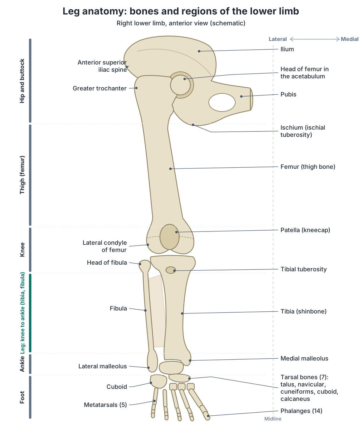
Bones of the lower limb
| Bone | Where | Number per limb | Key facts |
|---|---|---|---|
| Hip bone (ilium, ischium, pubis) | Hip and pelvis | 1 (not in the 30-bone count) | Its acetabulum receives the head of the femur at the hip joint |
| Femur | Thigh | 1 | Longest and strongest bone; greater trochanter palpable on the outer upper thigh |
| Patella | Front of the knee | 1 | Largest sesamoid bone; sits in the quadriceps tendon; articulates only with the femur |
| Tibia | Leg, medial side | 1 | Main weight-bearing bone of the leg; tibial tuberosity; medial malleolus |
| Fibula | Leg, lateral side | 1 | Does not bear weight; lateral malleolus |
| Tarsal bones | Ankle and back of the foot | 7 | Talus, calcaneus, navicular, cuboid, three cuneiforms |
| Metatarsal bones | Middle of the foot | 5 | Numbered I to V from the medial side |
| Phalanges | Toes | 14 | Two in the great toe, three in each other toe |
Sources: OpenStax Anatomy and Physiology 2e, 8.4; Kenhub.
Femur: the thigh bone
The femur is the single bone of the thigh and the longest and strongest bone of the body. Its rounded head fits into the acetabulum of the hip bone to form the hip joint. The greater trochanter can be felt just under the skin on the outer side of the upper thigh, and the narrow femoral neck is a common place for the femur to break. (OpenStax Anatomy and Physiology 2e, 8.4: Bones of the Lower Limb.)
The head and neck of the femur are supplied by the medial femoral circumflex artery, a branch of the deep femoral artery. Damage to this artery in a femoral neck fracture can cut off the blood supply to the head, which may then die (avascular necrosis). (TeachMeAnatomy: Arterial Supply to the Lower Limb.)
Head, neck, trochanters, linea aspera, condyles and fractures of the thigh bone. Femur.
Patella: the kneecap
The patella (kneecap) is the largest sesamoid bone in the body. It lies within the tendon of quadriceps femoris and articulates only with the lower end of the femur, never with the tibia. (OpenStax Anatomy and Physiology 2e, 8.4: Bones of the Lower Limb.)
Kenhub describes two jobs for the patella: it protects the front of the knee and acts as a pulley that improves the leverage of the quadriceps when they straighten the knee. (Kenhub: Lower leg and knee anatomy.)
Facets, attachments, development and injuries of the kneecap. Patella.
Tibia: the shinbone
The tibia (shinbone) is the medial bone of the leg and its main weight-bearing bone; it is the second-longest bone in the body. Its upper end carries the medial and lateral condyles, which articulate with the femoral condyles at the knee, and the tibial tuberosity, where the patellar ligament (patellar tendon) attaches. (OpenStax Anatomy and Physiology 2e, 8.4: Bones of the Lower Limb.)
The anterior border and medial surface of the tibial shaft lie directly under the skin, so you can feel the bone down the whole front and inner side of your shin. At its lower end the medial malleolus forms the bony bump on the inside of the ankle. (OpenStax Anatomy and Physiology 2e, 8.4: Bones of the Lower Limb.)
Plateau, tuberosity, shaft and medial malleolus of the shinbone. Tibia.
Fibula: the slender outer leg bone
The fibula is the slender bone on the lateral side of the leg, and it does not bear weight. Its head articulates with the underside of the lateral tibial condyle at the proximal tibiofibular joint, and a sheet of dense connective tissue, the interosseous membrane, joins its shaft to the tibia. Only its two ends can be felt; the lower end forms the lateral malleolus, the bump on the outside of the ankle. (OpenStax Anatomy and Physiology 2e, 8.4: Bones of the Lower Limb.)
The common fibular (peroneal) nerve winds around the neck of the fibula just below the knee, so a fracture there or a tight plaster cast can damage it. (TeachMeAnatomy: The Common Fibular Nerve.)
Head, shaft, lateral malleolus and tibiofibular joints. Fibula.
Joints of the leg: hip and knee
The hip joint is a multiaxial ball-and-socket joint between the head of the femur and the acetabulum, which a rim of fibrocartilage, the acetabular labrum, deepens further. Three ligaments, the iliofemoral, pubofemoral and ischiofemoral, tighten when you stand upright and pull the femoral head firmly into its socket. (OpenStax Anatomy and Physiology 2e, 9.6: Anatomy of Selected Synovial Joints.)
The knee is the largest joint of the body. It contains three articulations in one capsule (between the patella and femur, and between the medial and lateral condyles of the femur and tibia) and works mainly as a hinge. Two C-shaped fibrocartilage menisci sit on the tibia, the collateral ligaments lie outside the capsule, and the anterior and posterior cruciate ligaments cross inside it; the posterior cruciate is the stronger and stops the femur sliding forwards off the tibia. (OpenStax Anatomy and Physiology 2e, 9.6: Anatomy of Selected Synovial Joints.)
The tibia and fibula also meet at a superior tibiofibular joint just below the knee and an inferior tibiofibular joint just above the ankle. (Kenhub: Lower leg and knee anatomy.)
Bones, ligaments (ACL, PCL, MCL, LCL), menisci and bursae of the knee. Knee anatomy.
Ankle anatomy in brief
The ankle (talocrural) joint is where the talus fits between the lower ends of the tibia and fibula. It is a uniaxial hinge that allows only dorsiflexion and plantar flexion. The broad deltoid ligament supports its medial side; the anterior talofibular, calcaneofibular and posterior talofibular ligaments support the lateral side. (OpenStax Anatomy and Physiology 2e, 9.6: Anatomy of Selected Synovial Joints.)
OpenStax calls the ankle the most frequently injured joint in the body. The usual injury is an inversion sprain, which most often damages the anterior talofibular ligament, followed by the calcaneofibular ligament. (OpenStax Anatomy and Physiology 2e, 9.6: Anatomy of Selected Synovial Joints.)
Turning the sole inwards and outwards (inversion and eversion) happens at the subtalar (talocalcaneal) joint between the talus and calcaneus, below the ankle joint. (Kenhub: Lower limb anatomy.)
Talocrural and subtalar joints, malleoli and ankle ligaments. Ankle anatomy.
Thigh muscles: anterior, medial and posterior compartments
Fascia and intermuscular septa divide the thigh muscles into three compartments: the anterior compartment, mainly quadriceps femoris; the medial compartment, the adductor group; and the posterior compartment, the hamstrings. (Kenhub: Regions of the lower limb.)
The four parts of quadriceps femoris (rectus femoris and vastus lateralis, medialis and intermedius) share a tendon that inserts on the patella and continues below it as the patellar ligament; together they extend and stabilise the knee. The adductors pull the thigh towards the midline, and the hamstrings (biceps femoris, semitendinosus and semimembranosus) flex the knee. (OpenStax Anatomy and Physiology 2e, 11.6: Appendicular Muscles of the Pelvic Girdle and Lower Limbs.)
Each compartment has its own main nerve. The femoral nerve supplies the quadriceps and sartorius, the obturator nerve supplies gracilis, and the sciatic nerve supplies the hamstrings through its tibial part and, for the short head of biceps femoris, its common fibular part. (Kenhub: Lower leg and knee anatomy.)
The obturator nerve also supplies adductor longus, adductor brevis, adductor magnus and obturator externus, while the femoral nerve supplies pectineus as well as the anterior compartment. (TeachMeAnatomy: The Lumbar Plexus.)
Quadriceps, adductors and hamstrings: origins, insertions and nerves. Thigh muscles.
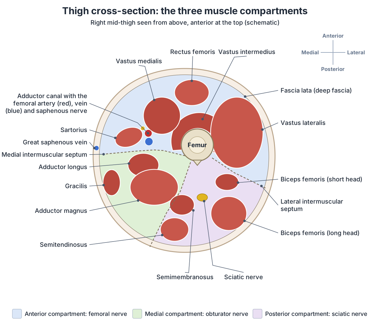
Gluteal and hip muscles
The gluteal group sits behind the hip. Gluteus maximus is the largest and moves the thigh backwards; gluteus medius lies deep to it and gluteus minimus is the smallest and deepest. At the front, the iliopsoas group (psoas major and iliacus) raises the knee by flexing the hip. (OpenStax Anatomy and Physiology 2e, 11.6: Appendicular Muscles of the Pelvic Girdle and Lower Limbs.)
The deep muscles of the gluteal region are piriformis, obturator internus, the superior and inferior gemelli and quadratus femoris, which lie under the gluteal muscles. (Kenhub: Regions of the lower limb.)
The superior gluteal nerve (L4 to S1) supplies gluteus medius, gluteus minimus and tensor fasciae latae, and the inferior gluteal nerve (L5 to S2) supplies gluteus maximus. Both are branches of the sacral plexus. (TeachMeAnatomy: The Sacral Plexus.)
Gluteal muscles, deep lateral rotators, hip flexors and adductors. Hip muscles.
Lower leg muscles: anterior, lateral and posterior compartments
Deep fascia divides the leg below the knee into anterior, lateral and posterior compartments, and the posterior compartment has superficial and deep layers. (OpenStax Anatomy and Physiology 2e, 11.6: Appendicular Muscles of the Pelvic Girdle and Lower Limbs.)
The anterior compartment holds tibialis anterior, extensor hallucis longus, extensor digitorum longus and fibularis tertius, which is not present in everyone. They dorsiflex the foot and extend the toes, tibialis anterior being the strongest dorsiflexor. All are supplied by the deep fibular nerve and the anterior tibial artery. (TeachMeAnatomy: Muscles of the Anterior Compartment of the Leg.)
The lateral compartment contains fibularis (peroneus) longus and brevis, which evert the foot and are supplied by the superficial fibular nerve. The posterior compartment forms the calf: gastrocnemius, soleus and plantaris in the superficial layer, and popliteus, tibialis posterior, flexor digitorum longus and flexor hallucis longus in the deep layer. Together they plantarflex and invert the foot and flex the toes, and the tibial nerve supplies them all. (Kenhub: Lower leg and knee anatomy.)
Gastrocnemius is the most visible calf muscle, and soleus lies deep to it. Both, with plantaris, insert through the strong calcaneal (Achilles) tendon on the heel bone. (OpenStax Anatomy and Physiology 2e, 11.6: Appendicular Muscles of the Pelvic Girdle and Lower Limbs.)
Calf and shin muscles by compartment, with actions and nerves. Leg muscles.
Muscle compartments of the thigh and leg
| Compartment | Muscles | Main action | Nerve |
|---|---|---|---|
| Thigh, anterior | Quadriceps femoris (rectus femoris, vastus lateralis, vastus medialis, vastus intermedius), sartorius | Extend the knee | Femoral nerve (L2–L4) |
| Thigh, medial | Adductor longus, adductor brevis, adductor magnus, gracilis, pectineus | Adduct the thigh | Obturator nerve (L2–L4); pectineus by the femoral nerve; hamstring part of adductor magnus by the sciatic nerve |
| Thigh, posterior | Biceps femoris, semitendinosus, semimembranosus | Flex the knee | Sciatic nerve (tibial part; common fibular part to the short head of biceps femoris) |
| Leg, anterior | Tibialis anterior, extensor hallucis longus, extensor digitorum longus, fibularis tertius | Dorsiflex the foot, extend the toes | Deep fibular nerve |
| Leg, lateral | Fibularis (peroneus) longus and brevis | Evert the foot | Superficial fibular nerve |
| Leg, posterior (superficial) | Gastrocnemius, soleus, plantaris | Plantarflex the foot | Tibial nerve |
| Leg, posterior (deep) | Popliteus, tibialis posterior, flexor digitorum longus, flexor hallucis longus | Plantarflex and invert the foot, flex the toes | Tibial nerve |
Sources: Kenhub; OpenStax Anatomy and Physiology 2e, 11.6; TeachMeAnatomy.
The foot: tarsals, metatarsals and toes
The foot is divided into the tarsus, metatarsus and phalanges, with an upper surface (dorsum) and a sole (plantar surface). Its seven tarsal bones are the calcaneus, talus, navicular, cuboid and the medial, intermediate and lateral cuneiforms. There are five metatarsals, numbered I to V, and 14 phalanges: three in each toe except the great toe, which has two. (Kenhub: Regions of the lower limb.)
Intrinsic foot muscles start and end within the foot and support the arches. The sole has four layers of muscles, and the plantar aponeurosis is the main support of the longitudinal arch. OpenStax counts a single muscle on the dorsum, extensor digitorum brevis, while Kenhub lists it with extensor hallucis brevis as two dorsal muscles. (OpenStax Anatomy and Physiology 2e, 11.6: Appendicular Muscles of the Pelvic Girdle and Lower Limbs.)
Bones, arches, sole, heel, toes and tendons of the foot. Foot anatomy.
Nerves of the leg: the lumbar and sacral plexuses
The lumbar plexus forms from the anterior rami of L1 to L4, with a contribution from T12, inside the psoas major muscle. Its two big branches for the limb are the femoral nerve (L2 to L4), which supplies the front of the thigh and the skin of the anterior thigh and medial leg, and the obturator nerve (L2 to L4), which supplies the adductors and the skin of the medial thigh. (TeachMeAnatomy: The Lumbar Plexus.)
The sacral plexus forms on the back wall of the pelvis, in front of piriformis, from S1 to S4 plus L4 and L5 through the lumbosacral trunk. Its branches are the superior and inferior gluteal nerves, the sciatic nerve, the posterior femoral cutaneous nerve and the pudendal nerve. (TeachMeAnatomy: The Sacral Plexus.)
The sciatic nerve (L4 to S3) is the largest nerve in the body, a flat band about 2 cm wide. It leaves the pelvis through the greater sciatic foramen below piriformis, runs down the back of the thigh deep to the long head of biceps femoris and splits into the tibial and common fibular nerves at the top of the popliteal fossa. In about 12% of people the two parts are already separate as they leave the pelvis. (TeachMeAnatomy: The Sciatic Nerve.)
The tibial nerve supplies the calf muscles and most of the small muscles of the sole, passes behind the medial malleolus through the tarsal tunnel and ends as the medial and lateral plantar nerves. (TeachMeAnatomy: The Tibial Nerve.)
The common fibular nerve wraps around the neck of the fibula into the lateral compartment and divides into the superficial fibular nerve, for fibularis longus and brevis and the skin of the anterolateral leg and dorsum of the foot, and the deep fibular nerve, for the anterior compartment and the skin of the first web space between the big and second toes. (TeachMeAnatomy: The Common Fibular Nerve.)
Lumbar and sacral plexuses, sciatic, tibial and fibular nerves in detail. Nerves of the leg.
Main nerves of the lower limb
| Nerve | Roots or origin | Muscles supplied | Skin supplied |
|---|---|---|---|
| Femoral | L2–L4 (lumbar plexus) | Iliacus, pectineus, sartorius, quadriceps femoris | Anterior thigh; medial leg (through the saphenous nerve) |
| Obturator | L2–L4 (lumbar plexus) | Obturator externus, adductor longus, brevis and magnus, gracilis | Medial thigh |
| Superior gluteal | L4–S1 (sacral plexus) | Gluteus medius, gluteus minimus, tensor fasciae latae | None (motor only) |
| Inferior gluteal | L5–S2 (sacral plexus) | Gluteus maximus | None (motor only) |
| Sciatic | L4–S3 (sacral plexus) | Hamstrings and hamstring part of adductor magnus; leg and foot through its branches | None directly |
| Tibial | Branch of the sciatic nerve | Posterior leg muscles; most intrinsic foot muscles | Back and outer side of the calf and lateral foot (through the sural nerve); heel and sole |
| Common fibular | Branch of the sciatic nerve | Lateral and anterior leg muscles, through the superficial and deep fibular nerves | Lateral leg and dorsum of the foot |
Source: TeachMeAnatomy (lumbar plexus, sacral plexus, sciatic, tibial and common fibular nerves).
Arteries of the leg and where to feel the pulses
The femoral artery continues the external iliac artery as it passes under the inguinal ligament into the femoral triangle. There it gives off the deep femoral artery (profunda femoris), whose perforating branches pierce adductor magnus to supply the medial and posterior thigh. The femoral artery itself runs down through the adductor canal and, after passing through the adductor hiatus, becomes the popliteal artery behind the knee. (TeachMeAnatomy: Arterial Supply to the Lower Limb.)
At the lower border of popliteus, the popliteal artery divides into the anterior tibial artery and the tibioperoneal trunk, which splits into the posterior tibial and fibular arteries. The anterior tibial artery passes forwards above the interosseous membrane and becomes the dorsalis pedis artery on the foot; the posterior tibial artery enters the sole through the tarsal tunnel and divides into the medial and lateral plantar arteries. The buttock gets its blood from the superior and inferior gluteal arteries, branches of the internal iliac artery. (TeachMeAnatomy: Arterial Supply to the Lower Limb.)
Four pulses are checked in the lower limb: the femoral pulse at the mid-inguinal point, halfway between the anterior superior iliac spine and the pubic symphysis; the popliteal pulse, the hardest to feel, deep behind a slightly bent knee; the dorsalis pedis pulse on the top of the foot just lateral to the extensor hallucis longus tendon; and the posterior tibial pulse behind and below the medial malleolus. (TeachMeAnatomy: Arterial Supply to the Lower Limb.)
Iliac, femoral, popliteal, tibial and foot arteries with a labelled tree. Arteries of the leg.
Veins and lymph drainage of the leg
The leg has deep and superficial veins. Deep veins lie under the deep fascia beside the arteries and share their names: the anterior and posterior tibial and fibular veins join to form the popliteal vein, which becomes the femoral vein and then the external iliac vein. Superficial veins run in the fat under the skin and drain into the deep system. (TeachMeAnatomy: Venous Drainage of the Lower Limb.)
The great saphenous vein starts on the top of the foot, passes in front of the medial malleolus, climbs the inner side of the leg and thigh and drains into the femoral vein just below the inguinal ligament; surgeons use it as a graft in coronary bypass operations. The small saphenous vein passes behind the lateral malleolus, climbs the back of the calf and drains into the popliteal vein. Valves stop blood flowing backwards; when they fail, the superficial veins become dilated and twisted (varicose veins). (TeachMeAnatomy: Venous Drainage of the Lower Limb.)
A clot in a deep leg vein (deep vein thrombosis) causes pain, swelling and tenderness, and its main danger is that it can travel to the lungs as a pulmonary embolism. (TeachMeAnatomy: Venous Drainage of the Lower Limb.)
Lymph vessels follow the veins. Superficial vessels along the great saphenous vein drain to the superficial (sub-inguinal) nodes in the groin, while deep vessels and those along the small saphenous vein drain first to the popliteal nodes (usually five to seven small nodes behind the knee) and then to the deep inguinal nodes. (TeachMeAnatomy: Lymphatic Drainage of the Lower Limb.)
Course, valves, saphenofemoral junction and varicose veins. Great saphenous vein.
Femoral triangle, adductor canal and popliteal fossa
The femoral triangle is a hollow at the top of the front of the thigh, bordered by the inguinal ligament above, sartorius laterally and adductor longus medially. From lateral to medial it contains the femoral nerve, femoral artery, femoral vein and the inguinal lymph nodes. (Kenhub: Regions of the lower limb.)
From the apex of the triangle, the adductor canal runs for about 15 cm to the adductor hiatus. Sartorius covers it in front, vastus medialis lies lateral to it and adductor longus and adductor magnus lie behind it. It carries the femoral artery and vein, the saphenous nerve and the nerve to vastus medialis. (TeachMeAnatomy: The Adductor Canal.)
The popliteal fossa is the hollow at the back of the knee. The hamstring tendons form its upper borders and the two heads of gastrocnemius its lower borders, and it contains the popliteal artery and vein and the tibial and common fibular nerves. (Kenhub: Regions of the lower limb.)
The artery behind the knee: relations, branches and pulse. Popliteal artery.
Common leg injuries and nerve problems
Because the sciatic nerve runs through the lower inner part of the buttock, intramuscular injections are given in the upper outer quadrant. Piriformis can occasionally compress the nerve (piriformis syndrome), causing buttock tenderness with pain, numbness or weakness down the leg. (TeachMeAnatomy: The Sciatic Nerve.)
Damage to the common fibular nerve at the fibular neck, usually from a fibular fracture or a tight plaster cast, paralyses the dorsiflexors. The foot then hangs pointing down (foot drop) and the person walks with a characteristic gait. Sensation is lost over the lateral leg and the top of the foot. (TeachMeAnatomy: The Common Fibular Nerve.)
Compression of the tibial nerve in the tarsal tunnel behind the medial malleolus (tarsal tunnel syndrome) alters sensation in the sole and, in severe cases, weakens the small muscles of the foot. (TeachMeAnatomy: The Tibial Nerve.)
Gluteal region landmarks, sciatic nerve path and the safe injection quadrant. Buttock anatomy.
Exploring leg anatomy in the 3D model
The 3D view uses the BodyParts3D adult male data set. It highlights the femur, patella, tibia, fibula, interosseous membrane, talus and calcaneus; the quadriceps, sartorius, adductor longus, gracilis and hamstrings; tibialis anterior, fibularis longus, gastrocnemius, soleus and the calcaneal tendon; and the femoral and popliteal vessels with both saphenous veins. The model has no peripheral nerves, knee ligaments or menisci, lymph nodes, deep femoral artery or fibular artery, so the diagrams and text above describe those.
Orient yourself with landmarks you can feel on your own leg: the greater trochanter on the outer side of the upper thigh, the anterior border of the tibia down the shin, and the bony bumps of the medial and lateral malleoli at the ankle. Only the upper and lower ends of the fibula can be felt. (OpenStax Anatomy and Physiology 2e, 8.4: Bones of the Lower Limb.)
Leg anatomy in the 3D model
- Adductor longus
- The most anterior adductor muscle; its medial edge forms the medial border of the femoral triangle.
- Calcaneal tendon
- The thick tendon joining gastrocnemius and soleus to the heel bone.
- Calcaneus
- The largest tarsal bone, forming the heel.
- Femoral artery
- The femoral artery is the main artery of the thigh, continuing the external iliac artery.
- Femoral vein
- The femoral vein is the main deep vein of the thigh and continues the popliteal vein.
- Femur
- The thigh bone, the longest, heaviest and strongest bone in the body.
- Fibula
- The slender lateral bone of the leg.
- Fibularis longus
- A long lateral leg muscle whose tendon crosses the sole of the foot.
- Gracilis
- A long, thin muscle on the medial side of the thigh.
- Great saphenous vein
- The great saphenous vein is the longest vein in the body and runs up the medial side of the leg and thigh.
- Interosseous membrane of leg
- A broad fibrous sheet joining the tibia and fibula.
- Lateral head of gastrocnemius
- The lateral head of the superficial calf muscle.
- Long head of biceps femoris
- The long, superficial head of biceps femoris, the lateral hamstring.
- Medial head of gastrocnemius
- The larger medial head of the superficial calf muscle.
- Patella
- The kneecap, a triangular sesamoid bone in the tendon of quadriceps femoris.
- Popliteal artery
- The continuation of the femoral artery behind the knee.
- Rectus femoris
- The only part of quadriceps femoris that crosses both the hip and knee.
- Sartorius
- The longest muscle in the body, a strap-like muscle crossing the thigh obliquely.
- Semimembranosus
- A hamstring muscle on the posteromedial thigh with a long flat tendon of origin.
- Semitendinosus
- A hamstring muscle with a long cord-like tendon on the posteromedial thigh.
- Small saphenous vein
- A superficial vein of the posterior leg.
- Soleus
- A broad flat calf muscle lying deep to gastrocnemius.
- Talus
- A tarsal bone that sits between the tibia and fibula above and the calcaneus below.
- Tibia
- The shinbone, the medial and main weight-bearing bone of the leg.
- Tibialis anterior
- The main dorsiflexor on the front of the leg.
- Vastus lateralis
- The largest quadriceps muscle on the lateral thigh.
- Vastus medialis
- The medial quadriceps muscle with oblique fibers near the patella.
Frequently asked questions
What are the parts of the leg called?
The lower limb is divided into the gluteal region (buttock), hip, thigh, knee, leg, ankle and foot. In strict anatomical use, 'leg' means only the segment between the knee and the ankle, which contains the tibia and fibula. The front of the leg is the shin and the back is the calf. The thigh, between hip and knee, contains the femur.
How many bones are in the leg?
Each lower limb has 30 bones: the femur, patella, tibia, fibula, seven tarsals, five metatarsals and 14 phalanges. Adding the hip bone that joins the limb to the trunk makes 31 per side. The leg in the strict sense, between knee and ankle, has only two bones: the tibia (shinbone) and the fibula.
What is the difference between the leg and the thigh?
In anatomy the thigh is the part of the lower limb between the hip and the knee, and it contains one bone, the femur. The leg is the part between the knee and the ankle, and it contains the tibia and fibula. In everyday speech 'leg' covers the whole limb, which is why the strict terms matter in exams.
What is the longest and strongest bone in the body?
The femur, the single bone of the thigh. Its head fits into the acetabulum of the hip bone to form the hip joint, and its lower end forms the knee with the tibia and patella. The tibia, the main weight-bearing bone of the leg below the knee, is the second-longest bone in the body.
What is the main nerve of the leg?
The sciatic nerve, from roots L4 to S3, is the largest nerve in the body. It leaves the pelvis below piriformis, runs down the back of the thigh and supplies the hamstrings, then divides near the back of the knee into the tibial and common fibular nerves, which supply every muscle below the knee. The femoral and obturator nerves supply the front and inner thigh.
What are the main muscle groups of the leg?
The thigh has three compartments: quadriceps and sartorius at the front, the adductors on the inner side and the hamstrings at the back. The leg below the knee has three more: the anterior compartment (tibialis anterior and toe extensors), the lateral compartment (fibularis longus and brevis) and the posterior compartment, the calf (gastrocnemius, soleus and the deep flexors).
Where can you feel pulses in the leg?
There are four standard pulse points. The femoral pulse is at the mid-inguinal point in the groin. The popliteal pulse lies deep behind the knee and is easiest to feel with the knee slightly bent. The dorsalis pedis pulse is on the top of the foot beside the big toe extensor tendon, and the posterior tibial pulse is behind the inner ankle bone.
References
- OpenStax. Anatomy and Physiology 2e, §8.4 Bones of the Lower Limb. Rice University, 2022.
- OpenStax. Anatomy and Physiology 2e, §9.6 Anatomy of Selected Synovial Joints. Rice University, 2022.
- OpenStax. Anatomy and Physiology 2e, §11.6 Appendicular Muscles of the Pelvic Girdle and Lower Limbs. Rice University, 2022.
- Kenhub. Regions of the lower limb.
- Kenhub. Lower limb anatomy.
- Kenhub. Lower leg and knee anatomy.
- TeachMeAnatomy. Arterial Supply to the Lower Limb.
- TeachMeAnatomy. Venous Drainage of the Lower Limb.
- TeachMeAnatomy. Lymphatic Drainage of the Lower Limb.
- TeachMeAnatomy. The Lumbar Plexus.
- TeachMeAnatomy. The Sacral Plexus.
- TeachMeAnatomy. The Sciatic Nerve.
- TeachMeAnatomy. The Tibial Nerve.
- TeachMeAnatomy. The Common Fibular Nerve.
- TeachMeAnatomy. Muscles of the Anterior Compartment of the Leg.
- TeachMeAnatomy. The Adductor Canal.
- Mitsuhashi N. et al. (2009). BodyParts3D: 3D structure database for anatomical concepts. Nucleic Acids Research 37, D782–D785. Data © The Database Center for Life Science, CC BY 4.0.
Related anatomy
- Thigh musclesThigh muscles explained: quadriceps and sartorius at the front, adductor muscles of the inner thigh, hamstrings at the…
- Leg musclesLeg muscle anatomy with a 3D model and labeled diagrams: all leg muscle groups, the calf muscles, tibialis anterior…
- FemurFemur bone labelled: head, neck, trochanters, linea aspera, condyles and epicondyles, with muscle attachments, blood…
- TibiaTibia anatomy explained: where the shinbone is, the proximal tibia, tibial plateau, shaft and medial malleolus, its…
- Knee jointKnee anatomy with labelled diagrams and a 3D model: femur, tibia and patella, the ACL, PCL, MCL and LCL, menisci…
- Foot anatomyFoot anatomy with labelled diagrams and a 3D model: the 26 bones, joints, arches, muscles, tendons, nerves, vessels…
Explore the whole body in the interactive 3D human anatomy model, or browse the A–Z list of human structures.