

Where is the small intestine?
The small intestine runs from the pylorus of the stomach to the ileocaecal junction, where it opens into the large intestine. The duodenum curves around the head of the pancreas in the upper abdomen; the jejunum lies mainly in the upper left quadrant and the ileum mainly in the lower right quadrant. (TeachMeAnatomy: The Small Intestine.)
The greater omentum hangs in front of all three parts, and the terminal ileum can reach down into the pelvic cavity. The duodenum is partly retroperitoneal, fixed against the back wall of the abdomen, while the jejunum and ileum are entirely intraperitoneal and hang from the mesentery. (Kenhub: Small intestine.)
Quadrants, regions and which organs lie in each. Abdomen anatomy.
Parts of the small intestine
From proximal to distal, the three parts are the duodenum, jejunum and ileum. The duodenum, the shortest part at about 25 cm, begins at the pyloric sphincter and curves around the head of the pancreas in four segments: superior, descending, horizontal and ascending. The jejunum and ileum follow with no clear boundary between them, and the ileum joins the caecum at the ileocaecal valve (sphincter). (OpenStax Anatomy and Physiology 2e, 23.5: The Small and Large Intestines.)
In clinical use, 'small bowel' often refers only to the jejunum and ileum. Of these two together, the jejunum makes up the proximal two-fifths and the ileum the distal three-fifths. (Radiopaedia: Small bowel.)
Parts of the small intestine at a glance
| Part | Length | Position | Peritoneum | Special features |
|---|---|---|---|---|
| Duodenum | About 20 to 30 cm (OpenStax: 25 cm) | C-shaped loop around the head of the pancreas, L1 to L3 | Mostly retroperitoneal; first 2 to 3 cm intraperitoneal | Receives bile and pancreatic juice at the major papilla; Brunner glands |
| Jejunum | Proximal two-fifths of the jejunoileum (OpenStax: 0.9 m in life; Radiopaedia: about 2.5 m) | Mainly upper left abdomen | Intraperitoneal, on the mesentery | Wider, thicker wall, prominent circular folds, long vasa recta |
| Ileum | Distal three-fifths (OpenStax: 1.8 m in life; Radiopaedia: 2 to 4 m) | Mainly lower right abdomen, reaching the pelvis | Intraperitoneal, on the mesentery | Many Peyer patches; absorbs vitamin B12 and bile salts; ends at the ileocaecal valve |
Compiled from OpenStax, TeachMeAnatomy, Kenhub and Radiopaedia. Lengths differ because the gut is longer after death, when its muscle relaxes.
How long is the small intestine?
OpenStax gives about 3.05 m (10 ft) for the small intestine of a living person, and about twice that in a cadaver, because the muscle in the wall loses its tone after death. For life it gives about 25 cm for the duodenum, 0.9 m for the jejunum and 1.8 m for the ileum, and a diameter of about 2.5 cm. (OpenStax Anatomy and Physiology 2e, 23.5: The Small and Large Intestines.)
Figures based on dissection are larger. TeachMeAnatomy gives about 6.5 m in the average person. (TeachMeAnatomy: The Small Intestine.)
Radiopaedia gives a range of 3 to 10 m for the jejunum and ileum together, with an average of 6 m. (Radiopaedia: Small bowel.)
Cleveland Clinic quotes about 22 ft (6.7 m) and a width of about 2 cm, and notes that food from one meal can spend up to 6 hours in the small intestine. (Cleveland Clinic: Small Intestine.)
For their 2014 calculation of the gut's surface area, Helander and Fändriks used a small intestine 291 cm long and 2.5 cm across, close to the in-life figures. (BioNumbers 111127: Average diameter and length of small and large intestine (Helander and Fändriks 2014).)
In short: about 3 m in a living adult, and roughly 6 to 7 m when measured after death, when the relaxed bowel lengthens.
Duodenum anatomy: the four parts
The duodenum is a C-shaped hollow organ 20 to 30 cm long that lies mostly to the right of the vertebral column at the level of L1 to L3. Its curve, the duodenal sweep, usually surrounds the head of the pancreas. Only the first 2 to 3 cm, the duodenal bulb or cap, is intraperitoneal; the rest is retroperitoneal. (Radiopaedia: Duodenum.)
Its name comes from Latin for a length of twelve fingers. The superior part (D1, at L1) rises from the pylorus and is joined to the liver by the hepatoduodenal ligament; it is the commonest site of duodenal ulcers. The descending part (D2, L1 to L3) curves down around the head of the pancreas, behind the transverse colon and in front of the right kidney, and receives bile and pancreatic juice at the major duodenal papilla. (TeachMeAnatomy: The Small Intestine.)
The inferior (horizontal) part (D3, at L3) runs to the left, crossing in front of the inferior vena cava and aorta, below the pancreas and behind the superior mesenteric artery and vein. The ascending part (D4) rises and then curves forwards to meet the jejunum at the duodenojejunal flexure. (TeachMeAnatomy: The Small Intestine.)
D1 is about 5 cm long, with the gallbladder and liver in front of it and the common bile duct, portal vein and gastroduodenal artery behind. D2 has the right kidney, right ureter and right adrenal gland behind it. D4 runs up in front of or to the left of the aorta as far as the lower border of the body of the pancreas. A minor duodenal papilla, also in D2, receives the accessory pancreatic duct. (Radiopaedia: Duodenum.)
At the major papilla (papilla of Vater) the common bile duct and pancreatic duct open through the ampulla of Vater, controlled by the sphincter of Oddi. Many people also have an accessory pancreatic duct opening at the minor papilla (papilla of Santorini). (Kenhub: Duodenum.)
The head of the pancreas sits in the curve of the duodenum. Pancreas anatomy.
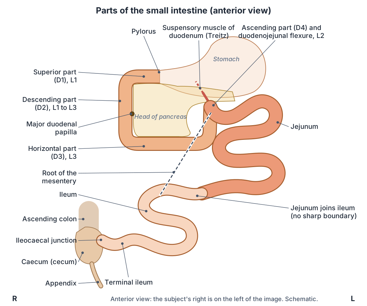
The four parts of the duodenum
| Part | Vertebral level | Course | Key relations |
|---|---|---|---|
| Superior (D1) | L1 | From the pylorus to the right, upwards and backwards; about 5 cm | Front: liver, gallbladder. Behind: common bile duct, portal vein, gastroduodenal artery |
| Descending (D2) | L1 to L3 | Down beside the head of the pancreas | Medial: pancreatic head. Behind: right kidney, ureter and adrenal gland. Major and minor duodenal papillae |
| Horizontal or inferior (D3) | L3 | Right to left across the vertebral column | Behind: inferior vena cava, aorta, right psoas. Front: superior mesenteric vessels and the root of the mesentery |
| Ascending (D4) | L3 to L2 | Up, in front of or to the left of the aorta | Ends at the duodenojejunal flexure, held by the suspensory muscle (ligament of Treitz) |
Compiled from TeachMeAnatomy and Radiopaedia.
Duodenojejunal flexure and the ligament of Treitz
At the duodenojejunal flexure, a slip of muscle, the suspensory muscle of the duodenum, attaches to the bowel. When it contracts, it widens the angle of the flexure and helps contents move on into the jejunum. (TeachMeAnatomy: The Small Intestine.)
The suspensory muscle is usually called the ligament of Treitz. In Treitz's 1853 description, an upper part arises from the right crus of the diaphragm and contains skeletal muscle, and a lower part, containing smooth muscle, runs behind the pancreas and splenic vein to the flexure. Its make-up is still debated, and parts of it may be rudimentary or absent. (Kenhub: Ligaments of the gastrointestinal tract.)
It is an important landmark. Bleeding above it is classed as upper GI bleeding and bleeding below it as lower GI bleeding, and its position guides the search for intestinal malrotation in children with unexplained recurrent vomiting. (Kenhub: Ligaments of the gastrointestinal tract.)
Jejunum and ileum
The jejunum and ileum make up the rest of the small intestine. Unlike most of the duodenum they are intraperitoneal, attached to the posterior abdominal wall by the mesentery, a double layer of peritoneum. The jejunum begins at the duodenojejunal flexure, and there is no clear external line where it becomes the ileum. The ileum ends at the ileocaecal junction, where it pushes into the caecum to form the ileocaecal valve. (TeachMeAnatomy: The Small Intestine.)
Jejunum means 'empty' in Latin. It averages about 2.5 m in length and lies in the left upper abdomen, where on a barium follow-through X-ray it has a delicate feathery pattern. It absorbs carbohydrates, amino acids and fatty acids. (Radiopaedia: Jejunum.)
The ileum is 2 to 4 m long. It contains abundant Peyer patches, unencapsulated clusters of lymphoid tissue found only on the antimesenteric border, the side of the bowel opposite the mesentery. (Radiopaedia: Ileum.)
Jejunum vs ileum: how to tell them apart
The jejunum is wider (under 3 cm across, against under 2 cm for the ileum) and has a thicker, more vascular wall with more numerous, deeper and thicker circular folds (2 to 3 mm thick, against 1 to 2 mm). Its mesentery has less fat, longer vasa recta and fewer arterial arcades. Peyer patches occur in both but are far more prominent in the ileum. (Radiopaedia: Jejunum vs ileum.)
At operation the colour helps too: the jejunum is red and the ileum pink. TeachMeAnatomy also contrasts the thick jejunal wall with the thin ileal wall, long straight vasa recta with short ones, and few arcades with many. (TeachMeAnatomy: The Small Intestine.)
One caution: OpenStax describes the ileum as 'thicker, more vascular' and more folded than the jejunum. That is the reverse of the radiology and anatomy references above, and OpenStax itself says the circular folds end near the middle of the ileum. The standard description, used by Radiopaedia, TeachMeAnatomy and Kenhub, is that the jejunum is the thicker, more folded part. (OpenStax Anatomy and Physiology 2e, 23.5: The Small and Large Intestines.)
Jejunum vs ileum
| Feature | Jejunum | Ileum |
|---|---|---|
| Share of the jejunoileum | Proximal two-fifths | Distal three-fifths |
| Usual position | Upper left abdomen | Lower right abdomen and pelvis |
| Diameter | Wider (under 3 cm) | Narrower (under 2 cm) |
| Wall | Thicker, more vascular | Thinner, less vascular |
| Colour in life | Red | Pink, lighter |
| Circular folds | Larger and more numerous, 2 to 3 mm thick | Smaller and fewer, 1 to 2 mm thick; end near the middle of the ileum |
| Fat in the mesentery | Less | More |
| Vasa recta | Long | Short |
| Arterial arcades | Few (often a single row) | Many (several rows) |
| Peyer patches | Few | Many, on the antimesenteric border |
| Main absorption | Most carbohydrates, amino acids and fats | Vitamin B12 and bile salts (terminal ileum) |
Compiled from Radiopaedia, TeachMeAnatomy, Kenhub and OpenStax. The change from jejunum to ileum is gradual, with no sharp boundary.
The mesentery of the small intestine
The mesentery is a fan-shaped double fold of peritoneum that suspends about 6 to 8 m of jejunum and ileum from a root only about 15 cm long. The root runs obliquely from the duodenojejunal flexure, to the left of the L2 vertebra, down to the right lower quadrant in front of the upper part of the right sacroiliac joint, so the intestinal edge of the mesentery is gathered into many pleats. (Radiopaedia: Small bowel mesentery.)
From left to right, the root crosses the horizontal (third) part of the duodenum, where the superior mesenteric vessels enter it, then the aorta, inferior vena cava, right ureter and right psoas muscle. Between its two layers run the jejunal and ileal branches of the superior mesenteric artery with their veins, nerves and lymphatics. (Radiopaedia: Small bowel mesentery.)
Wall of the small intestine: circular folds, villi and microvilli
Three features increase the absorptive surface of the small intestine more than 600-fold: circular folds, villi and microvilli. Circular folds (plicae circulares) are deep ridges of mucosa and submucosa that run from near the start of the duodenum to about the middle of the ileum. They make chyme spiral, slowing it so there is more time for absorption. (OpenStax Anatomy and Physiology 2e, 23.5: The Small and Large Intestines.)
Villi are finger-like projections of the mucosa, 0.5 to 1 mm long, with about 20 to 40 in each square millimetre. Each contains a capillary bed supplied by one arteriole and drained by one venule, and a lymphatic capillary called a lacteal, which takes up the products of fat digestion. The absorptive cells on the villi carry microvilli about 1 µm long, around 200 million per square millimetre, which form the brush border; enzymes on the brush border finish digesting carbohydrates and proteins. (OpenStax Anatomy and Physiology 2e, 23.5: The Small and Large Intestines.)
Kenhub's histology guide describes the lining as hills and valleys. The hills are the villi, each with a core of connective tissue, vessels, lymphatics and smooth muscle. The valleys are the intestinal glands (crypts of Lieberkühn), which contain absorptive enterocytes, mucus-secreting goblet cells, hormone-secreting enteroendocrine cells, Paneth cells at the base and stem cells that replace worn-out cells. The circular folds are permanent and do not flatten when the bowel is stretched. (Kenhub: Histology of the lower digestive tract.)
Paneth cells secrete lysozyme, an enzyme that kills bacteria. The crypts produce 0.95 to 1.9 litres of slightly alkaline intestinal juice a day. Duodenal (Brunner) glands, found only in the submucosa of the duodenum, secrete an alkaline mucus that helps neutralise stomach acid, and Peyer patches, aggregations of lymphoid tissue, are concentrated in the distal ileum. (OpenStax Anatomy and Physiology 2e, 23.5: The Small and Large Intestines.)
Under the microscope the three parts can be told apart: Brunner glands mark the duodenum, Peyer patches covered by M cells mark the distal ileum, and the villi become less prominent towards the end of the small intestine. (Kenhub: Histology of the lower digestive tract.)
The wall has the four layers found throughout the gut: mucosa, submucosa, muscularis (inner circular and outer longitudinal smooth muscle) and serosa. The submucosal (Meissner) plexus lies in the submucosa and the myenteric (Auerbach) plexus in the muscularis. (OpenStax Anatomy and Physiology 2e, 23.1: Overview of the Digestive System.)
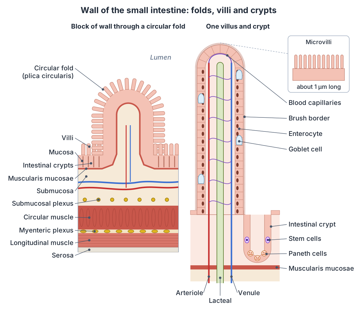
Small intestine surface area
OpenStax gives the absorptive surface of the small intestine as about 200 m², more than 100 times the area of the skin. (OpenStax Anatomy and Physiology 2e, 23.5: The Small and Large Intestines.)
A 2014 re-measurement by Helander and Fändriks, using quantitative microscopy, found much less: about 30 m² for the small intestine and about 32 m² for the whole digestive tract, with the large intestine adding only 1.9 m². In their calculation, based on a mean diameter of 2.5 cm, villi enlarge the surface of the small intestine about 6.5 times and microvilli about 13 times. (BioNumbers 111126: Average surface area of parts of the gastrointestinal tract (Helander and Fändriks 2014).)
What does the small intestine do?
Almost all ingested food, about 80% of electrolytes and 90% of water are absorbed in the small intestine. About 9 litres of fluid enter it each day, and about 90% of this water is absorbed there. (OpenStax Anatomy and Physiology 2e, 23.7: Chemical Digestion and Absorption: A Closer Look.)
The parts specialise. The duodenum mixes chyme with bile and pancreatic enzymes. Carbohydrates are absorbed mainly in the duodenum, and proteins and most fats in the jejunum. The ileum absorbs vitamin B12, bile salts and anything not absorbed earlier, and all three parts absorb water and electrolytes. (Kenhub: Small intestine.)
Iron and calcium are absorbed in the duodenum. Vitamin B12, bound to intrinsic factor secreted by the stomach, and bile salts are absorbed in the terminal ileum. Fats travel as chylomicrons into the lacteals rather than into the blood capillaries. (OpenStax Anatomy and Physiology 2e, 23.7: Chemical Digestion and Absorption: A Closer Look.)
Two kinds of movement handle chyme. Segmentation, local contractions of the circular muscle, mixes chyme with digestive juices and presses it against the lining; it happens about 12 times a minute in the duodenum and about 8 times a minute in the ileum. Once most nutrients have been absorbed, migrating motility complexes, triggered by the hormone motilin, sweep the remains along over 90 to 120 minutes. All the chyme leaves the small intestine in about 3 to 5 hours. (OpenStax Anatomy and Physiology 2e, 23.5: The Small and Large Intestines.)
The whole GI tract from mouth to anus, with the accessory organs. Digestive system anatomy.
Blood supply, lymph drainage and nerves
The duodenum lies on the border between two arterial territories. Above the major duodenal papilla it is supplied by the gastroduodenal artery, a branch of the common hepatic artery from the coeliac trunk; below the papilla, by the inferior pancreaticoduodenal artery from the superior mesenteric artery (SMA). This junction marks the embryonic change from foregut to midgut. (TeachMeAnatomy: The Small Intestine.)
In more detail, the first 2.5 cm (the duodenal cap) is supplied by the right gastric and right gastroepiploic arteries, the rest of D1 down to the middle of D2 by the superior pancreaticoduodenal artery, and the duodenum from mid-D2 to the ligament of Treitz by the inferior pancreaticoduodenal artery. (Radiopaedia: Duodenum.)
The jejunum and ileum are supplied by the SMA, which leaves the aorta at L1 just below the coeliac trunk. It gives about 20 branches, which join into loops called arcades; straight arteries, the vasa recta, run from the arcades to the bowel wall. (TeachMeAnatomy: The Small Intestine.)
The SMA supplies the bowel from the middle of the second part of the duodenum to the terminal ileum; its jejunal and ileal branches arise from the left side of its trunk, and their final branches in the wall are end arteries. Small bowel veins drain to the superior mesenteric vein and then the portal vein, and lymph passes to the superior mesenteric group of pre-aortic nodes. (Radiopaedia: Small bowel.)
The terminal ileum is supplied by the ileal branch of the ileocolic artery, another branch of the SMA. Nerves reach the jejunum and ileum through the superior mesenteric plexus: sympathetic fibres from spinal segments T9 and T10, and parasympathetic fibres from the vagus nerves. (Radiopaedia: Ileum.)
The foregut artery whose gastroduodenal branch supplies the upper duodenum. Celiac trunk anatomy.
Small intestine vs large intestine
The large intestine is about half as long as the small intestine but about three times as wide (about 7.6 cm against 2.5 cm). It has no circular folds or villi and few enzyme-secreting cells; its glands secrete mucus but no digestive enzymes. Its outer longitudinal muscle forms three bands, the teniae coli, which pucker the wall into pouches called haustra. (OpenStax Anatomy and Physiology 2e, 23.5: The Small and Large Intestines.)
In Helander and Fändriks' calculation, the small intestine averaged 2.5 cm across and 291 cm long, and the large intestine 4.8 cm across and 190 cm long. (BioNumbers 111127: Average diameter and length of small and large intestine (Helander and Fändriks 2014).)
Caecum, appendix, colon, rectum and anal canal. Large intestine anatomy.
Small intestine vs large intestine
| Feature | Small intestine | Large intestine |
|---|---|---|
| Parts | Duodenum, jejunum, ileum | Caecum and appendix, colon, rectum, anal canal |
| Length | About 3 m in life (2.9 m in Helander and Fändriks) | About half the small intestine (1.9 m in Helander and Fändriks) |
| Diameter | About 2.5 cm | About 4.8 to 7.6 cm |
| Lining | Circular folds, villi and microvilli | No circular folds or villi |
| Outer muscle | Continuous longitudinal layer | Three teniae coli that pucker the wall into haustra |
| Secretions | Intestinal juice; brush-border enzymes | Mucus, but no digestive enzymes |
| Main job | Completes digestion; absorbs most nutrients and water | Absorbs remaining water and salts; forms faeces |
| Time contents stay | About 3 to 5 hours | Usually 12 to 24 hours |
Compiled from OpenStax §23.5 and Helander and Fändriks (2014) via BioNumbers.
Development, Meckel diverticulum and duodenal variants
The midgut, which forms the jejunum and ileum, herniates into the umbilical cord during weeks 6 to 10 of gestation, then rotates 270 degrees as it returns to its final position in the abdomen. (Radiopaedia: Jejunum.)
A Meckel diverticulum is the commonest structural congenital anomaly of the gut, found in about 2% of people. It is a true diverticulum, with all layers of the bowel wall, left over from the vitelline (omphalomesenteric) duct. It arises from the antimesenteric border of the distal ileum, usually within 60 cm of the ileocaecal valve, and averages about 3 cm long. Bleeding is its commonest complication, followed by small bowel obstruction, and it is about twice as common in males. (Radiopaedia: Meckel diverticulum.)
Duodenal diverticula are most often found in the second or third part of the duodenum, and duplication cysts usually on the medial wall of D2 or D3. (Radiopaedia: Duodenum.)
Small intestine conditions linked to its anatomy
Duodenal ulcers occur most often in the first part of the duodenum and are mainly caused by Helicobacter pylori infection and long-term use of NSAIDs. A perforated ulcer can cause peritonitis or erode the gastroduodenal artery, causing heavy bleeding and hypovolaemic shock. (TeachMeAnatomy: The Small Intestine.)
Coeliac (celiac) disease, an immune reaction to gluten that affects about 1% of people, mainly involves the proximal small bowel, starting in the duodenum. The villi are blunted and can be lost completely while the crypts enlarge, which shrinks the absorptive surface. Duodenal biopsy is the gold-standard test, and treatment is a strict lifelong gluten-free diet. (Radiopaedia: Coeliac disease.)
The ileocaecal valve normally stops colonic contents flowing back into the ileum. If it stays competent when the large bowel is blocked, the colon becomes a closed loop that can perforate; if it is incompetent, pressure escapes into the small bowel and the obstruction progresses more slowly. (TeachMeAnatomy: The Small Intestine.)
Small intestine in the 3D model
- Distal part of ileum
- The distal part of the ileum is a segment of the lower small intestine.
- Distal part of jejunum
- The distal part of the jejunum is a segment of the middle small intestine.
- Duodenum
- The duodenum is the first and shortest part of the small intestine, about 20 to 25 cm long and C-shaped.
- Ileocecal junction
- The ileocecal junction is where the terminal ileum opens into the cecum of the large intestine.
- Mesentery of small intestine
- The mesentery is a broad double fold of peritoneum that suspends the jejunum and ileum from the posterior abdominal wall.
- Middle part of ileum
- The middle part of the ileum is a segment of the lower small intestine.
- Middle part of jejunum
- The middle part of the jejunum is a segment of the middle small intestine.
- Proximal part of ileum
- The proximal part of the ileum is a segment of the lower small intestine.
- Proximal part of jejunum
- The proximal part of the jejunum is a segment of the middle small intestine.
- Superior mesenteric artery
- An unpaired artery of the midgut.
- Superior mesenteric vein
- The vein draining the small intestine and right colon.
Frequently asked questions
How long is the small intestine?
In a living adult the small intestine is about 3 m (10 ft) long and about 2.5 cm wide. Measured after death, when its muscle has relaxed, it is roughly twice as long, which is why many books give 6 to 7 m. The duodenum is about 25 cm; the jejunum makes up about two-fifths and the ileum three-fifths of the rest.
What are the three parts of the small intestine?
The duodenum, jejunum and ileum. The duodenum is a short C-shaped loop around the head of the pancreas that receives bile and pancreatic juice. The jejunum, mainly in the upper left abdomen, absorbs most nutrients. The ileum, mainly in the lower right abdomen, absorbs vitamin B12 and bile salts and ends at the ileocaecal valve.
What is the difference between the jejunum and the ileum?
The jejunum is wider, with a thicker, redder, more vascular wall, more prominent circular folds, long vasa recta and little fat in its mesentery. The ileum is narrower and paler, with more mesenteric fat, short vasa recta, several rows of arterial arcades and many Peyer patches. The change is gradual, with no sharp boundary.
What are the four parts of the duodenum?
Superior (D1, at L1), descending (D2, L1 to L3), horizontal or inferior (D3, at L3) and ascending (D4, rising to about L2). D1 is the commonest site of duodenal ulcers, D2 receives bile and pancreatic juice at the major papilla, D3 crosses the aorta and inferior vena cava, and D4 ends at the duodenojejunal flexure.
What are villi and what do they do?
Villi are finger-like projections of the small intestine's lining, 0.5 to 1 mm long. Each contains blood capillaries and a lymphatic lacteal. Together with the circular folds and the microvilli on their cells, they greatly enlarge the surface for absorption: sugars and amino acids enter the capillaries, and the products of fat digestion enter the lacteals.
Which part of the small intestine absorbs vitamin B12?
The terminal ileum. Vitamin B12 binds to intrinsic factor, a protein made by parietal cells in the stomach, and the complex is absorbed at the end of the ileum, where bile salts are also reabsorbed. Iron and calcium, by contrast, are absorbed in the duodenum.
Is the small intestine longer than the large intestine?
Yes. The small intestine is about twice as long as the large intestine, roughly 3 m against 1.5 to 1.9 m in life, but much narrower: about 2.5 cm wide against 4.8 to 7.6 cm. The words small and large describe the width of the tube, not its length.
References
- OpenStax. Anatomy and Physiology 2e, §23.1 Overview of the Digestive System. Rice University, 2022.
- OpenStax. Anatomy and Physiology 2e, §23.5 The Small and Large Intestines. Rice University, 2022.
- OpenStax. Anatomy and Physiology 2e, §23.7 Chemical Digestion and Absorption: A Closer Look. Rice University, 2022.
- Jones O. The Small Intestine. TeachMeAnatomy.
- Small intestine. Kenhub anatomy library.
- Duodenum. Kenhub anatomy library.
- Histology of the lower digestive tract. Kenhub anatomy library.
- Ligaments of the gastrointestinal tract. Kenhub anatomy library.
- Small bowel. Radiopaedia reference article.
- Duodenum. Radiopaedia reference article.
- Jejunum. Radiopaedia reference article.
- Ileum. Radiopaedia reference article.
- Knipe H. Jejunum vs ileum. Radiopaedia reference article.
- Small bowel mesentery. Radiopaedia reference article.
- Meckel diverticulum. Radiopaedia reference article.
- Coeliac disease. Radiopaedia reference article.
- Small Intestine. Cleveland Clinic health library.
- Helander HF, Fändriks L. Surface area of the digestive tract, revisited. Scand J Gastroenterol. 2014;49(6):681-9. Via BioNumbers (Harvard Medical School), entry 111126.
- Helander HF, Fändriks L. Surface area of the digestive tract, revisited. Scand J Gastroenterol. 2014;49(6):681-9. Via BioNumbers (Harvard Medical School), entry 111127.
- Mitsuhashi N. et al. (2009). BodyParts3D: 3D structure database for anatomical concepts. Nucleic Acids Research 37, D782–D785. Data © The Database Center for Life Science, CC BY 4.0.
Related anatomy
- Digestive systemGI tract anatomy: each digestive system organ from mouth to anus in order, the accessory organs, wall layers and blood…
- Large intestineColon anatomy with labelled diagrams and a 3D model: caecum and appendix, each part of the colon, rectum, anal canal…
- DuodenumDuodenum: The duodenum is the first and shortest part of the small intestine, about 20 to 25 cm long and C-shaped. See…
- PancreasPancreas anatomy: head, uncinate process, neck, body and tail, ducts, acini and islets, blood supply and variants…
- StomachStomach anatomy with labelled diagrams and a 3D model: cardia, fundus, body, antrum and pylorus, curvatures, rugae…
- Celiac trunkCeliac trunk (celiac axis) anatomy: origin at T12, the left gastric, splenic and common hepatic arteries, their…
Explore the whole body in the interactive 3D human anatomy model, or browse the A–Z list of human structures.