

What is the large intestine? Parts and location
The large intestine is the last part of the digestive tract. It begins where the ileum of the small intestine opens into it at the ileocaecal valve, frames the small intestine on three sides and ends at the anus. Its four main regions are the caecum, the colon, the rectum and the anal canal. (OpenStax Anatomy and Physiology 2e, 23.5: The Small and Large Intestines.)
In order, residue passes through the caecum with the appendix attached, the ascending colon, the right colic flexure, the transverse colon, the left colic flexure, the descending colon, the sigmoid colon and the rectum. (Radiopaedia: Large intestine.)
The ascending colon runs up the right side of the abdomen to the underside of the liver, where it turns at the right colic (hepatic) flexure. The transverse colon crosses to the left and turns down at the left colic (splenic) flexure, just below the spleen. The descending colon runs down the left side of the posterior abdominal wall, and the S-shaped sigmoid colon curves towards the midline, where the rectum follows the front of the sacrum and coccyx. (OpenStax Anatomy and Physiology 2e, 23.5: The Small and Large Intestines.)
The 3D model above highlights the caecum, appendix, each part of the colon, the rectum, the three taeniae coli, the external anal sphincter and the colic arteries. Rotate it to see the ascending and descending colon lying flat against the back wall while the transverse and sigmoid colon hang forwards. BodyParts3D models the colon as one piece; for this site the caecum was split off below the ileocaecal junction, and the sigmoid colon where the colon crosses the left external iliac artery at the pelvic brim. There is no anal canal mesh, so the diagrams show it.
The whole GI tract from mouth to anus, with the accessory organs. Digestive system anatomy.
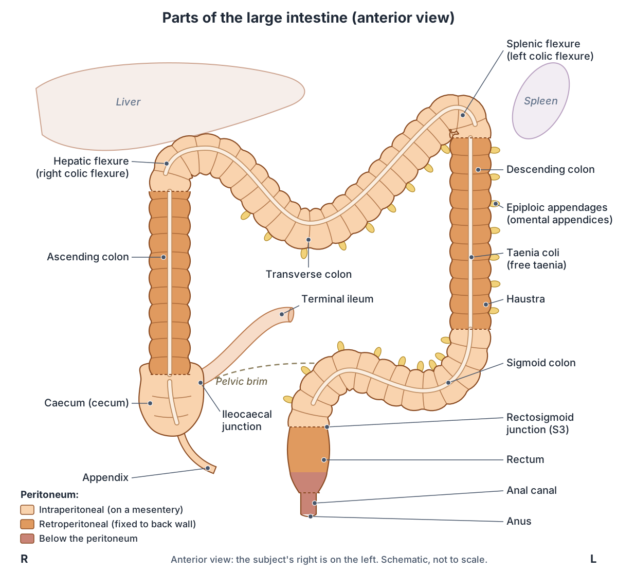
Parts of the large intestine at a glance
| Part | Where it lies | Peritoneum | Main arteries |
|---|---|---|---|
| Caecum (cecum) and appendix | Right iliac fossa, below the ileocaecal valve | Intraperitoneal; caecum mobile in about 15%; appendix on the mesoappendix | Anterior and posterior caecal and appendicular branches of the ileocolic artery (SMA) |
| Ascending colon | Right side, from the caecum to the hepatic flexure under the liver | Secondarily retroperitoneal (about 25% of people have a mesentery) | Ileocolic and right colic arteries (SMA) |
| Transverse colon | Across the upper abdomen, from the hepatic to the splenic flexure | Intraperitoneal, on the transverse mesocolon; the least fixed part | Middle colic artery (SMA); left colic artery (IMA) for the distal third |
| Descending colon | Left side, from the splenic flexure to the left iliac fossa | Secondarily retroperitoneal; front and sides covered | Left colic and sigmoid arteries (IMA) |
| Sigmoid colon | Left iliac fossa into the pelvis, to about S3 | Intraperitoneal, on the inverted-V sigmoid mesocolon | Two to four sigmoid arteries (IMA) |
| Rectum | In front of the sacrum and coccyx, from S3 to the anorectal junction | Upper third covered in front and at the sides, middle third in front only, lower third not at all | Superior rectal (IMA), middle rectal (internal iliac) and inferior rectal (internal pudendal) arteries |
| Anal canal and anus | Anal triangle of the perineum, between the ischioanal fossae | Below the peritoneum (extraperitoneal) | Superior rectal artery above the pectinate line, inferior rectal artery below |
SMA: superior mesenteric artery. IMA: inferior mesenteric artery. Compiled from TeachMeAnatomy, Radiopaedia, Kenhub and OpenStax.
Colon vs large intestine vs bowel: what the terms mean
The colon is one part of the large intestine, not all of it. 'Colon' is often used as a synonym for the large intestine (large bowel), but strictly it excludes the rectum, so the two words are not interchangeable. (Radiopaedia: Large intestine.)
Some sources also count the caecum as part of the colon. The US National Cancer Institute's SEER training module divides the colon into five segments, the caecum and the ascending, transverse, descending and sigmoid colon, and treats the rectum as a separate organ that stores stool. (NCI SEER Training Modules: Anatomy of Colon and Rectum.)
'Large colon' is not a separate part of the human gut. In horses it is a named, folded segment: the sacculated right ventral colon turns at the sternal flexure into the left ventral colon, which narrows and folds back at the pelvic flexure, the commonest site of impaction, into the dorsal colon, which has no sacculations. (Merck Veterinary Manual: Overview of Colic in Horses.)
The equine caecum, large colon and pelvic flexure in 3D. Horse anatomy.
How long is the large intestine and colon?
The colon of an adult is about 1.5 m (5 ft) long. TeachMeAnatomy gives an average of 150 cm for the colon, which arches around the small intestine in four parts. (TeachMeAnatomy: The Colon.)
The best summary of real measurements is a 2024 systematic review and meta-analysis of 31 studies covering 5,741 adults. Pooled across studies, the adult colon measured 148.3 cm on average (standard deviation 27.1 cm). The mean lengths of its parts were 22.8 cm for the ascending colon, 46.6 cm for the transverse colon, 25.8 cm for the descending colon, 41.4 cm for the sigmoid colon and 16.8 cm for the rectum. The transverse colon was the longest part and the sigmoid colon the most variable. (Alqarni F et al. Experimental Measurements of the Length of the Human Colon: A Systematic Review and Meta-Analysis. Diagnostics 2024.)
The same review found that several studies measured longer colons in men, by 4 to 18%, while others found no difference or the reverse, and that people with constipation have been reported to have longer colons. (Alqarni F et al. Experimental Measurements of the Length of the Human Colon: A Systematic Review and Meta-Analysis. Diagnostics 2024.)
Figures for the whole large intestine are larger because they include the caecum, rectum and anal canal. Cleveland Clinic gives about 1.8 m (6 ft) in length and about 8 cm (3 in) in diameter. (Cleveland Clinic: Large Intestine.)
OpenStax describes the large intestine as about half as long as the small intestine but about 7.6 cm (3 in) wide, more than twice as wide. In other words, it is called large because it is wider, not because it is longer. (OpenStax Anatomy and Physiology 2e, 23.5: The Small and Large Intestines.)
How long is each part of the large intestine? Sources compared
| Part | Meta-analysis, mean (range of study means) | Radiopaedia | Cleveland Clinic | Other sources |
|---|---|---|---|---|
| Caecum | Not pooled | 6 cm (abnormal if wider than 9 cm) | About 8 cm (3 in) | OpenStax: about 6 cm |
| Ascending colon | 22.8 cm (16.2 to 34.0) | 15 cm | About 20 cm (8 in) | |
| Transverse colon | 46.6 cm (27.8 to 66.3) | Up to 45 cm | More than 46 cm (18 in) | |
| Descending colon | 25.8 cm (16.7 to 42.9) | Up to 25 cm | About 15 cm (6 in) | |
| Sigmoid colon | 41.4 cm (15.0 to 80.9) | About 15 cm | 35 to 40 cm (14 to 16 in) | TeachMeAnatomy: about 40 cm |
| Rectum | 16.8 cm (10.7 to 23.9) | About 15 cm | 12 to 15 cm (5 to 6 in) | Kenhub: 12 to 16 cm; OpenStax: 20.3 cm |
| Anal canal | Not pooled | About 4 cm | About 5 cm (2 in, as 'anus') | OpenStax: 3.8 to 5 cm; Kenhub: 3 to 4 cm; TeachMeAnatomy: about 4 cm |
| Colon or whole large intestine | Colon 148.3 cm (93.5 to 210.8) | Large intestine about 1.5 m | Large intestine about 1.8 m (6 ft) | TeachMeAnatomy and SEER: colon about 150 cm |
Meta-analysis: Alqarni et al. 2024, 31 studies of 5,741 adults; its ranges are the lowest and highest study averages, not individual people. Lengths depend on the method (dissection, surgery, colonoscopy, CT or MRI) and differ widely between people; the sigmoid colon varies most.
Caecum (cecum) and appendix anatomy
The caecum (cecum) is the first part of the large intestine: a blind-ended pouch about 6 cm long in the right lower quadrant, below the ileocaecal valve, which marks its upper border. Above the valve it continues as the ascending colon. A caecum wider than 9 cm is considered abnormally dilated. (Radiopaedia: Caecum.)
Its name comes from the Latin caecus, blind, after its closed lower end. It lies in the right iliac fossa and, unlike the ascending colon, is intraperitoneal with a mesentery of variable size. It acts as a reservoir for chyme arriving from the ileum. The ileocaecal valve stops large-bowel contents flowing back into the ileum, but it is thought to work passively rather than as a true muscular sphincter. (TeachMeAnatomy: The Cecum.)
Radiopaedia describes the caecum as covered by peritoneum except at the back. In about 15% of people the mesentery of the right colon fails to fuse with the lateral peritoneum, leaving a mobile caecum. Behind the caecum lie iliacus and psoas; below it is the lateral third of the inguinal ligament. (Radiopaedia: Caecum.)
The vermiform appendix is a narrow, blind-ended tube attached to the posteromedial end of the caecum and suspended from the terminal ileum by a fold of peritoneum, the mesoappendix. It contains a lot of lymphoid tissue but has no known vital function. (TeachMeAnatomy: The Appendix.)
It arises about 2 to 3 cm below the ileocaecal valve, where the three taeniae coli converge, and its length is highly variable, from 2 to 20 cm. Its tip most often lies behind the caecum (retrocaecal, 65 to 70%) or hangs over the pelvic brim (pelvic, 25 to 30%); pre-ileal and post-ileal positions account for about 5%. (Radiopaedia: Appendix.)
OpenStax gives a typical length of 7.6 cm (3 in). It notes that the appendix is generally considered vestigial, although one report suggests it may act as a reservoir of gut bacteria after a diarrhoeal illness. (OpenStax Anatomy and Physiology 2e, 23.5: The Small and Large Intestines.)
Position also varies between populations. In 48 Kenyan cadavers the appendix averaged 7.65 cm (range 3.5 to 14.5 cm), and only 27% were retrocaecal and 25% pelvic. The authors contrast earlier studies finding 67% retrocaecal in Ghana and 68% in India, but a mainly pelvic position (43.6%) in Zambia. (Mwachaka P et al. Variations in the Position and Length of the Vermiform Appendix in a Black Kenyan Population. ISRN Anatomy 2014.)
The vermiform appendix highlighted on its own in the 3D model. Appendix.
Colon anatomy: ascending, transverse, descending and sigmoid colon
The colon has four parts that form an arch around the small intestine: the ascending colon on the right, the transverse colon across the upper abdomen, the descending colon on the left and the S-shaped sigmoid colon in the left lower quadrant and pelvis. (TeachMeAnatomy: The Colon.)
The ascending colon rises from the caecum to the right lobe of the liver, where it turns about 90 degrees at the right colic (hepatic) flexure. In front of it are the small intestine, greater omentum and anterior abdominal wall; behind it are iliacus, quadratus lumborum, the right kidney and the iliohypogastric and ilioinguinal nerves. (TeachMeAnatomy: The Colon.)
Radiopaedia gives the ascending colon a length of 15 cm and notes that, although it is secondarily retroperitoneal, about 25% of people have a mesentery on it. (Radiopaedia: Ascending colon.)
The transverse colon runs from the hepatic flexure to the spleen. It is the least fixed part of the colon: it hangs on the transverse mesocolon, and in tall, thin people it can dip into the pelvis. Behind it lie the duodenum, the head of the pancreas and loops of jejunum and ileum. (TeachMeAnatomy: The Colon.)
Radiopaedia gives it a length of up to 45 cm. It is almost completely covered by peritoneum, its mesocolon attaches to the inferior border of the pancreas, and the gastrocolic ligament joins it to the stomach. (Radiopaedia: Transverse colon.)
The descending colon runs down from the left colic flexure towards the pelvis. It is retroperitoneal in most people and lies in front of the left kidney, crossing its lateral border, before turning medially to become the sigmoid colon. (TeachMeAnatomy: The Colon.)
Radiopaedia gives the descending colon a length of up to 25 cm, with peritoneum on its front and sides; its lower part crosses the external iliac and gonadal vessels. (Radiopaedia: Descending colon.)
The sigmoid colon begins where the descending colon curves medially into the pelvis and gains a mesentery, the inverted V-shaped sigmoid mesocolon, and becomes the rectum at about S3, where it loses it. Behind it lie the left internal iliac vessels, left ureter, piriformis and sacrum; in front is the bladder (and in females the uterus). Radiopaedia gives about 15 cm but stresses that its length and course vary; its apex can reach the umbilicus. (Radiopaedia: Sigmoid colon.)
TeachMeAnatomy instead gives about 40 cm, from the left iliac fossa to the level of S3, and notes that the sigmoid mesocolon makes it particularly mobile. (TeachMeAnatomy: The Colon.)
The sigmoid colon highlighted on its own in the 3D model. Sigmoid colon.
Hepatic and splenic flexures (right and left colic flexures)
The colic flexures are the two bends where the transverse colon meets the ascending and descending colon. The right colic (hepatic) flexure lies at the right lobe of the liver. The left colic (splenic) flexure lies by the spleen, where the phrenicocolic ligament attaches the colon to the diaphragm. (TeachMeAnatomy: The Colon.)
The splenic flexure is higher, sharper and less mobile than the hepatic flexure, and it normally lies in front of the lower pole of the left kidney. (Nalamolu VKS, Chatterjee A. Splenodiaphragmatic interposition of the splenic flexure of colon. J Postgrad Med 2022.)
The splenic flexure is also where the territories of the superior and inferior mesenteric arteries meet. The arterial links there are often weak or absent, so it is a watershed area prone to ischaemia and infarction (see the blood supply sections below). (Radiopaedia: Marginal artery of Drummond.)
Taeniae coli, haustra and epiploic appendages
Three surface features set the colon apart from the small intestine. Its outer longitudinal muscle is gathered into three bands of smooth muscle, the taeniae coli; their tone puckers the wall into pouches called haustra; and small fat-filled sacs of peritoneum, the epiploic appendages, hang from the bands. The rectum and anal canal have neither taeniae nor haustra. (OpenStax Anatomy and Physiology 2e, 23.5: The Small and Large Intestines.)
The three bands are the free taenia (taenia libera), the mesocolic taenia (taenia mesocolica) and the omental taenia (taenia omentalis). On the ascending and descending colon the free taenia is in front, the mesocolic taenia posteromedial and the omental taenia posterolateral. The transverse colon is turned so that they lie below, behind and above. They converge on the caecum at the base of the appendix, and in the sigmoid colon they broaden and fuse into the complete longitudinal muscle layer of the rectum. (Radiopaedia: Taeniae coli.)
Kenhub names each band by what attaches to it: the mesocolon to the mesocolic taenia, the greater omentum to the omental taenia, and nothing to the free taenia, which is fully visible. Inside the colon, crescent-shaped semilunar folds are formed by contraction of the muscle and are movable rather than permanent. (Kenhub: Colon.)
Radiopaedia explains the pouching mechanically: the taeniae are about 30 cm shorter than the bowel wall, so the wall bunches into sacculations, with the haustra as incomplete rings between them. (Radiopaedia: Large intestine.)
There are 50 to 100 epiploic appendages, typically about 1.5 by 3.5 cm, in two rows along the free and omental taeniae (one row on the transverse colon). They run from the caecum, where they may be absent, to the rectosigmoid junction; the rectum has none. Each is fed by one or two small arteries that pierce the serosa, and colonic diverticula are thought to form where these arteries enter. (Radiopaedia: Epiploic appendages.)
The free taenia coli highlighted in the 3D model. Taenia libera.
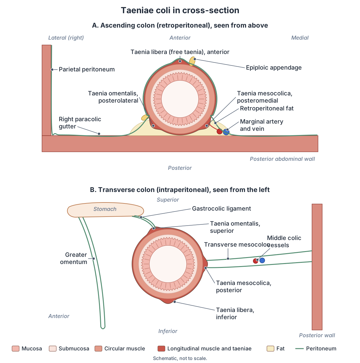
Which parts of the colon are intraperitoneal or retroperitoneal?
The ascending colon, descending colon and rectum are retroperitoneal: they lie wholly or partly behind the peritoneum, against the posterior wall. The transverse and sigmoid colon are held to the posterior abdominal wall by a mesocolon, a fold of peritoneum, which lets them move. (OpenStax Anatomy and Physiology 2e, 23.1: Overview of the Digestive System.)
Kenhub calls the ascending and descending colon secondarily retroperitoneal and the transverse and sigmoid colon intraperitoneal, each with its own mesocolon. (Kenhub: Colon.)
The rectum changes along its length: peritoneum covers the front and sides of its upper third, only the front of its middle third and none of its lower third. In males it then reflects onto the back of the bladder, forming the rectovesical pouch; in females it reflects onto the posterior vagina and cervix, forming the rectouterine pouch (pouch of Douglas). (TeachMeAnatomy: The Rectum.)
Between the ascending or descending colon and the side wall of the abdomen lie the paracolic gutters. Fluid or infected material from an inflamed organ can track along them to other parts of the abdomen. (TeachMeAnatomy: The Colon.)
Quadrants, regions and the organs that lie in each. Abdomen anatomy.
Rectum anatomy
The rectum is the last part of the large intestine before the anal canal. It continues from the sigmoid colon at the level of S3, follows the curve of the sacrum and coccyx as the most posterior organ of the pelvis, and ends by passing into the anal canal, which runs through the pelvic floor to the anus. (TeachMeAnatomy: The Rectum.)
Kenhub gives a length of about 12 to 16 cm and describes two bends: the sacral flexure, which follows the hollow of the sacrum, and the perineal (anorectal) flexure, where the puborectal sling of levator ani pulls the bowel forwards at the anorectal junction. (Kenhub: Rectum.)
Radiopaedia gives about 15 cm and places the change into the anal canal at the tip of the coccyx, where the rectum bends around the puborectalis sling. Behind it lie the sacrum, coccyx, median sacral artery and sacral sympathetic chain; below it is levator ani. (Radiopaedia: Rectum.)
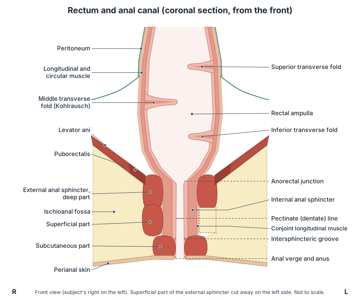
Unlike the colon, the rectum has no taeniae coli, haustra or omental appendices. Three lateral flexures, the superior, intermediate and inferior, are formed by transverse folds of its inner wall, and its lowest part, the ampulla, relaxes to store faeces until defecation. (TeachMeAnatomy: The Rectum.)
The three transverse folds are also called Houston's valves. The middle one, Kohlrausch's fold, is the strongest and lies about 7 cm from the anus; tumours below it may be felt on digital rectal examination. The highly stretchable ampulla lies between this fold and the anorectal junction. (Kenhub: Rectum.)
In males the bladder, prostate and seminal vesicles lie in front of the rectum; in females the vagina and cervix do. On rectal examination the prostate and seminal vesicles, or the cervix, can be felt through the front wall, and the sacrum and coccyx behind. (TeachMeAnatomy: The Rectum.)
The rectum highlighted on its own in the 3D model. Rectum.
Anal canal and anus anatomy
The anal canal is the last segment of the digestive tract, around 4 cm long. It continues from the rectum, runs downwards and backwards through the anal triangle of the perineum between the right and left ischioanal fossae, and ends at the anus. The perineal body lies in front of it and the anococcygeal ligament and coccyx behind. (TeachMeAnatomy: The Anal Canal.)
Lengths vary with the definition used. Radiopaedia distinguishes the anatomical anal canal, from the dentate line to the anal verge, from the surgical anal canal, from the anorectal sling of levator ani to the anal verge, and defines the anal margin as the 5 cm ring of skin around the anal verge. (Radiopaedia: Anal canal.)
Two sphincters close the canal. The internal anal sphincter is a thickening of the involuntary circular smooth muscle of the bowel wall around the upper two-thirds of the canal. The external anal sphincter is voluntary muscle around the lower two-thirds, overlapping the internal sphincter. Where the rectum meets the canal, the two sphincters and puborectalis fuse into the anorectal ring, which can be felt on rectal examination. (TeachMeAnatomy: The Anal Canal.)
The external sphincter is a short tube of skeletal muscle in three ill-defined parts: a deep part, some of whose fibres blend with puboanalis, part of levator ani; a superficial part, attached to the perineal body in front and through the anococcygeal ligament to the coccyx behind; and a subcutaneous part around the anal verge. A conjoint longitudinal muscle lies between the two sphincters. The external sphincter is supplied by the inferior anal (rectal) nerve, a branch of the pudendal nerve (S2 to S4). (Kenhub: External anal sphincter.)
The internal sphincter stays contracted through sympathetic tone and relaxes under parasympathetic influence, while the external sphincter and puborectalis are under voluntary control. (Kenhub: Anal canal.)
When puborectalis contracts, it pulls the anorectal junction forwards, making an angle of about 90 degrees between the rectum and the anal canal, so faeces cannot easily leave the rectum. It must relax for defecation. (Kenhub: Muscles of the pelvic floor.)
Inside, the upper canal has anal columns, long folds of mucosa joined at their lower ends by small folds called anal valves. Above the valves are small pouches, the anal sinuses, which hold mucus-secreting glands. (TeachMeAnatomy: The Anal Canal.)
Cleveland Clinic describes the anus as the last 3 or 4 cm of the large intestine, where the inner lining changes from mucosa to skin. The rectum just above it stores stool before a bowel movement, and the internal sphincter relaxes automatically as the rectum fills. (Cleveland Clinic: Anus.)
Levator ani, puborectalis, the anal triangle and the ischioanal fossa. Pelvic floor and perineum anatomy.
The pectinate (dentate) line
The pectinate (dentate) line is the irregular ring formed by the anal valves inside the anal canal. It divides the canal into an upper part, derived from the embryonic hindgut, and a lower part, derived from the ectoderm of the proctodeum, so the two parts differ in lining, blood supply, lymph drainage and nerves. (TeachMeAnatomy: The Anal Canal.)
Kenhub places the line at the junction of the upper two-thirds and lower third of the canal. Above it the lining is simple columnar epithelium; a transitional zone mixes columnar and stratified squamous cells; about 1 cm of anoderm below the line has non-keratinised stratified squamous epithelium; and the lowest zone is keratinised skin. (Kenhub: Anal canal.)
The pale, smooth lining just below the line is called the anal pecten. At the intersphincteric groove it changes into true skin, keratinised stratified squamous epithelium. (TeachMeAnatomy: The Anal Canal.)
Above the line the canal is supplied by the superior rectal artery and drained by the superior rectal vein to the inferior mesenteric vein and portal system. Below it, the inferior rectal artery supplies it, and the inferior and middle rectal veins drain through the internal pudendal vein to the internal iliac vein (systemic). Lymph from above the line drains to internal iliac nodes, and from below to superficial inguinal nodes. (Radiopaedia: Anal canal.)
The nerves explain why the two parts feel different. Above the line the mucosa is supplied by visceral nerves and is relatively insensitive; below it, somatic nerves make it very sensitive. (OpenStax Anatomy and Physiology 2e, 23.5: The Small and Large Intestines.)
Sources differ in details. Kenhub sends lymph from above the line to the lumbar (para-aortic) nodes rather than the internal iliac nodes, and lists the middle rectal artery with the inferior rectal artery as the supply below the line. It also calls the vascular cushions above the line the internal haemorrhoidal plexus and those below it the external plexus. (Kenhub: Anal canal.)
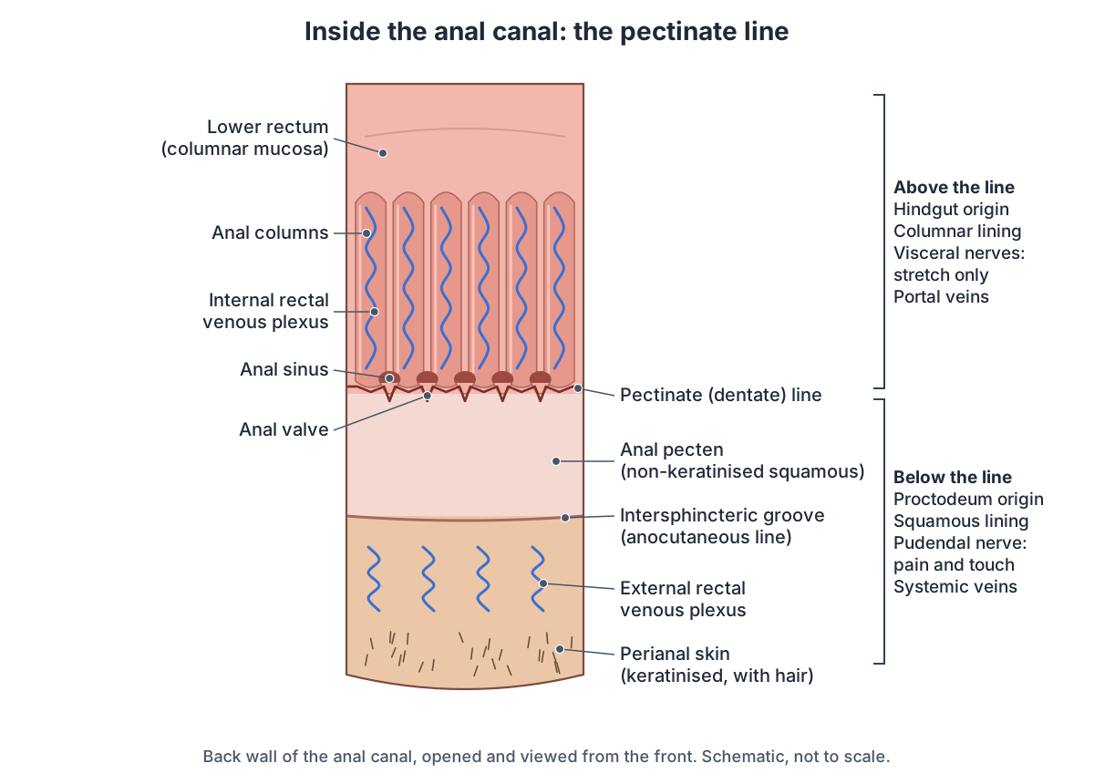
Above vs below the pectinate line
| Feature | Above the pectinate line | Below the pectinate line |
|---|---|---|
| Share of the anal canal | Upper two-thirds | Lower third |
| Embryonic origin | Hindgut (endoderm of the cloaca) | Proctodeum (ectoderm) |
| Lining | Columnar epithelium, as in the rectum | Non-keratinised stratified squamous (anal pecten), then skin below the intersphincteric groove |
| Arteries | Superior rectal artery (from the IMA) | Inferior rectal artery (from the internal pudendal) |
| Veins | Superior rectal vein to the inferior mesenteric vein (portal system) | Inferior and middle rectal veins to the internal pudendal and internal iliac veins (systemic) |
| Lymph nodes | Internal iliac nodes (Kenhub: lumbar nodes) | Superficial inguinal nodes |
| Nerves | Visceral (autonomic) nerves | Somatic: inferior rectal branches of the pudendal nerve |
| Sensation | Stretch; relatively insensitive | Pain, temperature, touch and pressure |
| Haemorrhoids that arise here | Internal haemorrhoids (bleed, prolapse) | External haemorrhoids (thrombose, painful) |
Compiled from TeachMeAnatomy, Radiopaedia, Kenhub, OpenStax and Cleveland Clinic. The middle rectal artery adds to the supply on both sides of the line.
Large intestine histology: the wall and its lining
The colon has no circular folds and no villi, and few enzyme-secreting cells. Outside the anal canal it is lined by simple columnar epithelium made mostly of absorptive enterocytes and goblet cells, and it has far more intestinal glands than the small intestine. The goblet cells secrete mucus that eases the movement of faeces and protects the wall from acids and gases made by bacteria. The anal canal is lined by stratified squamous epithelium. (OpenStax Anatomy and Physiology 2e, 23.5: The Small and Large Intestines.)
Its mucosa is pitted by crypts of Lieberkühn containing goblet cells and hormone-secreting enteroendocrine cells, and the lamina propria holds macrophages and plasma cells. The inner circular muscle layer is strongly developed, while the outer longitudinal muscle is found mainly in the taeniae. (Kenhub: Colon.)
Blood supply of the colon: SMA and IMA territories
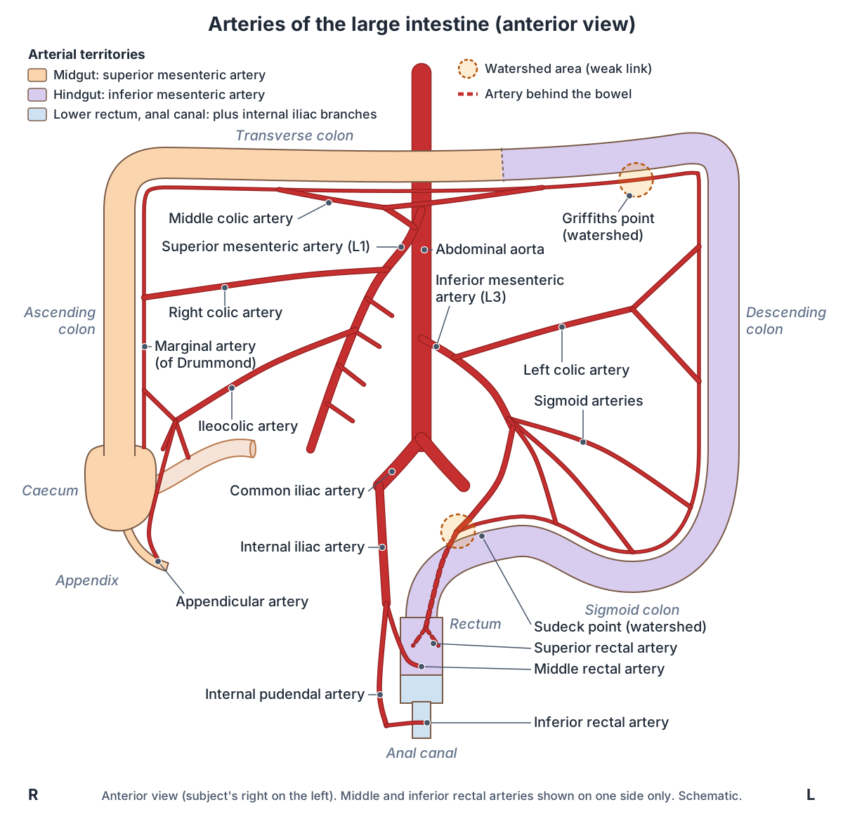
The arteries of the colon follow its development. The midgut part, the ascending colon and the proximal two-thirds of the transverse colon, is supplied by the superior mesenteric artery (SMA). The hindgut part, the distal third of the transverse colon, the descending colon and the sigmoid colon, is supplied by the inferior mesenteric artery (IMA). (TeachMeAnatomy: The Colon.)
From the SMA, the ileocolic artery (with colic, anterior caecal and posterior caecal branches) and the right colic artery supply the ascending colon, and the middle colic artery supplies the transverse colon. From the IMA, the left colic artery supplies the distal transverse and descending colon, and the sigmoid arteries supply the sigmoid colon. (TeachMeAnatomy: The Colon.)
The SMA leaves the front of the aorta at L1, usually just below the coeliac trunk, crosses in front of the third part of the duodenum and runs down to the right in the root of the mesentery. Its colic branches (ileocolic, right colic and middle colic) come from its right side. (Radiopaedia: Superior mesenteric artery.)
The IMA, the smallest of the three unpaired gut arteries, leaves the aorta at L3, at the lower edge of the third part of the duodenum. It runs down to the left, gives the left colic artery and two to four sigmoid arteries, crosses the left common iliac artery and continues as the superior rectal artery. Its territory runs from the splenic flexure to the upper two-thirds of the rectum. (Radiopaedia: Inferior mesenteric artery.)
The appendicular artery, from the ileocolic artery or its posterior caecal branch, is an end artery. The appendix has no collateral circulation, which is why an inflamed appendix can quickly become necrotic or rupture. (Radiopaedia: Appendix.)
The rectum has three arteries: the superior rectal artery, the end of the IMA; the middle rectal artery, from the internal iliac artery; and the inferior rectal artery, from the internal pudendal artery. (TeachMeAnatomy: The Rectum.)
The abdominal aorta and the origins of the mesenteric arteries. Aorta anatomy.
Marginal artery and watershed areas (Griffiths and Sudeck points)
The marginal artery (of Drummond) is a continuous arterial arcade along the inner border of the colon. It is formed by links between the terminal branches of the SMA (ileocolic, right colic and middle colic arteries) and the IMA (left colic and sigmoid arteries). Straight vessels, the vasa recta, run from it into the colon wall, and it can carry blood around a blocked or narrowed main artery. (Radiopaedia: Marginal artery of Drummond.)
Griffiths point is the weak link near the splenic flexure, between the ascending branch of the left colic artery and the marginal artery. In one angiographic series the connection there was present in 48% of people, poor in 9% and absent in 43%. The splenic flexure watershed is the commonest site of ischaemic colitis. (Radiopaedia: Griffiths point.)
Sudeck point is a second watershed at the rectosigmoid junction, where the last sigmoid branch of the IMA usually joins a branch of the superior rectal artery through a small, often single, connection. It is less vulnerable than Griffiths point because the superior, middle and inferior rectal arteries form a submucosal plexus in the rectal wall. (Radiopaedia: Sudeck point.)
In ischaemic colitis the left colon is affected in about 75% of cases, about a quarter of them involving the splenic flexure, and isolated right-sided ischaemia makes up about 10%. The rectum is usually spared because the middle and inferior rectal arteries give it a collateral supply. The marginal artery is absent at the splenic flexure in up to 5% of people and underdeveloped in the right colon in up to 50%. (Hung A et al. Ischaemic colitis: practical challenges and evidence-based recommendations for management. Frontline Gastroenterology 2021.)
Some people also have a more central SMA to IMA link, usually called the arc of Riolan, classically joining the middle colic and left colic arteries near the root of the mesentery. It is inconstant, and there is no agreement on exactly which vessel the name means. (Radiopaedia: Arc of Riolan.)
Veins and lymph drainage of the large intestine
The veins follow the arteries. The ileocolic, right colic and middle colic veins drain into the superior mesenteric vein, and the left colic and sigmoid veins into the inferior mesenteric vein. Both reach the hepatic portal vein, so substances absorbed from the colon pass through the liver first. Lymph from the ascending and transverse colon drains to the superior mesenteric nodes and from the descending and sigmoid colon to the inferior mesenteric nodes, then through the intestinal lymph trunks to the cisterna chyli and thoracic duct. (TeachMeAnatomy: The Colon.)
The inferior mesenteric vein runs up to the left of the inferior mesenteric artery and usually drains into the splenic vein; draining into the superior mesenteric vein or their junction is not uncommon. (Radiopaedia: Inferior mesenteric vein.)
In the rectum the portal and systemic veins meet. The superior rectal vein drains to the portal system and the middle and inferior rectal veins to systemic veins. In portal hypertension these portosystemic connections can dilate into anorectal varices, which may cause painless bleeding and are different from haemorrhoids. Pararectal nodes drain to the inferior mesenteric nodes, while lymph from the lower rectum drains directly to the internal iliac nodes. (TeachMeAnatomy: The Rectum.)
The caecum drains through the ileocolic vein to the superior mesenteric vein, and its lymph passes to the ileocolic nodes around the ileocolic artery. (TeachMeAnatomy: The Cecum.)
How the mesenteric veins carry blood from the gut to the liver. Portal vein.
Veins and lymph drainage of each part
| Part | Veins drain to | Lymph nodes |
|---|---|---|
| Caecum and appendix | Ileocolic vein (appendix through the appendicular vein), then superior mesenteric vein | Ileocolic nodes (appendix also to nodes in the mesoappendix), then superior mesenteric nodes |
| Ascending colon | Ileocolic and right colic veins, then superior mesenteric vein | Paracolic nodes, then superior mesenteric nodes |
| Transverse colon | Middle colic vein, then superior mesenteric vein | Superior mesenteric nodes |
| Descending colon | Left colic vein, then inferior mesenteric vein | Inferior mesenteric nodes |
| Sigmoid colon | Sigmoid veins, then inferior mesenteric vein | Inferior mesenteric nodes |
| Rectum | Superior rectal vein to the inferior mesenteric vein (portal); middle and inferior rectal veins to the internal iliac vein (systemic) | Upper third: inferior mesenteric nodes; lower two-thirds: internal iliac nodes |
| Anal canal | Above the pectinate line: portal; below: systemic | Above: internal iliac nodes; below: superficial inguinal nodes |
The superior and inferior mesenteric veins both end in the hepatic portal vein. Compiled from TeachMeAnatomy, Radiopaedia and Kenhub.
Nerve supply of the colon, rectum and anal canal
The midgut part of the colon receives sympathetic, parasympathetic and sensory fibres through the superior mesenteric plexus. The hindgut part receives its nerves through the inferior mesenteric plexus, with parasympathetic fibres from the pelvic splanchnic nerves and sympathetic fibres from the lumbar splanchnic nerves. (TeachMeAnatomy: The Colon.)
Kenhub places the changeover, the Cannon-Böhm point, at the left colic flexure. Before it the parasympathetic supply comes from the vagus nerve and the sympathetic supply from the superior mesenteric plexus; after it they come from the pelvic splanchnic nerves and the inferior mesenteric plexus. (Kenhub: Colon.)
Sympathetic sensory fibres from the appendix enter the spinal cord at T10, which is why the early pain of appendicitis is felt in the centre of the abdomen, around the umbilicus. (TeachMeAnatomy: The Appendix.)
The rectum receives sympathetic fibres through the lumbar splanchnic nerves and the superior and inferior hypogastric plexuses, and parasympathetic fibres from S2 to S4 through the pelvic splanchnic nerves. Its visceral sensory fibres follow the parasympathetic route. (TeachMeAnatomy: The Rectum.)
In the anal canal, the internal sphincter and the lining above the dentate line receive sympathetic fibres from the pelvic plexus and parasympathetic and sensory fibres from the pelvic splanchnic nerves. The external sphincter and the lining below the line are supplied by the inferior rectal branches of the pudendal nerve. (Radiopaedia: Anal canal.)
Sympathetic and parasympathetic pathways, including the splanchnic nerves. Autonomic nervous system.
What does the large intestine do?
The large intestine absorbs most of the water left after the small intestine has taken about 90% of it, along with salts and some vitamins, and turns liquid residue into semisolid faeces. Of every 500 mL of residue that enters the caecum each day, about 150 mL leaves as faeces. Residue usually stays in the large intestine for 12 to 24 hours. (OpenStax Anatomy and Physiology 2e, 23.5: The Small and Large Intestines.)
It secretes mucus but no digestive enzymes, so any digestion there is done by bacteria. Trillions of bacteria from more than 700 species ferment what is left, releasing hydrogen, carbon dioxide and methane (up to 1,500 mL of gas a day), and some make vitamins: biotin, pantothenic acid and vitamin K. (OpenStax Anatomy and Physiology 2e, 23.5: The Small and Large Intestines.)
Its movements are slow. Haustral contractions, mainly in the transverse and descending colon, occur about every 30 minutes and last about a minute. Three or four times a day, usually during or just after a meal, mass movements that start in the middle of the transverse colon push contents towards the rectum. Stretching of the stomach also increases colonic movement through the gastrocolic reflex. (OpenStax Anatomy and Physiology 2e, 23.5: The Small and Large Intestines.)
Kenhub gives about 1 litre of water absorbed by the colon each day, along with sodium, potassium and chloride; the colon can also secrete potassium. Its mostly anaerobic bacteria reach about 10¹¹ (100 billion) per gram. (Kenhub: Colon.)
When faeces stretch the rectal ampulla, stretch receptors signal the urge to defecate, and rising pressure relaxes the internal sphincter (the rectoanal inhibitory reflex). Defecation can be put off by tightening levator ani and the external sphincter, or started by relaxing them, helped by pressure from the diaphragm and abdominal muscles. (Kenhub: Rectum.)
The defecation reflex is a parasympathetic reflex through the spinal cord: it contracts the sigmoid colon and rectum, relaxes the internal sphincter and at first contracts the external sphincter. Putting off defecation lets more water be absorbed, which can lead to constipation. (OpenStax Anatomy and Physiology 2e, 23.5: The Small and Large Intestines.)
Large intestine vs small intestine (anatomy of the bowel)
Together the small and large intestines form the bowel. The small intestine is about 3 m long in life and about 2.5 cm wide; the large intestine is about half as long but more than twice as wide. The large intestine has no circular folds or villi and secretes no digestive enzymes, and its longitudinal muscle forms the taeniae coli instead of a continuous layer. (OpenStax Anatomy and Physiology 2e, 23.5: The Small and Large Intestines.)
Without villi, the large intestine adds little absorptive surface. Helander and Fändriks estimated about 1.9 m² for the large intestine against about 30 m² for the small intestine, assuming a large intestine 190 cm long and 4.8 cm wide whose microvilli enlarge its surface about 6.5 times. (BioNumbers 111126: Average surface area of parts of the gastrointestinal tract (Helander and Fändriks 2014).)
Duodenum, jejunum and ileum, villi and the mesentery. Small intestine anatomy.
Large intestine vs small intestine
| Feature | Small intestine | Large intestine |
|---|---|---|
| Parts | Duodenum, jejunum, ileum | Caecum and appendix, colon (ascending, transverse, descending, sigmoid), rectum, anal canal |
| Length in life | About 3 m (2.9 m in Helander and Fändriks) | About half as long: colon about 1.5 m (1.9 m for the whole large intestine in Helander and Fändriks) |
| Width | About 2.5 cm | About 4.8 to 7.6 cm |
| Inner surface | Circular folds, villi and microvilli | No circular folds or villi; semilunar folds between the haustra |
| Outer longitudinal muscle | Continuous layer | Three taeniae coli that pucker the wall into haustra |
| Fat tags on the surface | None | Epiploic appendages |
| Digestive enzymes | Secreted, and present on the brush border | None; mucus only, digestion by bacteria |
| Main job | Completes digestion; absorbs most nutrients and about 90% of water | Absorbs most of the remaining water and salts; bacterial fermentation; forms faeces |
| Time contents stay | About 3 to 5 hours | Usually 12 to 24 hours |
| Absorptive surface | About 30 m² | About 1.9 m² |
Compiled from OpenStax §23.5 and Helander and Fändriks (2014) via BioNumbers.
Colon and anal conditions linked to the anatomy
Colonic diverticulosis means many small pouches (diverticula) in the colon wall. Almost all are false diverticula: mucosa herniates through a gap in the muscle where nutrient arteries enter, so they are commoner on the mesenteric side. They are most common in the sigmoid colon; about 15% of patients have right-sided diverticula, more often in people from Asia. About 5% of people have them at 40, 30% at 60 and 50 to 80% by 80. Most cause no symptoms, but they can become inflamed (diverticulitis) or bleed. (Radiopaedia: Colonic diverticulosis.)
In sigmoid volvulus the sigmoid colon twists on its mesentery. About 60% of intestinal volvulus involves the sigmoid colon. It is commoner in older people and linked to chronic constipation; endoscopic untwisting succeeds in about 80% of patients, but the twist can cut off the blood supply. (Radiopaedia: Sigmoid volvulus.)
Caecal volvulus makes up about 10% of intestinal volvulus, the sigmoid colon being the commonest site. It causes colicky pain, distension and absolute constipation, and an X-ray shows a distended loop of large bowel arising from the right lower quadrant. (TeachMeAnatomy: The Cecum.)
Acute appendicitis has a lifetime incidence of about 7% and peaks around the age of 20, when the appendix holds most lymphoid tissue. It usually starts with blockage of the lumen by swollen lymphoid tissue (about 60%, mainly in the young) or an appendicolith (about a third). Pain is first referred around the umbilicus (T10) and moves to McBurney point within a day or two; 10 to 20% perforate. (Radiopaedia: Acute appendicitis.)
McBurney point lies one-third of the way from the right anterior superior iliac spine to the umbilicus; tenderness is greatest there in established appendicitis. The pain settles in the right lower quadrant once the inflamed appendix irritates the parietal peritoneum. (TeachMeAnatomy: The Appendix.)
Haemorrhoids (hemorrhoids) are normal cushions of vascular submucosal tissue, present in the anal canal from birth, that cause symptoms when they swell. Internal haemorrhoids lie above the dentate line and tend to bleed or prolapse; external haemorrhoids lie below it and tend to thrombose and hurt. Prevalence is about 20% (range 4 to 40%), most often between 45 and 65 years. (Radiopaedia: Haemorrhoids.)
The cushions help maintain continence. Constipation, prolonged straining and raised abdominal pressure (for example in pregnancy) are linked to enlarged haemorrhoids, which in the lithotomy position are classically found at 3, 7 and 11 o'clock. They are not the same as anorectal varices. (TeachMeAnatomy: The Anal Canal.)
An anal fissure is a tear in the lining of the anal canal. More than half of drained anal abscesses leave an anal fistula, a tunnel-like passage from an anal gland to the skin. (Cleveland Clinic: Anus.)
Venous drainage also shapes how rectal cancer spreads: Kenhub notes that cancers of the upper rectum tend to spread to the liver and those of the lower rectum to the lungs, matching the portal and systemic venous routes. (Kenhub: Rectum.)
Large intestine in the 3D model
- Appendicular artery
- The appendicular artery is the artery of the vermiform appendix.
- Appendix
- The vermiform appendix is a narrow blind-ended tube arising from the cecum.
- Ascending colon
- The ascending colon is the part of the large intestine rising from the cecum to the right colic (hepatic) flexure.
- Cecum
- The caecum is the first part of the large intestine, a blind-ending pouch in the right lower abdomen below the point where the small intestine joins the colon.
- Colic artery
- The colic arteries are the branches of the mesenteric arteries that supply the colon.
- Descending colon
- The descending colon passes down from the left colic (splenic) flexure to the sigmoid colon.
- External anal sphincter
- A ring of voluntary skeletal muscle surrounding the anal canal.
- Ileocecal junction
- The ileocecal junction is where the terminal ileum opens into the cecum of the large intestine.
- Ileocolic artery
- The ileocolic artery is the lowest branch from the right side of the superior mesenteric artery.
- Inferior mesenteric artery
- The inferior mesenteric artery supplies the hindgut.
- Marginal artery of colon
- A continuous arterial arcade along the mesenteric border of the colon linking the superior and inferior mesenteric arteries.
- Mesoappendix
- The mesoappendix is a triangular peritoneal fold that attaches the appendix to the mesentery of the terminal ileum.
- Middle colic artery
- A branch of the superior mesenteric artery supplying the transverse colon.
- Rectum
- The rectum is the final straight portion of the large intestine before the anal canal.
- Sigmoid artery
- Branches of the inferior mesenteric artery to the sigmoid colon.
- Sigmoid colon
- The sigmoid colon is the S-shaped last part of the colon. It runs from the end of the descending colon at the pelvic brim to the rectum.
- Superior mesenteric artery
- An unpaired artery of the midgut.
- Superior rectal artery
- The continuation of the inferior mesenteric artery to the rectum.
- Taenia libera
- The taenia libera is one of the three longitudinal bands of smooth muscle on the colon (taeniae coli).
- Taenia mesocolica
- The taenia mesocolica is one of the three taeniae coli, the longitudinal muscle bands of the colon.
- Taenia omentalis
- The taenia omentalis is one of the three taeniae coli, the longitudinal muscle bands of the colon.
- Transverse colon
- The transverse colon is the part of the large intestine that crosses the abdomen between the right and left colic flexures.
- Transverse mesocolon
- The transverse mesocolon is a peritoneal fold attaching the transverse colon to the posterior abdominal wall.
Frequently asked questions
What are the parts of the large intestine?
The large intestine has four main regions: the caecum (cecum) with the appendix, the colon, the rectum and the anal canal. The colon has four parts, the ascending, transverse, descending and sigmoid colon, joined by the hepatic (right colic) and splenic (left colic) flexures. Some sources, such as the SEER training module, count the caecum as a fifth part of the colon.
How long is the colon?
About 1.5 m (5 ft) in an adult. A 2024 meta-analysis of 31 studies found a mean of 148.3 cm, with the transverse colon the longest part (about 47 cm) and the sigmoid colon the most variable. Figures for the whole large intestine, including the caecum, rectum and anal canal, range from about 1.5 to 1.8 m.
What is the difference between the colon and the large intestine?
The colon is the longest part of the large intestine, not all of it. The large intestine also includes the caecum and appendix at its start and the rectum and anal canal at its end. People often say colon when they mean the whole large bowel, but strictly the colon excludes the rectum.
What is the difference between the rectum and the anus?
The rectum is the last part of the large intestine, about 12 to 16 cm long, lying in front of the sacrum and storing faeces until defecation. The anal canal, about 4 cm long, continues from it through the pelvic floor, and the anus is its opening to the outside. The anal canal is closed by the internal and external anal sphincters.
Where is the appendix, and is it part of the large intestine?
Yes. The appendix is a narrow, blind-ended tube attached to the caecum, the first part of the large intestine, in the right iliac fossa. Its base lies about 2 to 3 cm below the ileocaecal valve, where the three taeniae coli meet. Its tip most often lies behind the caecum, but its position and length (2 to 20 cm) vary widely.
Which parts of the colon are retroperitoneal?
The ascending and descending colon are secondarily retroperitoneal, fixed to the back wall of the abdomen, and so is the rectum. The transverse and sigmoid colon are intraperitoneal: each hangs on its own mesocolon and can move. The caecum is usually intraperitoneal, and the lower third of the rectum and the anal canal lie below the peritoneum.
What is the pectinate (dentate) line?
It is the wavy line formed by the anal valves inside the anal canal, at the junction of its upper two-thirds and lower third. Above it the lining is columnar, supplied by visceral nerves and drained to the portal veins. Below it the lining is squamous, supplied by the pudendal nerve, so it is sensitive to pain, and drained to systemic veins.
Why is the splenic flexure prone to ischaemia?
Because it lies where the territories of the superior and inferior mesenteric arteries meet. The marginal artery that links them is often thin or interrupted there, at Griffiths point, so this part of the colon has the weakest backup supply when blood flow falls. The splenic flexure is the commonest site of ischaemic colitis.
References
- OpenStax. Anatomy and Physiology 2e, §23.1 Overview of the Digestive System. Rice University, 2022.
- OpenStax. Anatomy and Physiology 2e, §23.5 The Small and Large Intestines. Rice University, 2022.
- Aubrey-Jones D. The Colon. TeachMeAnatomy.
- Aubrey-Jones D. The Cecum. TeachMeAnatomy.
- Jones O. The Appendix. TeachMeAnatomy.
- Theakston V. The Rectum. TeachMeAnatomy.
- Mahmood H. The Anal Canal. TeachMeAnatomy.
- Karunaharamoorthy A. Colon: anatomy, histology, composition, function. Kenhub.
- Karunaharamoorthy A. Rectum: anatomy, histology, function. Kenhub.
- Karunaharamoorthy A. Anal canal: anatomy, histology and function. Kenhub.
- Pirie E. External anal sphincter: origin, insertion, action. Kenhub.
- Muscles of the pelvic floor. Kenhub anatomy library.
- Gaillard F, Bell D, Hacking C, et al. Large intestine. Radiopaedia reference article.
- Caecum. Radiopaedia reference article.
- Appendix. Radiopaedia reference article.
- Ascending colon. Radiopaedia reference article.
- Transverse colon. Radiopaedia reference article.
- Descending colon. Radiopaedia reference article.
- Sigmoid colon. Radiopaedia reference article.
- Rectum. Radiopaedia reference article.
- Anal canal. Radiopaedia reference article.
- Taeniae coli. Radiopaedia reference article.
- Epiploic appendages. Radiopaedia reference article.
- Superior mesenteric artery. Radiopaedia reference article.
- Inferior mesenteric artery. Radiopaedia reference article.
- Inferior mesenteric vein. Radiopaedia reference article.
- Marginal artery of Drummond. Radiopaedia reference article.
- Griffiths point. Radiopaedia reference article.
- Sudeck point. Radiopaedia reference article.
- Arc of Riolan. Radiopaedia reference article.
- Colonic diverticulosis. Radiopaedia reference article.
- Sigmoid volvulus. Radiopaedia reference article.
- Acute appendicitis. Radiopaedia reference article.
- Haemorrhoids. Radiopaedia reference article.
- Large Intestine. Cleveland Clinic health library.
- Anus. Cleveland Clinic health library.
- National Cancer Institute. Anatomy of Colon and Rectum. SEER Training Modules.
- Alqarni F, Akbar T, Fatani H, et al. Experimental Measurements of the Length of the Human Colon: A Systematic Review and Meta-Analysis. Diagnostics (Basel). 2024;14(19):2190.
- Hung A, Calderbank T, Samaan MA, Plumb AA, Webster G. Ischaemic colitis: practical challenges and evidence-based recommendations for management. Frontline Gastroenterol. 2021;12(1):44-52.
- Mwachaka P, El-busaidy H, Sinkeet S, Ogeng'o J. Variations in the Position and Length of the Vermiform Appendix in a Black Kenyan Population. ISRN Anat. 2014;2014:871048.
- Nalamolu VKS, Chatterjee A. Splenodiaphragmatic interposition of the splenic flexure of colon. J Postgrad Med. 2022;68(2):122-123.
- Helander HF, Fändriks L. Surface area of the digestive tract, revisited. Scand J Gastroenterol. 2014;49(6):681-9. Via BioNumbers (Harvard Medical School), entry 111127.
- Helander HF, Fändriks L. Surface area of the digestive tract, revisited. Scand J Gastroenterol. 2014;49(6):681-9. Via BioNumbers (Harvard Medical School), entry 111126.
- Moore JN. Overview of Colic in Horses. Merck Veterinary Manual.
- Mitsuhashi N. et al. (2009). BodyParts3D: 3D structure database for anatomical concepts. Nucleic Acids Research 37, D782–D785. Data © The Database Center for Life Science, CC BY 4.0.
Related anatomy
- Small intestineSmall intestine anatomy: duodenum parts, jejunum vs ileum, villi and circular folds, length and blood supply, with…
- Digestive systemGI tract anatomy: each digestive system organ from mouth to anus in order, the accessory organs, wall layers and blood…
- Pelvic floor and perineumPerineum and pelvic floor anatomy with labelled diagrams and a 3D model: levator ani, coccygeus, perineal body, both…
- AbdomenAbdominal anatomy with labelled quadrant and nine-region diagrams and a 3D model: organs on each side, abdominal wall…
- Portal veinHepatic portal vein explained: how it forms, its tributaries, the porta hepatis, portal vs hepatic veins…
- Sigmoid colonSigmoid colon: The sigmoid colon is the S-shaped last part of the colon. It runs from the end of the descending colon…
Explore the whole body in the interactive 3D human anatomy model, or browse the A–Z list of human structures.