

What is an organ?
An organ is an anatomically distinct structure of the body made of two or more tissue types. A tissue is a group of many similar cells working together for a specific function, and an organ system is a group of organs that work together to perform major functions. (OpenStax Anatomy and Physiology 2e, 1.2: Structural Organization of the Human Body.)
The body is organised in six levels of increasing complexity: chemical, cellular, tissue, organ, organ system and the whole organism. (OpenStax Anatomy and Physiology 2e, 1.2: Structural Organization of the Human Body.)
Sorting organs into systems is not exact, because most organs contribute to more than one system. OpenStax lists the pancreas in the endocrine system as well as in digestion, and the testes and ovaries in both the endocrine and the reproductive systems. (OpenStax Anatomy and Physiology 2e, 1.2: Structural Organization of the Human Body.)
What counts as an organ is still argued about, which is why lists of organs differ. In 2016 a review argued that the mesentery has distinctive anatomical and functional features that justify calling it an organ. (Coffey JC, O'Leary DP. The mesentery: structure, function, and role in disease. Lancet Gastroenterol Hepatol. 2016.)
The mesentery is the band of tissue in front of the lumbar vertebrae that anchors the small intestine, apart from the duodenum. (OpenStax Anatomy and Physiology 2e, 23.1: Overview of the Digestive System.)
The four kinds of tissue that organs are built from are explained in Tissue types.
Organ systems and their organs
OpenStax describes eleven organ systems: integumentary (skin, hair, nails), skeletal (bones, joints, cartilage), muscular (skeletal muscles, tendons), nervous (brain, spinal cord, nerves), endocrine (pituitary, thyroid, pancreas, adrenal glands, testes, ovaries), cardiovascular (heart, blood vessels), lymphatic (thymus, spleen, lymph vessels and nodes), respiratory (nasal passages, trachea, lungs), digestive (stomach, liver, gallbladder, intestines), urinary (kidneys, bladder) and reproductive (testes, epididymis, mammary glands, ovaries, uterus). (OpenStax Anatomy and Physiology 2e, 1.2: Structural Organization of the Human Body.)
Labelled whole-body diagrams of each organ system, the skeleton and the muscles are collected in Human body diagrams.
Organ systems of the human body
| Organ system | Main organs | Guide |
|---|---|---|
| Integumentary | Skin, hair, nails | Layers of the skin |
| Skeletal | Bones, joints, cartilage | Skeletal system |
| Muscular | Skeletal muscles, tendons | Muscular system |
| Nervous | Brain, spinal cord, peripheral nerves | Brain anatomy; spinal cord |
| Endocrine | Pituitary, thyroid, pancreas, adrenal glands, testes, ovaries | Thyroid gland; adrenal gland |
| Cardiovascular | Heart, blood vessels | Circulatory system; heart |
| Lymphatic | Thymus, spleen, lymph vessels, lymph nodes | Lymphatic system |
| Respiratory | Nasal passages, trachea, lungs | Respiratory system; lungs |
| Digestive | Stomach, liver, gallbladder, small and large intestines | Digestive system |
| Urinary | Kidneys, urinary bladder | Urinary system |
| Reproductive | Testes, epididymis, mammary glands, ovaries, uterus | Male and female reproductive systems |
Organ lists from OpenStax Anatomy and Physiology 2e, section 1.2. Many organs belong to more than one system.
Where are the organs? The body cavities
Most organs sit in body cavities. The posterior (dorsal) cavity holds the nervous system: the cranial cavity houses the brain and the spinal (vertebral) cavity encloses the spinal cord, and the two are continuous. (OpenStax Anatomy and Physiology 2e, 1.6: Anatomical Terminology.)
The anterior (ventral) cavity is divided by the diaphragm. Above it, the thoracic cavity, enclosed by the rib cage, holds the lungs in the pleural cavities and the heart in the pericardial cavity within the mediastinum. Below it, the abdominopelvic cavity is the largest body cavity: its upper abdominal part houses the digestive organs and its lower pelvic part the reproductive organs, with no membrane between them. (OpenStax Anatomy and Physiology 2e, 1.6: Anatomical Terminology.)
The organs of the anterior cavity are covered by serous membranes. Each has two layers, a parietal layer lining the cavity wall and a visceral layer covering the organ, with a thin film of fluid between them that reduces friction as the organs move. These membranes are the pleura around the lungs, the pericardium around the heart and the peritoneum around the abdominopelvic organs. (OpenStax Anatomy and Physiology 2e, 1.6: Anatomical Terminology.)
Some abdominal organs lie behind the peritoneum, against the back wall: among the digestive organs these retroperitoneal organs are the duodenum, pancreas, ascending and descending colon and rectum. (OpenStax Anatomy and Physiology 2e, 23.1: Overview of the Digestive System.)
Regional terms, body cavities and surface landmarks are explained in the guide to Body regions.
Major internal organs and where they are
| Organ | Body cavity | Where exactly | Size or key fact |
|---|---|---|---|
| Brain | Cranial cavity | Inside the skull | Continuous with the spinal cord |
| Thyroid gland | Neck | Below the larynx, in front of the trachea | Butterfly-shaped, two lobes and an isthmus |
| Heart | Thoracic (mediastinum) | Behind the sternum, two-thirds left of the midline | About the size of a fist |
| Lungs | Thoracic (pleural cavities) | Either side of the heart, on the diaphragm | Right lung 3 lobes, left lung 2 |
| Oesophagus | Thoracic (mediastinum) | Behind the trachea, through the diaphragm to the stomach | About 25 cm long |
| Thymus | Thoracic (mediastinum) | Between the sternum and the aorta, above the heart | Shrinks after puberty |
| Liver | Abdominal | Right upper quadrant, under the diaphragm | Largest gland, about 1.4 kg |
| Gallbladder | Abdominal | Under the right lobe of the liver | 8 to 10 cm long |
| Stomach | Abdominal | Left upper quadrant, below the oesophagus | Holds up to about 4 litres |
| Spleen | Abdominal | Left upper quadrant, under ribs 9 to 11 | About 12 cm long |
| Pancreas | Abdominal (retroperitoneal) | Behind the stomach, from the duodenum to the spleen | Exocrine and endocrine |
| Small intestine | Abdominal | Centre of the abdomen | About 3 m long in life |
| Large intestine | Abdominal and pelvic | Frames the small intestine | About 7.6 cm wide |
| Kidneys | Abdominal (retroperitoneal) | Either side of the spine, T12 to L3 | 11 to 14 cm long |
| Adrenal glands | Abdominal (retroperitoneal) | On top of each kidney | Endocrine glands |
| Urinary bladder | Pelvic | Behind the pubic bone, in front of the rectum | Ureters about 30 cm long |
Compiled from OpenStax Anatomy and Physiology 2e, TeachMeAnatomy and Radiopaedia (see references).
Torso anatomy: what is inside the trunk?
The trunk, also called the torso, is the body without the head and limbs. (Taber's Medical Dictionary: Trunk.)
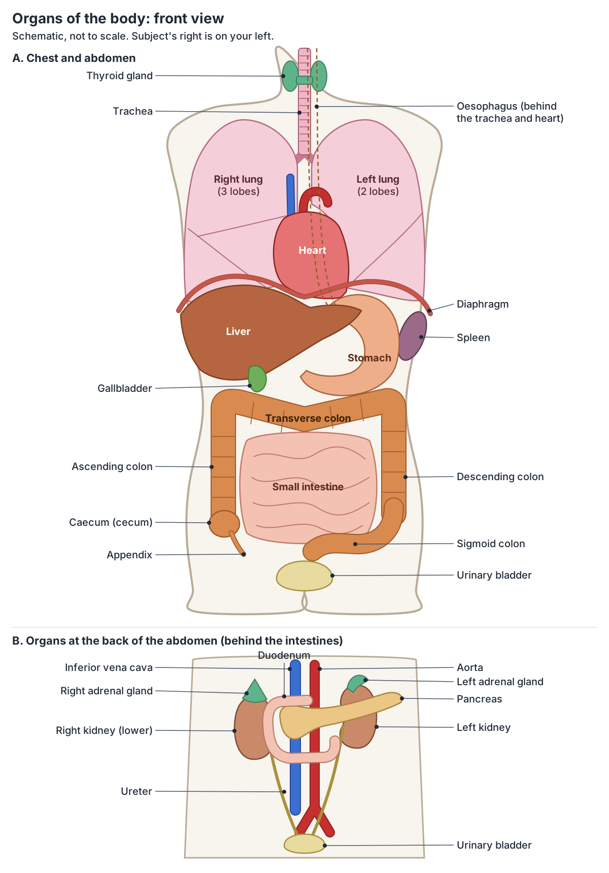
Inside the torso are the two parts of the anterior body cavity: the thoracic cavity, enclosed by the rib cage, and the abdominopelvic cavity below the diaphragm. In OpenStax's summary the lungs and heart fill the thoracic cavity, the digestive organs the abdominal cavity and the reproductive organs the pelvic cavity. (OpenStax Anatomy and Physiology 2e, 1.6: Anatomical Terminology.)
The diagram below shows the torso organs from the front in two layers. Organs near the back, such as the kidneys, adrenal glands, pancreas and duodenum, are hidden behind the stomach and intestines in a real body, so they are drawn in a second panel as if those organs had been lifted away.
The chest wall, breast region, lungs, heart and diaphragm are covered together in Chest anatomy.
Brain: the organ inside the skull
The brain lies in the cranial cavity of the skull. It is continuous with the spinal cord, which runs down the spinal cavity inside the vertebral column. (OpenStax Anatomy and Physiology 2e, 1.6: Anatomical Terminology.)
The brain, spinal cord and peripheral nerves are the organs of the nervous system. (OpenStax Anatomy and Physiology 2e, 1.2: Structural Organization of the Human Body.)
The lobes, brainstem, cerebellum and deep structures of the brain are described in Brain anatomy.
Thyroid gland: the main organ in the front of the neck
The thyroid gland is a butterfly-shaped organ in the front of the neck, just below the larynx and in front of the trachea. A narrow middle part, the isthmus, joins its left and right lobes, and the small parathyroid glands are embedded mainly in the back of the lobes. (OpenStax Anatomy and Physiology 2e, 17.4: The Thyroid Gland.)
Lobes, blood supply, relations and the parathyroid glands are covered in the guide to the Thyroid gland.
Heart
The heart is about the size of a fist and lies in the mediastinum, the space between the lungs, behind the sternum. Its four chambers pump blood to the lungs and to the rest of the body. (OpenStax Anatomy and Physiology 2e, 19.1: Heart Anatomy.)
It is not in the middle: about two-thirds of the heart lies left of the midline and one-third to the right, with its apex pointing down and to the left. (Radiopaedia: Heart.)
Chambers, valves, wall layers, great vessels and the conduction system are covered in Heart anatomy.
Lungs
The lungs are pyramid-shaped, paired organs connected to the trachea by the right and left bronchi. They rest on the diaphragm. The right lung has three lobes and the left has two, and the left lung has a dent on its surface, the cardiac notch, that makes room for the heart. (OpenStax Anatomy and Physiology 2e, 22.2: The Lungs.)
Each lung is wrapped in pleura: the visceral pleura covers the lung and the parietal pleura lines the chest wall, mediastinum and diaphragm, with lubricating pleural fluid in the pleural cavity between them. (OpenStax Anatomy and Physiology 2e, 22.2: The Lungs.)
Lobes, fissures, the hilum and the bronchopulmonary segments are covered in Lung anatomy.
Trachea and bronchi
The trachea (windpipe) divides into the right and left bronchi, which connect it to the two lungs. The oesophagus runs behind it. (OpenStax Anatomy and Physiology 2e, 22.2: The Lungs.)
The whole airway from the nose to the alveoli is described in the guide to the Respiratory system.
Oesophagus (esophagus)
The oesophagus is a muscular tube about 25.4 cm (10 in) long. It runs behind the trachea, straight down through the mediastinum, passes through the diaphragm at the oesophageal hiatus and ends at the stomach. (OpenStax Anatomy and Physiology 2e, 23.3: The Mouth, Pharynx, and Esophagus.)
Its parts, constrictions, sphincters and blood supply are described on the page about the Oesophagus.
Thymus and diaphragm
The thymus lies in the space between the sternum and the aorta, above the heart. It is a lymphatic organ, and it shrinks after puberty, losing about 3 percent of its tissue a year until age 35 to 45 and about 1 percent a year after that. (OpenStax Anatomy and Physiology 2e, 21.1: Anatomy of the Lymphatic and Immune Systems.)
The diaphragm forms the floor of the thoracic cavity and separates it from the abdominopelvic cavity below. (OpenStax Anatomy and Physiology 2e, 1.6: Anatomical Terminology.)
The openings in the diaphragm and how it moves during breathing are explained on the page about the Diaphragm.
Liver
The liver lies under the diaphragm in the right upper quadrant of the abdomen, protected by the ribs. It is the largest gland in the body and weighs about 3 pounds (about 1.4 kg) in an adult. It has a large right lobe and a much smaller left lobe. (OpenStax Anatomy and Physiology 2e, 23.6: Accessory Organs in Digestion: The Liver, Pancreas, and Gallbladder.)
TeachMeAnatomy calls the liver the largest visceral structure in the abdominal cavity. It lies mainly in the right hypochondrium and epigastric regions and reaches across into the left hypochondrium. (TeachMeAnatomy: The Liver.)
Lobes, Couinaud segments, the porta hepatis and the liver's double blood supply are covered in Liver anatomy.
Gallbladder
The gallbladder is a small sac, 8 to 10 cm (about 3 to 4 in) long, nested in a shallow hollow on the posterior aspect of the right lobe of the liver. (OpenStax Anatomy and Physiology 2e, 23.6: Accessory Organs in Digestion: The Liver, Pancreas, and Gallbladder.)
With the liver and pancreas it is an accessory digestive organ: it releases bile into the small intestine. (OpenStax Anatomy and Physiology 2e, 23.1: Overview of the Digestive System.)
Its parts, the bile ducts and gallstones are covered on the page about the Gallbladder.
Stomach
The stomach lies just below the oesophagus. Its rounded top, the fundus, sits under the diaphragm, above and to the left of the cardia where the oesophagus enters. Empty, it is only about the size of a fist, but it can stretch to hold as much as 4 litres. Its four regions are the cardia, fundus, body and pylorus. (OpenStax Anatomy and Physiology 2e, 23.4: The Stomach.)
On the four-quadrant map of the abdomen, the stomach lies in the left upper quadrant, together with the lower left ribs and part of the transverse colon. (OpenStax Anatomy and Physiology 2e, 1.6: Anatomical Terminology.)
Its parts, curvatures, wall layers and blood supply are described in Stomach anatomy.
Spleen
The spleen lies in the upper left quadrant of the abdomen, under the diaphragm and protected by ribs 9 to 11. It is roughly the size of a clenched fist and is almost entirely wrapped in peritoneum. (TeachMeAnatomy: The Spleen.)
It is about 12 cm long, attached to the stomach, and works as a filter of the blood, removing microbes and worn-out red blood cells. (OpenStax Anatomy and Physiology 2e, 21.1: Anatomy of the Lymphatic and Immune Systems.)
Where the spleen is, what it does and splenic injury are covered on the page about the Spleen.
Pancreas
The pancreas lies across the back of the abdomen behind the stomach, in the retroperitoneum. Its head sits in the C-shaped curve of the duodenum and its tail tapers to the hilum of the spleen. It is both exocrine, making digestive enzymes, and endocrine, releasing hormones into the blood. (OpenStax Anatomy and Physiology 2e, 23.6: Accessory Organs in Digestion: The Liver, Pancreas, and Gallbladder.)
Its parts, ducts, islets and relations are covered on the page about the Pancreas.
Small intestine
The small intestine is the longest part of the digestive tract: about 3.05 m (10 ft) long in a living person and about 2.54 cm (1 in) wide. It has three parts: the duodenum, about 25.4 cm long, the jejunum, about 0.9 m, and the ileum, about 1.8 m. After death, when its muscle relaxes, it measures about twice as long. (OpenStax Anatomy and Physiology 2e, 23.5: The Small and Large Intestines.)
The duodenum, jejunum and ileum and how they differ are covered in the guide to the Small intestine.
Large intestine and appendix
The large intestine is about half the length of the small intestine but more than twice as wide, about 7.62 cm (3 in). It frames the small intestine: the caecum (cecum) with the appendix in the lower right, the ascending colon up the right side, the transverse colon across from right to left, the descending colon down the left side, and the S-shaped sigmoid colon into the pelvis, followed by the rectum in front of the sacrum and the anal canal. (OpenStax Anatomy and Physiology 2e, 23.5: The Small and Large Intestines.)
The appendix arises from the back and inner (posteromedial) side of the caecum, and its most common position is behind the caecum (retrocaecal). Appendicitis pain settles in the right lower quadrant, with tenderness at McBurney's point, one-third of the way from the right anterior superior iliac spine to the umbilicus. (TeachMeAnatomy: The Appendix.)
The caecum, colon, rectum and anus are described in the guide to the Large intestine.
Kidneys
The kidneys lie on either side of the spine in the retroperitoneal space, against the back wall of the abdomen. The left kidney sits at about the level of T12 to L3, and the right is slightly lower because the liver pushes it down. Each is about 11 to 14 cm long and weighs about 125 to 175 g in males and 115 to 155 g in females. (OpenStax Anatomy and Physiology 2e, 25.3: Gross Anatomy of the Kidney.)
The inside of the kidney, its hilum, coverings and blood supply are covered on the page about Kidney anatomy.
Adrenal glands
An adrenal (suprarenal) gland sits on the upper end of each kidney. (OpenStax Anatomy and Physiology 2e, 25.3: Gross Anatomy of the Kidney.)
The adrenal glands are endocrine organs, listed by OpenStax with the pituitary, thyroid, pancreas, testes and ovaries. (OpenStax Anatomy and Physiology 2e, 1.2: Structural Organization of the Human Body.)
The cortex, medulla and relations of each gland are covered on the page about the Adrenal gland.
Urinary bladder and ureters
Each ureter is about 30 cm long and carries urine from a kidney down the pelvic wall to the bladder. The urinary bladder sits in the pelvis behind the pubic bone and in front of the rectum; in females it lies in front of the uterus, and in males the prostate lies below it. The urethra is about 20 cm long in males and about 4 cm in females. (OpenStax Anatomy and Physiology 2e, 25.2: Gross Anatomy of Urine Transport.)
Its parts, wall, trigone and relations in men and women are covered on the page about the Urinary bladder.
Female reproductive organs
The ovaries lie in the pelvic cavity, each attached to the uterus by the ovarian ligament, with a uterine tube close to it. The uterus is held in the pelvis by several ligaments and, when not pregnant, is about 5 cm wide and 7 cm long. (OpenStax Anatomy and Physiology 2e, 27.2: Anatomy and Physiology of the Ovarian Reproductive System.)
The ovaries, uterine tubes, uterus and vagina are covered in the guide to the Female reproductive system.
Male reproductive organs
The testes hang outside the main body cavity in the scrotum, because sperm production works best 2 to 4 °C below core body temperature. The prostate, about the size of a walnut, lies below the bladder and in front of the rectum, wrapped around the urethra. (OpenStax Anatomy and Physiology 2e, 27.1: Anatomy and Physiology of the Testicular Reproductive System.)
The testes, ducts, glands and penis are covered in the guide to the Male reproductive system.
Skin: the largest organ system
The skin is easy to forget as an organ, but with its accessory structures it is the largest organ system in the body. In an adult it makes up about 16 percent of body weight and covers 1.5 to 2 square metres. (OpenStax Anatomy and Physiology 2e, 5: The Integumentary System (Introduction).)
The epidermis, dermis and hypodermis are shown in 3D in Layers of the skin.
Which organs are on the left and right side of the body?
In the abdomen, the right upper quadrant holds the right side of the liver and of the transverse colon, and the left upper quadrant holds the stomach and the upper left part of the transverse colon. The right lower quadrant holds the right half of the small intestine and the ascending colon; the left lower quadrant the left half of the small intestine. (OpenStax Anatomy and Physiology 2e, 1.6: Anatomical Terminology.)
The large intestine also has a clear side for each part: the ascending colon runs up the right side, the transverse colon crosses from right to left, and the descending colon runs down the left side. (OpenStax Anatomy and Physiology 2e, 23.5: The Small and Large Intestines.)
In the chest, the right lung has three lobes and the left only two, because the left lung gives up space to the heart at the cardiac notch. (OpenStax Anatomy and Physiology 2e, 22.2: The Lungs.)
Paired organs (lungs, kidneys, adrenal glands, ovaries and testes) have one on each side, but even these are not mirror images: the right lung has an extra lobe and the right kidney sits lower than the left. Single organs in the midline, such as the trachea, oesophagus, bladder and uterus, belong to neither side.
The four quadrants and nine regions of the abdomen and the organs in each are mapped in Abdominal anatomy.
Organs on the right side and left side of the body
| Region | Right side | Left side | Midline or both sides |
|---|---|---|---|
| Chest | Right lung (3 lobes) | Left lung (2 lobes), most of the heart | Trachea, oesophagus, thymus |
| Upper abdomen | Liver (most of it), gallbladder | Stomach, spleen, tail of the pancreas | Pancreas (head in the duodenal curve), transverse colon |
| Back of the abdomen | Right kidney (slightly lower) | Left kidney | Adrenal glands on both kidneys |
| Lower abdomen | Caecum, appendix, ascending colon | Descending colon, sigmoid colon | Small intestine |
| Pelvis | Right ovary (female) | Left ovary (female) | Bladder, rectum, uterus or prostate |
Typical positions in an adult; organs move with breathing, posture and filling. Compiled from the sources cited in the sections above.
The organs in the 3D model
The 3D model on this page comes from BodyParts3D, a whole-body model of an adult human male released as an anatomical reference database under a CC BY licence. (BodyParts3D, Database Center for Life Science.)
It highlights the heart, the five lobes of the lungs, the trachea and main bronchi, oesophagus, thymus, stomach, the liver segments, gallbladder, pancreas, spleen, duodenum, jejunum, ileum, caecum, appendix, ascending, transverse, descending and sigmoid colon, rectum, kidneys, adrenal glands, ureters and bladder. Switch off the lungs, skin and skeleton and rotate the body to see which organs lie at the back. The model has no thyroid gland, so the diagram shows it. Use the Female switch to see the uterus, ovaries and breasts.
Organs of the body in the 3D model
- Adrenal gland
- The adrenal (suprarenal) gland is a small endocrine gland on top of each kidney.
- Appendix
- The vermiform appendix is a narrow blind-ended tube arising from the cecum.
- Ascending colon
- The ascending colon is the part of the large intestine rising from the cecum to the right colic (hepatic) flexure.
- Caudate lobe of liver
- The caudate lobe (Couinaud segment I) is a small liver lobe on the posterior surface between the inferior vena cava and the fissure for the ligamentum venosum.
- Cecum
- The caecum is the first part of the large intestine, a blind-ending pouch in the right lower abdomen below the point where the small intestine joins the colon.
- Descending colon
- The descending colon passes down from the left colic (splenic) flexure to the sigmoid colon.
- Distal part of ileum
- The distal part of the ileum is a segment of the lower small intestine.
- Distal part of jejunum
- The distal part of the jejunum is a segment of the middle small intestine.
- Duodenum
- The duodenum is the first and shortest part of the small intestine, about 20 to 25 cm long and C-shaped.
- Esophagus
- The esophagus is a muscular tube about 22 to 25 cm long that carries swallowed food from the pharynx to the stomach.
- Gallbladder
- The gallbladder is a pear-shaped sac that stores and concentrates bile.
- Hepatovenous segment II
- Couinaud segment II is the superior part of the left lateral section of the liver.
- Hepatovenous segment III
- Couinaud segment III is the inferior part of the left lateral section of the liver.
- Hepatovenous segment IV
- Couinaud segment IV is the left medial section of the liver, also called the quadrate lobe in its inferior part.
- Hepatovenous segment V
- Couinaud segment V is the inferior part of the right anterior section of the liver.
- Hepatovenous segment VI
- Couinaud segment VI is the inferior part of the right posterior section of the liver.
- Hepatovenous segment VII
- Couinaud segment VII is the superior part of the right posterior section of the liver.
- Hepatovenous segment VIII
- Couinaud segment VIII is the superior part of the right anterior section of the liver.
- Kidney
- The kidney is a bean-shaped organ that filters blood to form urine and regulate fluid, electrolytes and blood pressure.
- Lobe of thymus
- The thymus lobes are the two lobes of the thymus gland, where T lymphocytes mature.
- Lower lobe of left lung
- The lower lobe of the left lung lies below and behind the oblique fissure. It forms the back and the base of the left lung, resting on the left dome of the diaphragm.
- Lower lobe of right lung
- The lower lobe of the right lung lies below and behind the oblique fissure. It forms the back and the base of the right lung, resting on the right dome of the diaphragm above the liver.
- Main bronchus
- The left main (primary) bronchus, one of the two airways formed by the division of the trachea.
- Main bronchus proper
- The right main (primary) bronchus, the extrapulmonary airway from the carina to the hilum of the right lung.
- Middle lobe of right lung
- The middle lobe is the wedge of the right lung that lies between the horizontal and oblique fissures, at the front and side of the lower chest. Only the right lung has one.
- Middle part of ileum
- The middle part of the ileum is a segment of the lower small intestine.
- Middle part of jejunum
- The middle part of the jejunum is a segment of the middle small intestine.
- Pancreas
- The pancreas is a soft, elongated gland with both exocrine and endocrine functions.
- Proximal part of ileum
- The proximal part of the ileum is a segment of the lower small intestine.
- Proximal part of jejunum
- The proximal part of the jejunum is a segment of the middle small intestine.
- Rectum
- The rectum is the final straight portion of the large intestine before the anal canal.
- Sigmoid colon
- The sigmoid colon is the S-shaped last part of the colon. It runs from the end of the descending colon at the pelvic brim to the rectum.
- Spleen
- The spleen is the largest lymphoid organ, filtering blood and supporting immune responses.
- Stomach
- The stomach is a J-shaped, expandable part of the digestive tract that mixes food with acid and enzymes.
- Trachea
- The trachea (windpipe) is a flexible air tube extending from the larynx to the bronchi.
- Transverse colon
- The transverse colon is the part of the large intestine that crosses the abdomen between the right and left colic flexures.
- Upper lobe of left lung
- The upper lobe of the left lung is one of the left lung's two lobes. It lies above and in front of the oblique fissure, forms the apex of the lung and includes the lingula.
- Upper lobe of right lung
- The upper lobe of the right lung is the top of the right lung's three lobes. It lies above the horizontal fissure and above the upper part of the oblique fissure, and forms the apex of the lung.
- Ureter
- The ureter is a muscular tube carrying urine from the renal pelvis to the urinary bladder.
- Urinary bladder
- The urinary bladder is a hollow muscular organ that stores urine.
- Wall of atrium
- The muscular walls of the right and left atria, the thin-walled receiving chambers of the heart.
- Wall of ventricle
- The muscular wall of a ventricle of the heart.
Frequently asked questions
What is an organ?
An organ is a distinct structure of the body made of two or more types of tissue working together for a common purpose. The heart, for example, combines cardiac muscle, a lining of endothelium and a framework of connective tissue. Groups of organs that work together, such as the stomach, intestines and liver in digestion, form an organ system.
How many organ systems are there in the human body?
OpenStax describes eleven: integumentary, skeletal, muscular, nervous, endocrine, cardiovascular, lymphatic, respiratory, digestive, urinary and reproductive. Many organs belong to more than one system: the pancreas makes digestive enzymes and hormones, and the testes and ovaries are both endocrine and reproductive organs. The table of organ systems on this page lists the main organs of each.
What is the largest organ of the body?
The skin, with its hair, nails and glands, is the largest organ system: in an adult it makes up about 16 percent of body weight and covers about 1.5 to 2 square metres. The largest internal organ is the liver, the largest gland in the body, which weighs about 1.4 kg and fills the right upper part of the abdomen.
What organs are on the right side of the body?
On the right side are the right lung (with three lobes), most of the liver, the gallbladder, the right kidney, which sits a little lower than the left, and in the lower abdomen the caecum, appendix and ascending colon. In women the right ovary and uterine tube are also on the right.
What organs are on the left side of the body?
On the left side are most of the heart, the left lung (with two lobes), the stomach, the spleen, the tail of the pancreas, the left kidney, and in the lower abdomen the descending and sigmoid colon. The left part of the liver also reaches across the midline into the upper left abdomen.
What organs are in the torso?
The torso (trunk) is the body without the head and limbs. Its chest holds the heart, lungs, trachea, oesophagus and thymus. Below the diaphragm, the abdomen holds the stomach, liver, gallbladder, pancreas, spleen, small and large intestines, kidneys and adrenal glands, and the pelvis holds the bladder, rectum and internal reproductive organs.
Which organs are retroperitoneal?
Retroperitoneal organs lie behind the peritoneum, against the back wall of the abdomen, rather than hanging inside the peritoneal cavity. They include the kidneys and adrenal glands, and among the digestive organs the duodenum, the pancreas, the ascending and descending colon and the rectum.
References
- OpenStax. Anatomy and Physiology 2e, §1.2 Structural Organization of the Human Body. Rice University, 2022.
- OpenStax. Anatomy and Physiology 2e, §1.6 Anatomical Terminology. Rice University, 2022.
- OpenStax. Anatomy and Physiology 2e, Chapter 5 The Integumentary System, Introduction. Rice University, 2022.
- OpenStax. Anatomy and Physiology 2e, §17.4 The Thyroid Gland. Rice University, 2022.
- OpenStax. Anatomy and Physiology 2e, §19.1 Heart Anatomy. Rice University, 2022.
- OpenStax. Anatomy and Physiology 2e, §21.1 Anatomy of the Lymphatic and Immune Systems. Rice University, 2022.
- OpenStax. Anatomy and Physiology 2e, §22.2 The Lungs. Rice University, 2022.
- OpenStax. Anatomy and Physiology 2e, §23.1 Overview of the Digestive System. Rice University, 2022.
- OpenStax. Anatomy and Physiology 2e, §23.3 The Mouth, Pharynx, and Esophagus. Rice University, 2022.
- OpenStax. Anatomy and Physiology 2e, §23.4 The Stomach. Rice University, 2022.
- OpenStax. Anatomy and Physiology 2e, §23.5 The Small and Large Intestines. Rice University, 2022.
- OpenStax. Anatomy and Physiology 2e, §23.6 Accessory Organs in Digestion: The Liver, Pancreas, and Gallbladder. Rice University, 2022.
- OpenStax. Anatomy and Physiology 2e, §25.2 Gross Anatomy of Urine Transport. Rice University, 2022.
- OpenStax. Anatomy and Physiology 2e, §25.3 Gross Anatomy of the Kidney. Rice University, 2022.
- OpenStax. Anatomy and Physiology 2e, §27.1 Anatomy and Physiology of the Testicular Reproductive System. Rice University, 2022.
- OpenStax. Anatomy and Physiology 2e, §27.2 Anatomy and Physiology of the Ovarian Reproductive System. Rice University, 2022.
- Jones O. The Liver. TeachMeAnatomy.
- Jones O. The Spleen. TeachMeAnatomy.
- Jones O. The Appendix. TeachMeAnatomy.
- Heart. Radiopaedia reference article.
- Trunk. In: Taber's Medical Dictionary. F.A. Davis, via Nursing Central.
- Coffey JC, O'Leary DP. The mesentery: structure, function, and role in disease. Lancet Gastroenterol Hepatol. 2016;1(3):238-247. doi:10.1016/S2468-1253(16)30026-7
- BodyParts3D. Database Center for Life Science, Japan.
- Mitsuhashi N. et al. (2009). BodyParts3D: 3D structure database for anatomical concepts. Nucleic Acids Research 37, D782–D785. Data © The Database Center for Life Science, CC BY 4.0.
- Radiopaedia. Oblique fissure (surface anatomy used to place the oblique fissures in the 3D lungs).
- Radiopaedia. Horizontal fissure (surface anatomy used to place the horizontal fissure in the 3D right lung).
Related anatomy
- HeartHeart anatomy with labelled diagrams and a 3D model: location, pericardium, wall layers, four chambers, valves, great…
- LungsLung anatomy with labelled diagrams and a 3D model of all five lobes: fissures, hilum, segments, pleura, blood supply…
- AbdomenAbdominal anatomy with labelled quadrant and nine-region diagrams and a 3D model: organs on each side, abdominal wall…
- Digestive systemGI tract anatomy: each digestive system organ from mouth to anus in order, the accessory organs, wall layers and blood…
- Urinary systemUrinary anatomy: each organ of the urinary tract, the path of urine, male vs female urethra and the nerves of…
- Chest anatomyChest anatomy with labelled diagrams and a 3D model: chest wall layers, ribs and sternum, chest muscles, heart, lungs…
Explore the whole body in the interactive 3D human anatomy model, or browse the A–Z list of human structures.