

Organs of the urinary system and the path of urine
The urinary system has four kinds of organ: the kidneys, ureters, urinary bladder and urethra. It is also called the excretory system. Together these organs filter the blood, remove waste, form urine and carry it out of the body. (Kenhub: Urinary system.)
Urine is made in the nephrons of the kidney. Collecting ducts from many nephrons merge into about 30 terminal ducts that open at the tip of each renal papilla. (OpenStax Anatomy and Physiology 2e, 25.4: Microscopic Anatomy of the Kidney.)
From the papillae, urine drains into the minor calyces, then the major calyces, then the renal pelvis, and leaves the kidney in the ureter. (TeachMeAnatomy: The Kidneys.)
The ureters carry it to the bladder, which stores it until it leaves through the urethra. The kidneys, ureters and bladder all lie in the abdomen and pelvis. (Cleveland Clinic: Urinary System.)
In the 3D model, click any highlighted part to read about it. The kidneys, renal vessels and ureters are BodyParts3D meshes shared by both bodies; the female bladder, its trigone and neck and the female urethra come from the Human Reference Atlas Visible Human Female model.
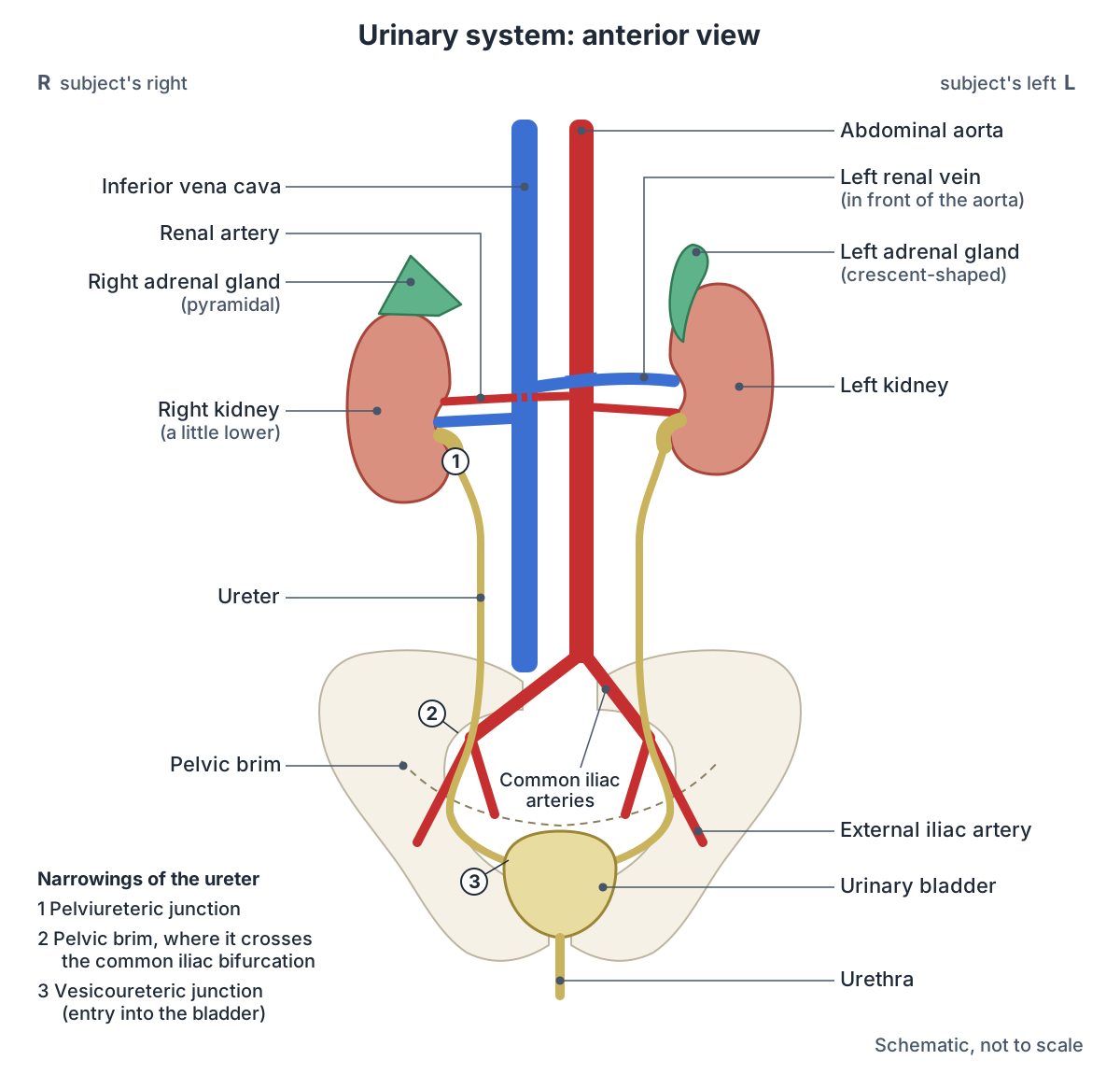
Organs of the urinary system: where they are, size and job
| Organ | Where it is | Size in an adult | Main job |
|---|---|---|---|
| Kidneys (2) | Back wall of the abdomen beside the spine, about T12 to L3, behind the peritoneum | 11 to 14 cm long, 6 cm wide, 4 cm thick; 115 to 175 g | Filter blood and make urine; balance water, salts and pH; help control blood pressure; make erythropoietin and activate vitamin D |
| Ureters (2) | From each renal pelvis down on the psoas major, over the pelvic brim, into the back of the bladder | 25 to 30 cm long, about 3 mm wide | Carry urine to the bladder by peristalsis |
| Urinary bladder (1) | Pelvis, behind the pubic symphysis | Comfortably holds about 300 to 400 mL; up to 500 to 600 mL | Stores urine and empties it when the detrusor muscle contracts |
| Urethra (1) | From the bladder neck to the external urethral orifice | About 4 cm (female); about 18 to 20 cm (male) | Carries urine out of the body; in males also carries semen |
Compiled from OpenStax (25.2, 25.3), Kenhub, Radiopaedia and TeachMeAnatomy (see references). Sizes vary between people and sources.
What does the urinary system do?
The urinary system removes waste and keeps the body's water, electrolytes and acid-base (pH) balance steady. The kidneys also help control blood volume and blood pressure through renin, stimulate red blood cell production with erythropoietin, and convert vitamin D into its active form, calcitriol. (Kenhub: Urinary system.)
Urine is made in three steps: filtration, reabsorption and secretion. The glomeruli filter about 180 litres a day in men and 150 litres in women, but 99% of this filtrate is returned to the blood, leaving only about 1 to 2 litres of urine a day. (OpenStax Anatomy and Physiology 2e, 25.5: Physiology of Urine Formation.)
OpenStax gives the normal daily urine volume as 750 to 2,000 mL. Less than 500 mL a day is called oliguria, less than 50 mL anuria, and more than 2.5 litres polyuria; about 500 mL a day is the minimum needed to clear the body's wastes. (OpenStax Anatomy and Physiology 2e, 25.1: Physical Characteristics of Urine.)
Consumer health sources quote a smaller filtered volume: the kidneys filter 120 to 150 quarts (about 114 to 142 litres) of blood a day to make 1 to 2 quarts of urine. (NIDDK: The Urinary Tract & How It Works.)
Kidneys: where urine is made
The kidneys lie on either side of the spine in the retroperitoneal space. The left kidney sits at about T12 to L3 and the right is a little lower because the liver displaces it. Each is about 11 to 14 cm long, 6 cm wide and 4 cm thick, and weighs 125 to 175 g in males and 115 to 155 g in females. (OpenStax Anatomy and Physiology 2e, 25.3: Gross Anatomy of the Kidney.)
Their position changes with posture: Basic Human Anatomy places the kidneys opposite the first four lumbar vertebrae when standing, rising by about one vertebra when lying down. (Basic Human Anatomy (O'Rahilly et al.), Dartmouth, chapter 29: The kidneys, ureters and suprarenal glands.)
Each kidney contains about 1.3 million nephrons, its working units, and at rest the two kidneys receive about 25% of the cardiac output. (OpenStax Anatomy and Physiology 2e, 25.3: Gross Anatomy of the Kidney.)
The OpenStax section on urine formation gives a slightly lower share: about 20% of a resting cardiac output of 5 litres a minute, or roughly 1 litre of blood a minute. (OpenStax Anatomy and Physiology 2e, 25.5: Physiology of Urine Formation.)
Location on the back, coverings, relations, calyces, renal pelvis and hilum. Kidney anatomy.
How is urine made? The nephron
Each nephron starts with a renal corpuscle: a tuft of capillaries (the glomerulus) inside Bowman's capsule, which catches the filtrate. The filtrate then passes through the proximal convoluted tubule, the loop of Henle, the distal convoluted tubule and finally a collecting duct shared with other nephrons. (OpenStax Anatomy and Physiology 2e, 25.4: Microscopic Anatomy of the Kidney.)
The proximal convoluted tubule is the main site of reabsorption and secretion. In the juxtaglomerular apparatus, macula densa cells in the wall of the distal tubule sense sodium and fluid flow, and juxtaglomerular cells in the afferent arteriole release renin. (OpenStax Anatomy and Physiology 2e, 25.4: Microscopic Anatomy of the Kidney.)
Filtration is driven by a net filtration pressure of about 10 mm Hg. A mean arterial pressure above about 60 mm Hg is generally enough to keep the kidneys filtering. (OpenStax Anatomy and Physiology 2e, 25.5: Physiology of Urine Formation.)
A labelled nephron diagram, step by step from glomerulus to collecting duct. Nephron anatomy.
Ureters and their three narrowings
Each ureter is 25 to 30 cm long and about 3 mm wide. It has an abdominal part from the renal pelvis to the pelvic brim, a pelvic part from the brim to the bladder, and an intravesical part inside the bladder wall. (Radiopaedia: Ureter.)
The ureters are completely retroperitoneal. They do not simply drain: waves of peristalsis in their smooth muscle push urine down. Each ureter pierces the bladder wall obliquely, which forms a one-way valve that stops urine flowing back up towards the kidney. (OpenStax Anatomy and Physiology 2e, 25.2: Gross Anatomy of Urine Transport.)
The ureter narrows at three points, the commonest places for kidney stones to stick: the pelviureteric junction, the pelvic brim where it crosses the bifurcation of the common iliac artery, and the vesicoureteric junction where it enters the bladder. (Radiopaedia: Ureter.)
TeachMeAnatomy calls this three-point model historical and notes newer evidence that stones lodge mainly in the upper ureter and at the vesicoureteric junction. Pain fibres from the ureter enter the spinal cord at T11 to L2, so ureteric pain is felt in those dermatomes. (TeachMeAnatomy: The Ureters.)
Course and relations of each ureter, the narrowings and ureter vs urethra. Ureter anatomy.
Urinary bladder: where urine is stored
The bladder lies behind the pubic bone. In females it is in front of the uterus and vagina; in males it is in front of the rectum with the prostate below it. It is retroperitoneal, with peritoneum only over its dome. Its volume in adults ranges from nearly zero to 500 to 600 mL. (OpenStax Anatomy and Physiology 2e, 25.2: Gross Anatomy of Urine Transport.)
Radiopaedia gives a normal functional adult volume of 300 to 400 mL. As the bladder fills it becomes ovoid and rises out of the pelvis into the abdomen. (Radiopaedia: Urinary bladder.)
The wall is mainly the detrusor, a smooth muscle with criss-crossing bundles. Inside the base, the two ureteric openings and the internal urethral orifice mark the corners of a smooth triangle, the trigone. The urge to void arises at about 150 mL. (OpenStax Anatomy and Physiology 2e, 25.2: Gross Anatomy of Urine Transport.)
Apex, body, fundus, neck and trigone, the detrusor and the sphincters. Bladder anatomy.
Male vs female urethra
The female urethra is about 4 cm long, and its external opening lies below the clitoris, in front of the vagina. The male urethra averages 20 cm and has four regions: preprostatic, prostatic, membranous and spongy (penile). (OpenStax Anatomy and Physiology 2e, 25.2: Gross Anatomy of Urine Transport.)
TeachMeAnatomy describes three male parts and notes that some authors count the segment in the bladder neck as a fourth. The prostatic urethra receives the ejaculatory and prostatic ducts and is the widest part; the membranous urethra, in the pelvic floor, is the narrowest and least dilatable; the penile urethra runs through the corpus spongiosum and widens into the navicular fossa in the glans. (TeachMeAnatomy: The Urethra.)
Radiopaedia gives the male urethra a length of 18 to 20 cm, with a prostatic part of about 3 cm, a membranous part of about 2 cm and a penile part of about 16 cm, and calls the external urethral meatus the narrowest part of the whole urethra. (Radiopaedia: Male urethra.)
The female urethra starts at the bladder neck, is embedded in the anterior vaginal wall and runs through the urogenital hiatus of the pelvic floor. It ends in the vaginal vestibule about 2.5 cm below the clitoris, and paraurethral (Skene) glands open beside its outer opening. (Radiopaedia: Female urethra.)
NCI SEER gives a slightly shorter female urethra, 3 to 4 cm, and about 20 cm for the male urethra, which carries both urine and semen. (NCI SEER Training Modules: Urethra.)
The male urethra in the 3D body, with its neighbours. Urethra in the 3D model.
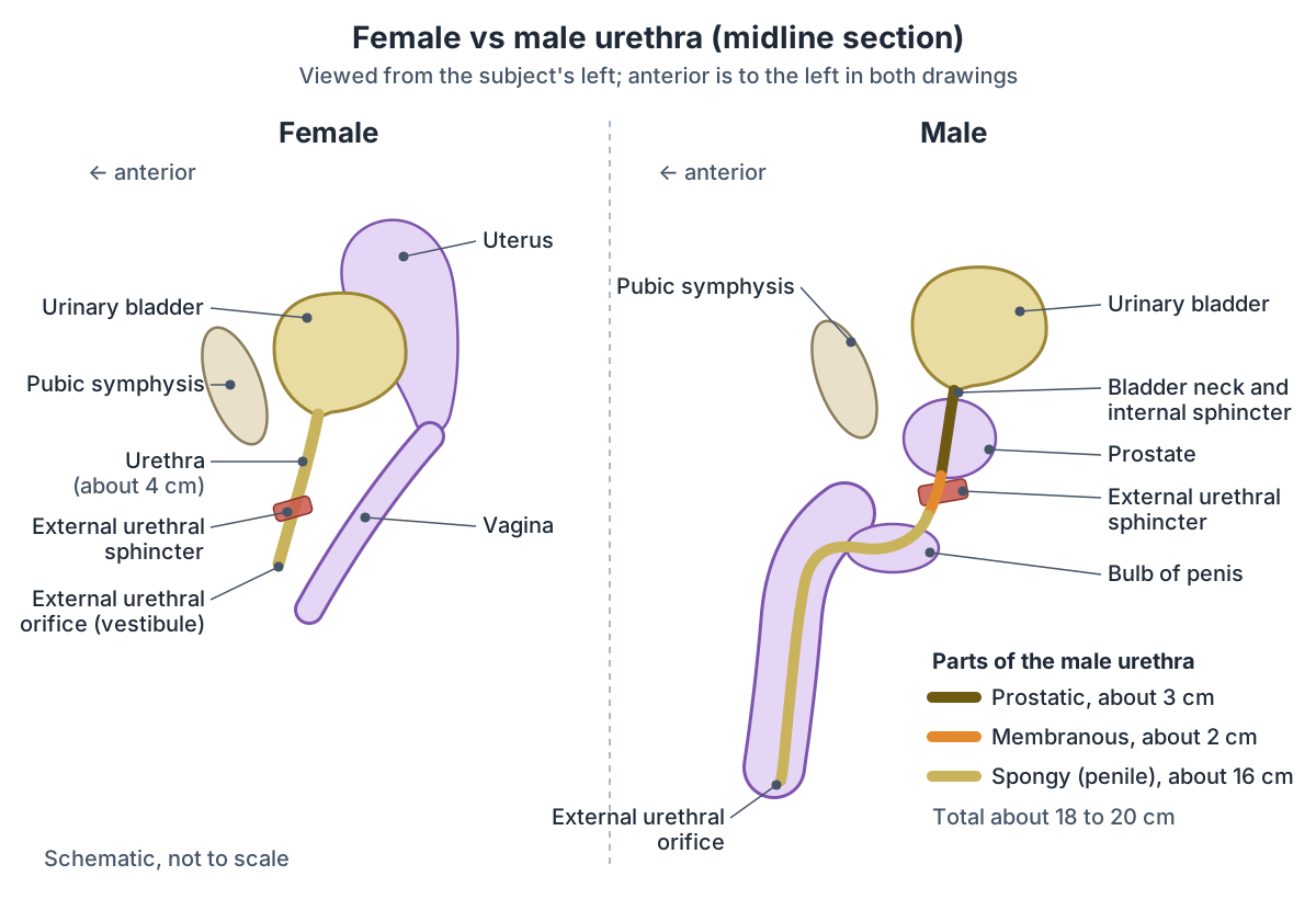
Female vs male urethra
| Feature | Female urethra | Male urethra |
|---|---|---|
| Length | About 4 cm (3 to 4 cm in some sources) | About 20 cm (15 to 20 cm or 18 to 20 cm in some sources) |
| Parts | One short tube fused with the front wall of the vagina | Prostatic (about 3 cm), membranous (about 2 cm) and spongy or penile (about 16 cm); some authors add a preprostatic part |
| Opening | In the vestibule, in front of the vaginal opening and 2 to 3 cm behind the clitoris | At the tip of the glans penis; the external orifice is the narrowest point |
| Carries | Urine | Urine and semen |
| Glands opening into it | Paraurethral (Skene) glands near the external orifice | Prostatic ducts and ejaculatory ducts (prostatic part); bulbourethral glands (spongy part) |
| Sphincters | Internal sphincter thought to be functional only; external sphincter of skeletal muscle | Smooth-muscle internal sphincter at the bladder neck; external sphincter around the membranous urethra |
Compiled from OpenStax 25.2, TeachMeAnatomy, Radiopaedia, NCI SEER and Basic Human Anatomy (see references).
Urethral sphincters: how urine is held in
Two sphincters close the urethra. The internal urethral sphincter is smooth muscle under involuntary, autonomic control. The external urethral sphincter is skeletal muscle under voluntary control, supplied by the pudendal nerve from S2 to S4. (OpenStax Anatomy and Physiology 2e, 25.2: Gross Anatomy of Urine Transport.)
In males the internal sphincter is a ring of circular smooth muscle thought to stop semen flowing back into the bladder during ejaculation. In females it is thought to be functional only, formed by the bladder neck and upper urethra without a separate sphincter muscle. The external sphincter has the same basic structure in both sexes. (TeachMeAnatomy: The Bladder.)
The pelvic floor muscles can be squeezed together with the external sphincter to stop leaks. Between toilet visits the bladder wall stays relaxed and the sphincters stay closed. (NIDDK: The Urinary Tract & How It Works.)
The female urethra in the 3D model, from bladder neck to vestibule. Female urethra.
Nerves of micturition: what controls urination?
Three sets of nerves control the lower urinary tract: sacral parasympathetic fibres in the pelvic nerves, thoracolumbar sympathetic fibres in the hypogastric nerves, and sacral somatic fibres in the pudendal nerves. The sympathetic fibres come from T11 to L2; the parasympathetic and pudendal fibres come from S2 to S4. (Fowler CJ, Griffiths D, de Groat WC. The neural control of micturition. Nat Rev Neurosci. 2008.)
During filling, sympathetic activity contracts the bladder outlet and stops the detrusor contracting, and the pudendal nerve keeps the external sphincter tight. This guarding reflex is organised mainly in the spinal cord. (Fowler CJ, Griffiths D, de Groat WC. The neural control of micturition. Nat Rev Neurosci. 2008.)
Voiding is switched on by the pontine micturition centre (Barrington's nucleus) in the brainstem. It first relaxes the urethra and, a few seconds later, activates the sacral parasympathetic outflow that contracts the bladder. (Fowler CJ, Griffiths D, de Groat WC. The neural control of micturition. Nat Rev Neurosci. 2008.)
Stretch receptors in the bladder wall start a spinal reflex. Children learn to override this reflex during toilet training, and voluntary control needs an intact spinal cord and a working pudendal nerve. (OpenStax Anatomy and Physiology 2e, 25.2: Gross Anatomy of Urine Transport.)
Sympathetic and parasympathetic control of the organs. Autonomic nervous system.
Nerves that control the bladder and urethra
| Pathway | Spinal origin and nerve | Main effect |
|---|---|---|
| Sympathetic | T11 to L2 (T12 to L2 in TeachMeAnatomy), through the hypogastric nerves | Relaxes the detrusor and contracts the bladder neck: storage |
| Parasympathetic | S2 to S4, through the pelvic (pelvic splanchnic) nerves | Contracts the detrusor and relaxes the urethral smooth muscle: emptying |
| Somatic | S2 to S4 (Onuf's nucleus), through the pudendal nerves | Contracts the external urethral sphincter: voluntary holding |
| Brainstem | Pontine micturition centre (Barrington's nucleus) in the pons | Switches the bladder from storage to voiding |
From Fowler, Griffiths and de Groat (2008), with TeachMeAnatomy for the alternative sympathetic range.
Blood supply of the urinary tract
The renal arteries leave the abdominal aorta just below the superior mesenteric artery. The right renal artery is longer and passes behind the inferior vena cava. The renal veins drain straight into the inferior vena cava; the left renal vein is longer and crosses in front of the aorta below the origin of the superior mesenteric artery. (TeachMeAnatomy: The Kidneys.)
The ureter picks up small arteries all along its course: from the renal, gonadal and vesical arteries, the aorta and the common iliac artery, and in females the uterine artery. The bladder is supplied by the superior and inferior vesical arteries and drained by the vesical venous plexus. (Kenhub: Urinary system.)
In both sexes the superior vesical artery supplies the upper bladder. The lower bladder is supplied by the inferior vesical artery in males and the vaginal artery in females, all from the anterior division of the internal iliac artery. (Radiopaedia: Urinary bladder.)
In males the prostatic urethra is supplied by the inferior vesical artery, the membranous urethra by the artery of the bulb and the penile urethra by branches of the internal pudendal artery. The female urethra is supplied by the internal pudendal and vaginal arteries. (TeachMeAnatomy: The Urethra.)
The large vein that receives the renal veins. Inferior vena cava.
Lining of the urinary tract: urothelium
The ureters and bladder are lined by transitional epithelium (urothelium). In the bladder it looks columnar when the organ is empty and flattens to a squamous shape when stretched, which lets the wall expand without tearing. (OpenStax Anatomy and Physiology 2e, 25.2: Gross Anatomy of Urine Transport.)
The urethral lining changes along its length. The upper female urethra is transitional epithelium and its end is non-keratinised stratified squamous epithelium. In males, a stretch of pseudostratified columnar epithelium lies between the two. (OpenStax Anatomy and Physiology 2e, 25.2: Gross Anatomy of Urine Transport.)
Radiopaedia describes the male anterior urethra as pseudostratified columnar, becoming squamous at its very end, and the posterior urethra as transitional. (Radiopaedia: Male urethra.)
Adrenal glands: on top of the kidneys
The adrenal (suprarenal) glands sit over the medial part of the upper pole of each kidney, between the kidney and the diaphragm. The same renal (perinephric) fascia encloses the adrenal gland and the kidney, and a thin layer of fibrous tissue separates the two. (TeachMeAnatomy: The Adrenal Glands.)
The adrenals are endocrine glands, not urinary organs, but they influence the kidney: aldosterone from the adrenal cortex makes the kidney reabsorb more sodium. (OpenStax Anatomy and Physiology 2e, 25.3: Gross Anatomy of the Kidney.)
Right vs left gland, cortex zones, medulla, hormones and blood supply. Adrenal gland anatomy.
Common urinary tract problems and their anatomy
More than 60% of females have at least one urinary tract infection in their lifetime, about 1 in 10 people get kidney stones, and more than half of those who have given birth have urinary incontinence at some point. (Cleveland Clinic: Urinary System.)
The short female urethra is why women are more prone to urinary tract infections, which usually show as cystitis with painful, frequent and urgent urination. (TeachMeAnatomy: The Urethra.)
Urinary retention is usually caused by a blockage from the bladder neck downwards; in males the commonest cause is an enlarged prostate (benign prostatic hyperplasia). In chronic retention the bladder can hold more than 1 to 1.5 litres. (TeachMeAnatomy: The Bladder.)
Near the ischial spine the ureter lies about 2 cm from the cervix of the uterus, where it can be injured during a hysterectomy. (Basic Human Anatomy (O'Rahilly et al.), Dartmouth, chapter 29: The kidneys, ureters and suprarenal glands.)
In males, the lowest part of the membranous urethra is the part most liable to rupture or to be pierced by a catheter. (Basic Human Anatomy (O'Rahilly et al.), Dartmouth, chapter 33: The ureter, bladder and urethra.)
Urinary system in the 3D model
- Adrenal gland
- The adrenal (suprarenal) gland is a small endocrine gland on top of each kidney.
- Female urethra
- The female urethra is a short tube, about 4 cm long, that carries urine from the bladder to the outside.
- Kidney
- The kidney is a bean-shaped organ that filters blood to form urine and regulate fluid, electrolytes and blood pressure.
- Neck of urinary bladder
- The neck is the lowest part of the bladder, where it joins the urethra.
- Renal artery
- A large paired branch of the abdominal aorta supplying the kidney.
- Renal vein
- A large vein draining the kidney into the inferior vena cava.
- Trigone of urinary bladder
- The trigone is the smooth triangular area on the inner surface of the bladder base.
- Ureter
- The ureter is a muscular tube carrying urine from the renal pelvis to the urinary bladder.
- Urinary bladder
- The urinary bladder is a hollow muscular organ that stores urine.
Frequently asked questions
What are the 4 organs of the urinary system?
The kidneys, ureters, urinary bladder and urethra. The two kidneys filter blood and make urine, the two ureters carry it down to the bladder, the bladder stores it, and the single urethra carries it out of the body. The adrenal glands sit on top of the kidneys but are endocrine glands, not part of the urinary system.
What is the path of urine through the urinary system?
Urine forms in the nephrons and drains through collecting ducts to the renal papillae, then into the minor calyces, major calyces and renal pelvis. It leaves each kidney in a ureter, which carries it by peristalsis to the bladder. The bladder stores it until the detrusor contracts and urine leaves through the urethra.
How long is the male urethra compared with the female urethra?
The female urethra is about 4 cm long and runs in the front wall of the vagina to the vestibule. The male urethra is about 18 to 20 cm long and runs through the prostate, the pelvic floor and the penis in three parts: prostatic, membranous and spongy (penile). It carries semen as well as urine.
How much urine can the bladder hold?
Sources differ. Radiopaedia gives a normal functional volume of 300 to 400 mL, while OpenStax says adult volumes range from nearly zero to 500 to 600 mL. The urge to urinate starts at about 150 mL and is easy to ignore at first; the bladder can stretch far more in chronic urinary retention.
How much urine do the kidneys make each day?
About 1 to 2 litres. The glomeruli filter around 180 litres of fluid a day in men and 150 litres in women, but 99% of it is reabsorbed back into the blood. OpenStax gives 750 to 2,000 mL as the normal daily range; below 500 mL a day the body cannot clear all its wastes.
Are the adrenal glands part of the urinary system?
No. The adrenal (suprarenal) glands sit on top of the kidneys inside the same renal fascia, but they are endocrine glands. Their cortex makes hormones such as aldosterone and cortisol, and their medulla makes adrenaline. Aldosterone does act on the kidney, making it keep more sodium.
What controls urination?
Sympathetic nerves from T11 to L2 relax the bladder wall and close its neck while it fills. Parasympathetic nerves from S2 to S4 contract the detrusor to empty it, and the pudendal nerve (S2 to S4) works the voluntary external sphincter. The pontine micturition centre in the brainstem switches the bladder from storing to emptying.
References
- OpenStax. Anatomy and Physiology 2e, §25.1 Physical Characteristics of Urine. Rice University, 2022.
- OpenStax. Anatomy and Physiology 2e, §25.2 Gross Anatomy of Urine Transport. Rice University, 2022.
- OpenStax. Anatomy and Physiology 2e, §25.3 Gross Anatomy of the Kidney. Rice University, 2022.
- OpenStax. Anatomy and Physiology 2e, §25.4 Microscopic Anatomy of the Kidney. Rice University, 2022.
- OpenStax. Anatomy and Physiology 2e, §25.5 Physiology of Urine Formation. Rice University, 2022.
- McLaren N. Urinary system. Kenhub anatomy library, reviewed 2023.
- The Kidneys. TeachMeAnatomy.
- The Ureters. TeachMeAnatomy.
- The Bladder. TeachMeAnatomy.
- The Urethra. TeachMeAnatomy.
- The Adrenal Glands. TeachMeAnatomy.
- Ureter. Radiopaedia reference article.
- Urinary bladder. Radiopaedia reference article.
- Male urethra. Radiopaedia reference article.
- Female urethra. Radiopaedia reference article.
- Urethra. NCI SEER Training Modules, National Cancer Institute.
- Urinary System. Cleveland Clinic health library.
- National Institute of Diabetes and Digestive and Kidney Diseases. The Urinary Tract & How It Works. NIH, 2020.
- Fowler CJ, Griffiths D, de Groat WC. The neural control of micturition. Nat Rev Neurosci. 2008;9(6):453-466. doi:10.1038/nrn2401.
- O'Rahilly R, Müller F, Carpenter S, Swenson R. Basic Human Anatomy, chapter 29: The kidneys, ureters and suprarenal glands. Dartmouth Medical School.
- O'Rahilly R, Müller F, Carpenter S, Swenson R. Basic Human Anatomy, chapter 33: The ureter, bladder and urethra. Dartmouth Medical School.
- Mitsuhashi N. et al. (2009). BodyParts3D: 3D structure database for anatomical concepts. Nucleic Acids Research 37, D782–D785. Data © The Database Center for Life Science, CC BY 4.0.
- Browne K., Schlehlein H. 3D Reference Organ Set for United, Female (Visible Human Female). Human Reference Atlas 3D Reference Object Library, HuBMAP / NIH. Data from the NLM Visible Human Project. CC BY 4.0.
Related anatomy
- KidneyWhere are your kidneys? Kidney location on the back (T12 to L3), coverings, relations, cortex, medulla, calyces, renal…
- UreterWhat do the ureters do? Ureter function, course, relations, the three narrowings where kidney stones lodge, blood…
- Urinary bladderBladder anatomy with a labelled diagram and 3D model: apex, body, fundus, neck and trigone, detrusor and sphincters…
- Adrenal glandAnatomy of the adrenal glands with diagrams and a 3D model: right vs left shape, cortex zones and medulla, hormones…
- Male reproductive systemMale reproductive anatomy with labelled diagrams: scrotum, testes, epididymis, ductus deferens, seminal gland…
- Female reproductive systemLabelled female reproductive system diagrams: uterus anatomy and layers, uterine tube parts, ovaries, ligaments…
Explore the whole body in the interactive 3D human anatomy model, or browse the A–Z list of human structures.