

What does female anatomy include?
Female anatomy has external and internal genital organs. The external genitalia are together called the vulva. The paired ovaries are the female gonads and lie in the pelvic cavity, and the breasts are counted as accessory organs of the female reproductive system. (OpenStax Anatomy and Physiology 2e, 27.2: Anatomy and Physiology of the Ovarian Reproductive System.)
Kenhub groups the vagina, uterus, uterine (fallopian) tubes and ovaries as the internal genitalia, and the mons pubis, labia majora and labia minora, clitoris, vestibule, vestibular glands and vestibular bulbs as the external genitalia. (Kenhub: Female reproductive organs.)
Together these organs make eggs and hormones, receive sperm, carry a pregnancy and deliver a baby. The ovaries produce eggs and hormones, fertilisation normally happens in the fallopian tube, the fertilised egg implants in the thickened lining of the uterus, and the vagina is the muscular canal that joins the cervix to the outside of the body. (Cleveland Clinic: Female Reproductive System.)
In the 3D model, the Female body swaps in the female pelvis, reproductive organs, breasts, bladder, urethra and skin. The skeleton above the pelvis, the muscles and the heart, lungs, digestive organs, kidneys and brain are the same meshes as in the Male body, which reflects how much anatomy the two share. The skin starts hidden; raise the Skin slider to see the outline of the female body, lower the Muscles slider to see the pelvic organs more clearly, or click any highlighted part to open its page.
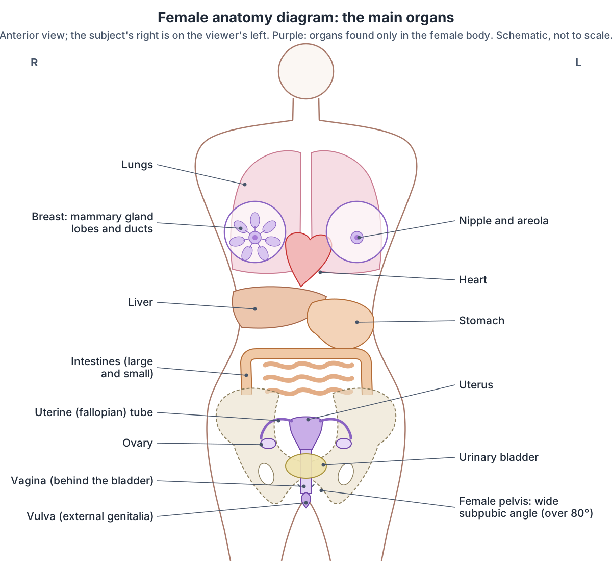
Female organs at a glance
| Organ | Where it is | Main role | Typical size |
|---|---|---|---|
| Ovaries (2) | Pelvic cavity, either side of the uterus | Release eggs; make estradiol, progesterone and inhibin | 2 to 3 cm long |
| Uterine (fallopian) tubes (2) | From the upper uterus towards each ovary | Catch the egg; site of fertilisation (ampulla) | About 10 to 12 cm long |
| Uterus | Centre of the pelvis, behind the bladder | Implantation, pregnancy, menstruation | About 7 cm long, 5 cm wide |
| Vagina | From the cervix to the vulva, between bladder and rectum | Intercourse, birth canal, menstrual flow | About 6 to 10 cm (sources differ) |
| Vulva | External genitalia below the pubic bone | Sensation, directing urine, protection | Varies widely |
| Breasts (2) | Front of the chest, 2nd to 6th costal cartilages | Milk production (lactation) | Varies; size does not affect milk supply |
| Urethra | In front of the vagina | Carries urine from the bladder | About 4 cm long |
Compiled from OpenStax 27.2, TeachMeAnatomy, Radiopaedia (vagina, fallopian tube), Luo et al. 2016 and Cleveland Clinic (see references).
Female vs male anatomy: what is different?
The difference starts with the sex chromosomes: XX individuals are female and XY individuals male. A gene on the Y chromosome, SRY, steers the embryo towards male development. Without it, the Müllerian ducts develop into the uterus, uterine tubes and part of the vagina, and the embryonic cells that would form the glans of the penis form the clitoris instead. (OpenStax Anatomy and Physiology 2e, 27.3: Development of the Male and Female Reproductive Systems.)
Because male and female genitals come from the same embryonic tissue, each female structure has a male counterpart (homologue): the clitoris corresponds to the penis, the vestibular bulbs to the bulb and corpus spongiosum of the penis, the greater vestibular (Bartholin) glands to the bulbourethral glands, and the paraurethral (Skene) glands to the prostate. (Kenhub: External female genitalia.)
The labia majora are homologous to the scrotum, and the ovaries are the female equivalent of the testes. (Kenhub: Female reproductive organs.)
The female urethra is about 4 cm long, against about 15 to 20 cm in males. Its short length is one reason women are more prone to urinary tract infections such as cystitis. (TeachMeAnatomy: The Urethra.)
Puberty adds the secondary sex characteristics. In females these include breast development, fat deposited mainly in the breasts and hips, and broadening of the pelvis. (OpenStax Anatomy and Physiology 2e, 27.3: Development of the Male and Female Reproductive Systems.)
The male body, its reproductive organs and how it differs from the female body. Male anatomy.
Female vs male anatomy: key differences and counterparts
| Feature | Female | Male |
|---|---|---|
| Sex chromosomes | XX | XY (with the SRY gene) |
| Gonads | Ovaries, in the pelvic cavity | Testes |
| Erectile organ | Clitoris | Penis |
| Erectile bulbs | Vestibular bulbs | Bulb and corpus spongiosum of the penis |
| Mucous glands at the opening | Greater vestibular (Bartholin) glands | Bulbourethral glands |
| Glands beside the urethra | Paraurethral (Skene) glands | Prostate |
| Outer skin folds | Labia majora | Scrotum |
| Urethra length | About 4 cm | About 15 to 20 cm |
| Pelvic inlet | Round or oval | Heart-shaped |
| Subpubic angle | More than 80 degrees | Less than 70 degrees |
| Lesser (true) pelvis | Shorter and wider | Longer and narrower |
From OpenStax 27.3 and 8.3, Kenhub (female reproductive organs, external female genitalia) and TeachMeAnatomy (urethra).
Female internal organs: ovaries, uterine tubes and uterus
The ovaries are paired, oval organs about 2 to 3 cm long, about the size of an almond, attached to the uterus by the ovarian ligament. As well as releasing eggs, they produce the hormones estradiol (a type of oestrogen, US estrogen), progesterone and inhibin. (OpenStax Anatomy and Physiology 2e, 27.2: Anatomy and Physiology of the Ovarian Reproductive System.)
Each uterine tube ends near its ovary in a wide infundibulum with finger-like fimbriae that sweep the released egg into the tube. Its middle part, the ampulla, is where fertilisation usually happens. (OpenStax Anatomy and Physiology 2e, 27.2: Anatomy and Physiology of the Ovarian Reproductive System.)
Each tube is about 10 to 12 cm long and 1 to 4 mm wide, and has five parts from the ovary inwards: fimbriae, infundibulum, ampulla, isthmus and the intramural part inside the wall of the uterus. (Radiopaedia: Fallopian tube.)
The uterus is about 7 cm long and 5 cm wide outside pregnancy. The part above the openings of the tubes is the fundus, the middle section is the body, and the narrow lower part, the cervix, projects into the vagina. Its wall has three layers: the outer perimetrium, the thick muscular myometrium and the inner endometrium, which thickens and is shed in each menstrual cycle. (OpenStax Anatomy and Physiology 2e, 27.2: Anatomy and Physiology of the Ovarian Reproductive System.)
The uterus normally tilts forwards (anteverted and anteflexed) and is held by the broad, round, cardinal, uterosacral and pubocervical ligaments. The uterine artery, a branch of the internal iliac artery, supplies it, while each ovary receives an ovarian artery straight from the abdominal aorta. (Kenhub: Female reproductive organs.)
Labelled diagrams of the uterus, uterine tubes, ovaries and their ligaments. Female reproductive system.
The vagina: the internal canal
The vagina is a stretchy (distensible) muscular tube that runs upwards and backwards from the vaginal opening to the cervix. It receives the penis and semen during intercourse, is the birth canal, and carries menstrual fluid out of the body. (TeachMeAnatomy: The Vagina.)
In front of the vagina are the bladder and urethra; behind it are the rectouterine pouch, the rectum and the anal canal; at its sides are the ureters and the levator ani muscles. (TeachMeAnatomy: The Vagina.)
Sources give different lengths. Radiopaedia describes a midline fibromuscular tube 6 to 8 cm long that passes through the urogenital diaphragm from the vestibule to the uterus. (Radiopaedia: Vagina.)
OpenStax gives a length of about 10 cm. (OpenStax Anatomy and Physiology 2e, 27.2: Anatomy and Physiology of the Ovarian Reproductive System.)
Part of the difference is that the two walls are not equal. An MRI study of 80 women measured the front (anterior) wall at 63 mm on average and the back (posterior) wall at 98 mm, with the canal widest near the cervix and narrowest at the opening. (Luo J, Betschart C, Ashton-Miller JA, DeLancey JOL. Quantitative analyses of variability in normal vaginal shape and dimension on MR images. Int Urogynecol J. 2016.)
Length, walls, fornices, layers, supports, vessels and nerves of the vagina, in 3D. Vagina anatomy.
External female anatomy: the vulva
The vulva is the external female genitalia: the mons pubis, labia majora and minora, the vestibule containing the vaginal and urethral openings, the greater vestibular (Bartholin) glands and the clitoris. It provides sensation during intercourse, directs the flow of urine and helps protect the internal reproductive tract from infection. (TeachMeAnatomy: The Vulva.)
The mons pubis is a pad of fat over the pubic bone. The labia majora are folds of hair-covered skin that begin just behind it, the labia minora inside them protect the urethra and the entrance to the reproductive tract, and the front ends of the labia minora come together around the clitoris. (OpenStax Anatomy and Physiology 2e, 27.2: Anatomy and Physiology of the Ovarian Reproductive System.)
Only the glans of the clitoris, about 5 to 8 mm long, is visible; about four-fifths of the organ lies internally, where its two crura attach to the ischiopubic rami of the pelvis. (Kenhub: Clitoris.)
All vulvas have the same parts but no two look alike. The labia come in many shapes and sizes, and the inner lips may be symmetrical or not and may sit inside or extend beyond the outer lips. (Cleveland Clinic: Vulva.)
Labia, clitoris, vestibule, hymen and glands, with labelled diagrams. Vulva anatomy.
Female urethra and bladder: how many openings are there?
Between the legs the female body has three openings: the urethral opening, through which urine leaves, the vaginal opening and the anus. All three pass through the pelvic floor muscles. (Cleveland Clinic: Pelvic Floor Muscles.)
From front to back the order is urethra, vagina, anus: the vaginal opening lies between the opening of the urethra and the anus. (OpenStax Anatomy and Physiology 2e, 27.2: Anatomy and Physiology of the Ovarian Reproductive System.)
The external urethral orifice opens in the vestibule, in front of the vaginal opening and 2 to 3 cm behind the clitoris. Two paraurethral (Skene) glands lie on either side of the lower urethra. (TeachMeAnatomy: The Urethra.)
The adult bladder holds up to about 400 to 600 mL. In females, the bladder neck is thought to have no true internal sphincter muscle, only a functional one, and the vaginal arteries help supply the bladder. (TeachMeAnatomy: The Bladder.)
Because the bladder rests on the front wall of the vagina, weakness there lets the bladder bulge into the vagina (a cystocele). Anterior vaginal wall prolapse is the most common type of pelvic organ prolapse. (Cleveland Clinic: Pelvic Organ Prolapse.)
Bladder anatomy, capacity and how it differs in females and males. Urinary bladder.
Breasts (mammary glands)
The breasts lie on the front of the chest over the pectoralis major and serratus anterior muscles. They reach from the side of the sternum to the mid-axillary line and from the 2nd to the 6th costal cartilages, and an axillary tail extends towards the armpit. (TeachMeAnatomy: The Breasts.)
Milk is made by the mammary glands, which are modified sweat glands. Clusters of milk-secreting alveoli drain into 15 to 20 lactiferous ducts, each widening into a lactiferous sinus before opening on the nipple. The pigmented areola, 25 to 100 mm across, carries small areolar glands, and bands of connective tissue called suspensory ligaments support the breast. Breast size does not affect how much milk is produced. (OpenStax Anatomy and Physiology 2e, 27.2: Anatomy and Physiology of the Ovarian Reproductive System.)
About 75% of breast lymph drains to the axillary lymph nodes, 20% to the parasternal nodes and 5% to the posterior intercostal nodes. Milk production is controlled by the pituitary hormones prolactin and oxytocin, not by the breast's nerves. (TeachMeAnatomy: The Breasts.)
The 3D model shows each breast as separate layers: the lobes of the mammary gland, the lactiferous ducts and sinuses, the fat (adipose tissue), the suspensory ligaments, the nipple, the areola and the areolar tubercles.
Lobes, ducts, ligaments, lymph drainage and blood supply of the breast. Breast anatomy.
The female pelvis
Because it is adapted for childbirth, the female pelvis is wider than the male pelvis. Its bones are lighter and thinner, the pelvic inlet is round or oval rather than heart-shaped, the lesser (true) pelvis is shorter and wider, and the subpubic angle is greater than 80 degrees, against less than 70 degrees in males. The ischial tuberosities are further apart, so the pelvic outlet is larger. (OpenStax Anatomy and Physiology 2e, 8.3: The Pelvic Girdle and Pelvis.)
TeachMeAnatomy adds that the ischial spines are less prominent, giving a wider distance between them, and that under the influence of progesterone the sacrotuberous and sacrospinous ligaments can stretch to enlarge the outlet further. (TeachMeAnatomy: The Pelvic Girdle.)
Sources disagree about the female sacrum: OpenStax describes it as wider, shorter and less curved, while TeachMeAnatomy describes it as shorter and more curved with a less prominent promontory, so treat the curvature with caution. (TeachMeAnatomy: The Pelvic Girdle.)
In the Female body of the 3D model, the hip bones, sacrum and coccyx come from the Visible Human Female and were fitted to the shared spine and thigh bones, so you can compare the wide female pelvis with the Male body by switching between them.
Bones of the pelvis and a labelled male vs female pelvis comparison. Pelvis anatomy.
Pelvic floor in women
The pelvic floor (pelvic diaphragm) is a funnel-shaped sheet of muscle that separates the pelvic cavity from the perineum below. Its main part, levator ani, has three parts (puborectalis, pubococcygeus and iliococcygeus), and coccygeus lies behind it. The urethra and vagina pass through a gap at the front, the urogenital hiatus, and the anal canal through the rectal hiatus; the perineal body lies between them. (TeachMeAnatomy: The Pelvic Floor.)
The pelvic floor muscles hold up the bladder, urethra, bowel and rectum, and in females also the vagina and uterus. They carry the weight of the growing fetus in pregnancy and take part in vaginal delivery, and they can weaken during pregnancy. (Cleveland Clinic: Pelvic Floor Muscles.)
When the support fails, pelvic organs can drop into the vagina (pelvic organ prolapse), which affects about 3% to 11% of women. Vaginal childbirth is the most common cause, and lower oestrogen after menopause weakens the supporting connective tissue. (Cleveland Clinic: Pelvic Organ Prolapse.)
Levator ani, coccygeus, the perineal body and the perineum. Pelvic floor and perineum.
Female hormones and the glands that make them
Female reproductive development is driven by a chain of hormones: gonadotropin-releasing hormone (GnRH) from the hypothalamus makes the anterior pituitary release luteinising hormone (LH) and follicle-stimulating hormone (FSH), and these make the ovaries release oestrogen. (OpenStax Anatomy and Physiology 2e, 27.3: Development of the Male and Female Reproductive Systems.)
The ovaries produce estradiol as the follicles grow and large amounts of progesterone after ovulation. A third ovarian hormone, inhibin, takes part in a negative feedback loop to the pituitary gland. (OpenStax Anatomy and Physiology 2e, 27.2: Anatomy and Physiology of the Ovarian Reproductive System.)
Cleveland Clinic names FSH, LH, oestrogen and progesterone as the four main hormones of the menstrual cycle. An average cycle lasts about 28 days: the follicular phase starts on the first day of a period, ovulation follows about 14 days later, and most people have their next period 10 to 16 days after ovulation. (Cleveland Clinic: Female Reproductive System.)
Menstruation itself lasts 2 to 7 days, about five on average, and in a typical 28-day cycle ovulation falls on day 14. (OpenStax Anatomy and Physiology 2e, 27.2: Anatomy and Physiology of the Ovarian Reproductive System.)
The gland that releases LH, FSH, prolactin and oxytocin. Pituitary gland.
How female anatomy changes through life
Puberty begins at around 8 or 9 years, when LH production becomes detectable. The first visible change is usually breast development, followed by axillary and pubic hair. A growth spurt starts at about 9 to 11 years and can add 3 inches (about 7.5 cm) of height a year for two years or more, and menarche, the first period, comes next. The average age of menarche fell from about 17 years in 1860 to about 12.75 years in 1960, and body fat appears to play a strong role in its timing. (OpenStax Anatomy and Physiology 2e, 27.3: Development of the Male and Female Reproductive Systems.)
The number of egg cells falls throughout life. OpenStax gives one to two million primary oocytes in an infant, about 400,000 at puberty and none by the end of menopause. (OpenStax Anatomy and Physiology 2e, 27.2: Anatomy and Physiology of the Ovarian Reproductive System.)
Cleveland Clinic gives different numbers: about 6 million eggs during fetal development, about 1 million at birth and about 300,000 by puberty. It counts the reproductive years from the first cycles, anywhere from 11 to 16 years of age, and defines menopause as a full year without a menstrual period. (Cleveland Clinic: Female Reproductive System.)
Menopause is the end of the menstrual cycle that follows the loss of the ovarian follicles. OpenStax gives an average age of 50 to 52 years worldwide and says it is mainly the lack of oestrogen that causes its symptoms. (OpenStax Anatomy and Physiology 2e, 27.2: Anatomy and Physiology of the Ovarian Reproductive System.)
The NHS says menopause usually happens between 45 and 55 years of age, although it can happen earlier. (NHS: Menopause.)
In the vulva, falling oestrogen at menopause can bring vaginal dryness, thinner pubic hair and less fatty tissue. (Cleveland Clinic: Vulva.)
The vagina shortens after menopause and its fornices almost disappear. (Radiopaedia: Vagina.)
The ovary in 3D, with its ligaments and blood supply. Ovary.
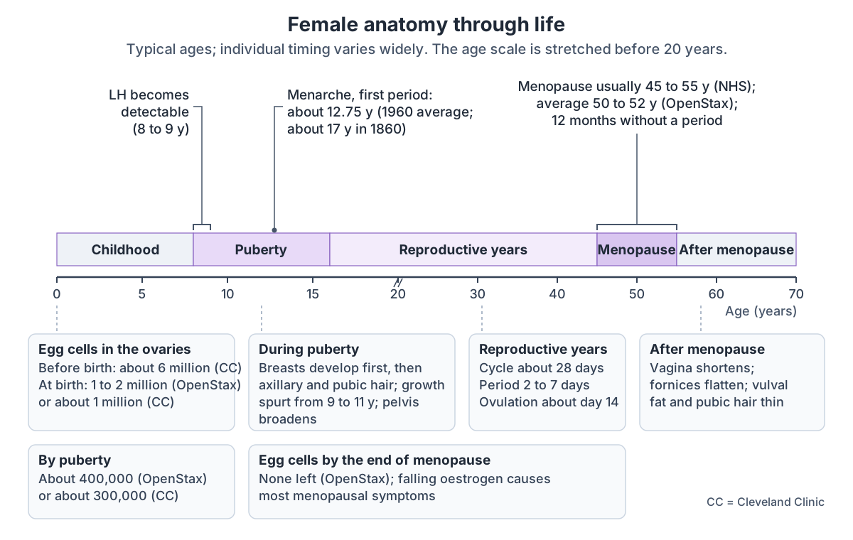
Female life stages
| Stage | Typical age | Main anatomical changes |
|---|---|---|
| Before birth | Embryo and fetus | Without SRY, Müllerian ducts form the uterus, tubes and upper vagina; about 6 million egg cells in the fetus (Cleveland Clinic) |
| Puberty | From about 8 to 9 years | LH rises; breast development, then pubic and axillary hair; growth spurt from 9 to 11; broadening of the pelvis; fat in breasts and hips |
| Menarche | About 12.75 years (1960 average) | First menstrual period |
| Reproductive years | From about 11 to 16 years until menopause | Cycles of about 28 days with ovulation around day 14 |
| Menopause | Usually 45 to 55; average 50 to 52 | No period for 12 months; follicles exhausted; oestrogen falls |
| After menopause | From the early 50s | Vagina shortens and fornices flatten; vulval fat and pubic hair thin; pelvic floor support weakens |
From OpenStax 27.2 and 27.3, Cleveland Clinic, the NHS and Radiopaedia. Timing varies widely between people.
Female anatomy in the 3D model
- Breast
- The breast contains the mammary gland, a modified sweat gland that produces milk, embedded in fat and fibrous tissue.
- Broad ligament of uterus
- The broad ligament is a double fold of peritoneum that hangs from the sides of the uterus to the side walls and floor of the pelvis.
- Female urethra
- The female urethra is a short tube, about 4 cm long, that carries urine from the bladder to the outside.
- Hip bone
- The large irregular bone of the pelvis formed by the fusion of the ilium, ischium and pubis.
- Ovary
- The ovary is the female gonad, which produces oocytes and the hormones oestrogen and progesterone.
- Round ligament of uterus
- The round ligament is a fibromuscular cord from the uterus to the labium majus.
- Sacrum
- A triangular bone of five fused sacral vertebrae.
- Urinary bladder
- The urinary bladder is a hollow muscular organ that stores urine.
- Uterine tube
- The uterine tube is a muscular tube, about 10–12 cm long, that carries the oocyte from the ovary to the uterus.
- Uterus
- The uterus is a hollow, pear-shaped muscular organ in which a fetus develops.
- Vagina
- The vagina is a fibromuscular canal from the cervix to the vestibule of the vulva.
Frequently asked questions
What are the parts of female anatomy?
Female anatomy includes the external genitalia, or vulva (mons pubis, labia majora and minora, clitoris, vestibule, urethral and vaginal openings), the internal reproductive organs in the pelvis (vagina, uterus with its cervix, two uterine tubes and two ovaries), the breasts, a wider pelvis and a short urethra about 4 cm long.
What organs do only females have?
Only the female body has ovaries, uterine (fallopian) tubes, a uterus with a cervix, a vagina and a vulva with a clitoris, vestibular bulbs and Bartholin glands. Each of these has a male counterpart that develops from the same embryonic tissue, for example the ovaries and testes, or the clitoris and penis. Breasts with functional mammary glands are accessory organs of the female reproductive system.
What is the difference between the vulva and the vagina?
The vulva is the external genital area that can be seen: the mons pubis, labia, clitoris and the vestibule with the urethral and vaginal openings. The vagina is the internal muscular canal that runs from the vaginal opening up to the cervix. In everyday speech people often call the whole area the vagina, but anatomically they are different structures.
How many openings does a woman have between the legs?
Three. From front to back they are the urethral opening, which passes urine, the vaginal opening, which leads to the vagina, and the anus at the end of the digestive tract. The urethral opening lies 2 to 3 cm behind the clitoris, and all three openings pass through the muscles of the pelvic floor.
How is the female pelvis different from the male pelvis?
The female pelvis is adapted for childbirth. Its bones are lighter, its inlet is round or oval rather than heart-shaped, the true pelvis is shorter and wider, the subpubic angle is more than 80 degrees instead of less than 70, and the ischial tuberosities are further apart, which makes the outlet larger.
Which hormones control the female reproductive system?
GnRH from the hypothalamus triggers the release of LH and FSH from the anterior pituitary gland. These act on the ovaries, which produce oestrogen (mainly estradiol), progesterone after ovulation and inhibin. FSH, LH, oestrogen and progesterone are the four main hormones of the roughly 28-day menstrual cycle.
At what age does menopause happen?
Menopause usually happens between 45 and 55 years of age, and the average age is about 50 to 52 worldwide, though it can occur earlier. It is defined as 12 months without a period and follows the loss of the ovarian follicles. Falling oestrogen afterwards can shorten the vagina and thin the tissues of the vulva.
References
- OpenStax. Anatomy and Physiology 2e, §27.2 Anatomy and Physiology of the Ovarian Reproductive System. Rice University, 2022.
- OpenStax. Anatomy and Physiology 2e, §27.3 Development of the Male and Female Reproductive Systems. Rice University, 2022.
- OpenStax. Anatomy and Physiology 2e, §8.3 The Pelvic Girdle and Pelvis. Rice University, 2022.
- TeachMeAnatomy. The Pelvic Girdle.
- Thompson L. The Vagina. TeachMeAnatomy, updated 2025.
- Thompson L. The Vulva. TeachMeAnatomy, updated 2025.
- TeachMeAnatomy. The Urethra.
- TeachMeAnatomy. The Bladder.
- TeachMeAnatomy. The Breasts.
- TeachMeAnatomy. The Pelvic Floor.
- Sendić G. Female reproductive organs. Kenhub, reviewed 2023.
- External female genitalia. Kenhub anatomy library, reviewed 2022.
- Clitoris. Kenhub anatomy library.
- Vagina. Radiopaedia reference article.
- Fallopian tube. Radiopaedia reference article.
- Cleveland Clinic. Female Reproductive System: Structure and Function.
- Cleveland Clinic. Vulva: Location, Anatomy, Function, Conditions and Care.
- Cleveland Clinic. Pelvic Floor Muscles: Anatomy, Function and Conditions.
- Cleveland Clinic. Pelvic Organ Prolapse: Types, Causes and Treatment.
- NHS. Menopause.
- Luo J, Betschart C, Ashton-Miller JA, DeLancey JOL. Quantitative analyses of variability in normal vaginal shape and dimension on MR images. Int Urogynecol J. 2016;27(7):1087-1095.
- Mitsuhashi N. et al. (2009). BodyParts3D: 3D structure database for anatomical concepts. Nucleic Acids Research 37, D782–D785. Data © The Database Center for Life Science, CC BY 4.0.
- Browne K., Schlehlein H. 3D Reference Organ Set for United, Female (Visible Human Female). Human Reference Atlas 3D Reference Object Library, HuBMAP / NIH. Data from the NLM Visible Human Project. CC BY 4.0.
Related anatomy
- Female reproductive systemLabelled female reproductive system diagrams: uterus anatomy and layers, uterine tube parts, ovaries, ligaments…
- VaginaVagina anatomy with labelled diagrams and a 3D model: vaginal canal length, walls, fornices, rugae, wall layers…
- VulvaVulva anatomy with labelled diagrams: mons pubis, labia, clitoris (glans, body, crura, bulbs), vestibule, hymen…
- Breast anatomyBreast anatomy with labelled diagrams and a 3D model: lobes, milk ducts, Cooper's ligaments, nipple, areola, blood…
- PelvisPelvis and hip bone anatomy in 3D: ilium, ischium and pubis, ASIS and PSIS, ligaments, female pelvic anatomy and a…
- Pelvic floor and perineumPerineum and pelvic floor anatomy with labelled diagrams and a 3D model: levator ani, coccygeus, perineal body, both…
Explore the whole body in the interactive 3D human anatomy model, or browse the A–Z list of human structures.