

Vagina at a glance
- What it is
- The vagina is a fibromuscular canal from the cervix to the vestibule of the vulva.
- Location
- It runs upward and backward from the vaginal orifice, behind the bladder and urethra and in front of the rectum, and passes through the urogenital hiatus of the pelvic floor.
- Function
- It receives the penis and sperm during intercourse, forms the lower birth canal and carries menstrual flow out of the body.
- Body system
- Reproductive · single, midline structure · female body
- FMA ID
- FMA19949 (Foundational Model of Anatomy)
Key facts
- It is about 6–10 cm long (sources vary); the posterior wall is longer than the anterior wall.
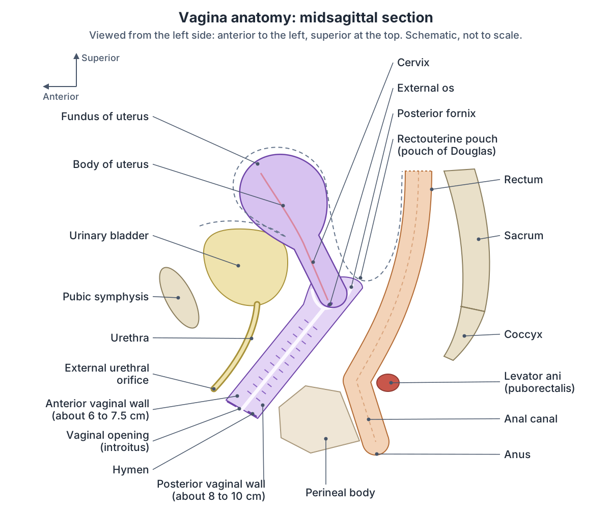
- The cervix projects into its top, creating anterior, posterior and lateral fornices; the posterior fornix is the deepest and lies just in front of the rectouterine pouch.
- Its lining is non-keratinised stratified squamous epithelium in folds called rugae; it has no glands.
- Lactobacilli keep its pH acidic, about 4.
Clinical relevance
Weakness of its supports causes pelvic organ prolapse: the bladder (cystocele) or rectum (rectocele) bulges into the vaginal wall.
Where is the vagina? The vaginal canal and its course
The vagina runs from the true pelvis, through the pelvic floor, to the perineum. The cervix protrudes into its upper end and its lower end opens into the vestibule of the vulva. It tilts backwards between the urethra in front and the rectum behind, and the urethra is bound to its front wall. (Kenhub: Vagina.)
On its way down the vagina passes, with the urethra, through the urogenital hiatus, the gap at the front of the levator ani muscles. (TeachMeAnatomy: The Pelvic Floor.)
Kenhub gives the angle of the vagina as about 45 degrees to the uterus and about 60 degrees to the horizontal when standing, varying with anatomy and with how full the bladder and bowel are. (Kenhub: Vagina.)
Radiopaedia gives a different figure, about 90 degrees between the vagina and the uterus. (Radiopaedia: Vagina.)
The canal is not straight. On MRI of women lying down, the axis of its lower part measured about 90 degrees to the horizontal reference line, its middle part about 72 degrees and its upper part about 41 degrees, giving a mid-sagittal profile the authors compared to a curved spoon. (Luo J, Betschart C, Ashton-Miller JA, DeLancey JOL. Quantitative analyses of variability in normal vaginal shape and dimension on MR images. Int Urogynecol J. 2016.)
In the 3D model the vagina is highlighted from the left side with the cervix above it, the urethra and bladder in front and the rectum behind. Turn the model to look from below to see how it passes between the two parts of levator ani shown.
How long is the vagina? Length, width and shape
At rest the vaginal canal is about 6 to 10 cm long, depending on the source and on which wall is measured, because the back wall is longer than the front. Kenhub gives 8 to 10 cm for the posterior wall and about 7.5 cm for the anterior wall. (Kenhub: Vagina.)
Radiopaedia gives a length of 6 to 8 cm. (Radiopaedia: Vagina.)
OpenStax describes the vagina as a muscular canal about 10 cm long. (OpenStax Anatomy and Physiology 2e, 27.2: Anatomy and Physiology of the Ovarian Reproductive System.)
Cleveland Clinic gives an unaroused depth of about 3 to 4 inches (7 to 10 cm), rising to about 4 to 8 inches during arousal. It notes that vaginas vary in length, width, colour and discharge, and that there is no single normal appearance. (Cleveland Clinic: Vagina.)
The most detailed measurements come from MRI of 80 women aged 28 to 70 with normal pelvic support. The anterior wall averaged 63 mm (range 44 to 84 mm) and the posterior wall 98 mm (range 51 to 144 mm). Width rose from 17 mm near the hymen to 45 mm at the top, and the inner surface area averaged 72 square centimetres. Age, height, weight, BMI and number of births together explained no more than 16% of the variation in any measurement. (Luo J, Betschart C, Ashton-Miller JA, DeLancey JOL. Quantitative analyses of variability in normal vaginal shape and dimension on MR images. Int Urogynecol J. 2016.)
How long is the vagina? Measurements by source
| Source | Method | Length | Other measurements |
|---|---|---|---|
| Radiopaedia | Reference text | 6 to 8 cm | Forms about a 90 degree angle with the uterus |
| Kenhub | Reference text | Posterior wall 8 to 10 cm; anterior wall about 7.5 cm | About 45 degrees to the uterus and 60 degrees to the horizontal when standing |
| OpenStax | Textbook | About 10 cm | pH below 4.5 |
| Cleveland Clinic | Patient information | About 7 to 10 cm unaroused; about 4 to 8 inches aroused | No single normal size or appearance |
| Luo et al. 2016 | MRI, 80 women aged 28 to 70 | Anterior wall 63 mm (44 to 84); posterior wall 98 mm (51 to 144) | Width 17 mm at the hymen to 45 mm at the top; surface area 72 cm² |
Different sources measure different things (whole canal, single walls, at rest or aroused), so their figures differ.
Parts of the vagina: walls, fornices, rugae and opening
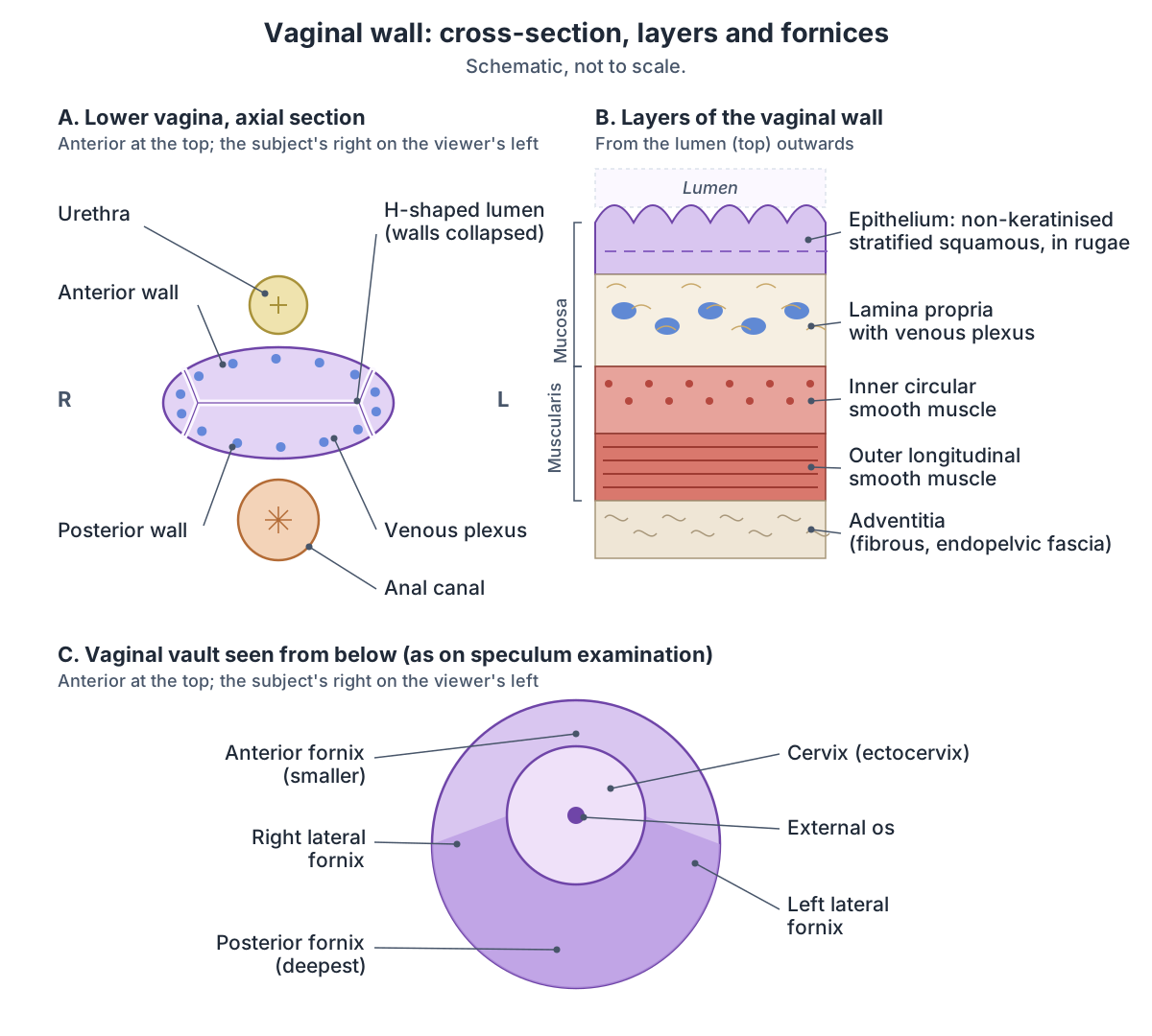
The front and back walls of the vagina normally lie against each other, so in cross-section the canal looks like an H lying on its side rather than a round tunnel. (TeachMeAnatomy: The Vagina.)
Radiopaedia divides the vagina into the vaginal orifice, which the hymen incompletely covers, the vault at its upper end, and the fornices, the recesses formed where the vagina surrounds the cervix. The walls diverge at the vault, and the posterior fornix is covered by the peritoneum of the rectouterine pouch. (Radiopaedia: Vagina.)
The fornix (vaginal vault) has a large posterior part behind the cervix, a smaller anterior part and two small lateral parts. Semen is usually deposited in the posterior fornix during intercourse. (Kenhub: Vaginal fornix.)
The posterior fornix acts as a reservoir for semen, which liquefies there over about 20 to 30 minutes, making it easier for sperm to pass through the cervical canal. (TeachMeAnatomy: The Vagina.)
Two longitudinal ridges run along the inner walls, the anterior and posterior vaginal columns, and from them the transverse folds called vaginal rugae spread out. The lower end of the anterior column forms the urethral carina, the ridge under the urethra. (Kenhub: Vagina.)
Layers of the vaginal wall
TeachMeAnatomy describes four layers from the inside out: a protective stratified squamous epithelium; an elastic lamina propria of dense connective tissue, which holds the larger veins; a fibromuscular layer of inner circular and outer longitudinal smooth muscle; and a fibrous adventitia that binds the vagina to its neighbours. (TeachMeAnatomy: The Vagina.)
Radiopaedia counts three layers, because it treats the epithelium and lamina propria together as the mucosa: a hormone-sensitive mucosa of non-keratinised stratified squamous epithelium, a muscularis with outer longitudinal and inner circular layers, and an adventitia of endopelvic fascia joining the vagina to the surrounding pelvic structures. (Radiopaedia: Vagina.)
The vagina has no glands of its own. It is moistened mainly by mucus from the cervix, with a small contribution from the greater vestibular glands. In women of childbearing age its pH is 3.8 to 4.5, because Döderlein bacilli (lactobacilli) turn glycogen in the lining into lactic acid, which discourages harmful microbes. (Kenhub: Vagina.)
OpenStax also names Lactobacillus as the main vaginal bacterium and gives a pH below 4.5. The transverse rugae of the epithelium increase the surface area and let the vagina stretch. (OpenStax Anatomy and Physiology 2e, 27.2: Anatomy and Physiology of the Ovarian Reproductive System.)
What lies around the vagina? Relations to the bladder, urethra and rectum
In front of the vagina are the bladder and urethra. Behind it are the rectouterine pouch, the rectum and the anal canal. At its sides are the ureters and the levator ani muscles. (TeachMeAnatomy: The Vagina.)
Radiopaedia lists the cervix, bladder and urethra in front, the rectouterine pouch (pouch of Douglas) and perineal body behind, and levator ani, the pelvic fascia and the ureters at the sides. On ultrasound a full bladder does not move the vagina, so it is a useful landmark even when the uterus is displaced. (Radiopaedia: Vagina.)
Relations of the vagina
| Direction | Structures |
|---|---|
| Anterior (front) | Cervix (at the top), urinary bladder, urethra bound to the front wall |
| Posterior (back) | Rectouterine pouch (behind the posterior fornix), rectum, anal canal, perineal body |
| Lateral (sides) | Ureters, levator ani, pelvic fascia |
| Superior (top) | Cervix projecting into the vault, surrounded by the fornices |
| Inferior (bottom) | Vaginal opening in the vestibule, partly covered by the hymen |
From TeachMeAnatomy, Radiopaedia and Kenhub.
What holds the vagina in place? The three levels of support
Radiopaedia lists the supports of the vagina as levator ani, the transverse cervical (cardinal), pubocervical and uterosacral ligaments, the perineal membrane and the perineal body. (Radiopaedia: Vagina.)
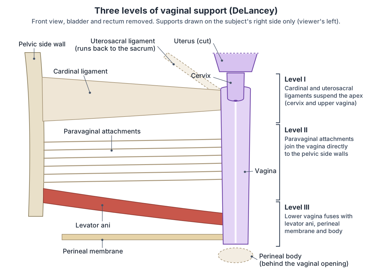
Gynaecologists group these supports into three levels described by DeLancey. Level I, at the top, has long mesenteric attachments, the cardinal and uterosacral ligaments, that suspend the apex. Level II has more direct connections to the pelvic side walls (the paravaginal attachments). At level III the vagina fuses directly with levator ani, the perineal membrane and the perineal body, so on MRI the boundary between vagina and muscle disappears. (Huebner M, DeLancey JOL. Levels of pelvic floor support: what do they look like on magnetic resonance imaging? Int Urogynecol J. 2019.)
Levator ani, the urogenital hiatus and the perineal body that support the lower vagina. Pelvic floor.
Blood supply, nerves and lymph drainage of the vagina
The vagina is supplied by the vaginal and uterine arteries, branches of the anterior division of the internal iliac artery. They join to form longitudinal azygos arteries in front of and behind the vagina. Venous plexuses in its muscular and mucosal layers drain to the internal iliac veins. (Kenhub: Vagina.)
Radiopaedia adds branches of the internal pudendal and middle rectal arteries, also from the internal iliac artery. Lymph from the upper three-quarters drains to the internal and external iliac and sacral nodes, and lymph from below the hymen to the superficial inguinal nodes. (Radiopaedia: Vagina.)
TeachMeAnatomy divides lymph drainage into thirds: the upper third to the external iliac nodes, the middle third to the internal iliac nodes and the lower third to the superficial inguinal nodes. Its nerve supply is mainly autonomic, from the uterovaginal plexus, and only the lowest fifth has somatic sensation, through the deep perineal nerve, a branch of the pudendal nerve. (TeachMeAnatomy: The Vagina.)
Kenhub gives a larger somatic territory: the upper two-thirds receive sympathetic fibres from the lumbar splanchnic nerves and parasympathetic fibres from the pelvic splanchnic nerves (S2 to S4), while the lower third also receives somatic sensory fibres through the pudendal nerve. Radiopaedia gives the pudendal territory as the lowest 2 to 3 cm, so sources describe somatic sensation as limited to roughly the lowest fifth to third of the canal. (Kenhub: Vagina.)
Blood supply, nerves and lymph of the vagina
| Supply | Details | Where sources differ |
|---|---|---|
| Arteries | Vaginal and uterine arteries from the internal iliac artery, joined as anterior and posterior azygos arteries | Radiopaedia adds the internal pudendal and middle rectal arteries |
| Veins | Vaginal venous plexus to the internal iliac veins | None |
| Autonomic nerves | Uterovaginal plexus: sympathetic (lumbar splanchnic) and parasympathetic (pelvic splanchnic, S2 to S4) fibres | None |
| Somatic (skin-type) nerves | Pudendal nerve: deep perineal and posterior labial branches | Lowest fifth (TeachMeAnatomy), lowest third (Kenhub), lowest 2 to 3 cm (Radiopaedia) |
| Lymph, upper part | External iliac nodes (upper third) | Radiopaedia: internal and external iliac and sacral nodes for the upper three-quarters |
| Lymph, middle part | Internal iliac nodes | None |
| Lymph, lower part | Superficial inguinal nodes | Radiopaedia: only the part below the hymen |
From TeachMeAnatomy, Kenhub and Radiopaedia (see references).
How the vagina develops: embryology and congenital variants
The upper two-thirds of the vagina develop, together with the cervix and uterus, from the paired Müllerian (paramesonephric) ducts. The lower third develops from the two sinovaginal bulbs of the urogenital sinus. Congenital conditions include vaginal atresia, vaginal stenosis and uterovaginal anomalies. (Radiopaedia: Vagina.)
In Mayer-Rokitansky-Küster-Hauser (MRKH) syndrome, the Müllerian ducts stop developing at about 7 weeks after fertilisation, and the uterus and upper two-thirds of the vagina are absent. It affects about 1 in 4,000 to 5,000 live female births. In the typical form (type A) the ovaries and uterine tubes are normal, and kidney anomalies are seen in 30% to 40% of those affected. (Radiopaedia: Mayer-Rokitansky-Küster-Hauser syndrome.)
Cleveland Clinic lists vaginal agenesis (an absent or underdeveloped vagina) and a vaginal septum, a wall of tissue dividing the canal, among congenital variants, and notes that the hymen at the opening varies in size, shape and thickness. (Cleveland Clinic: Vagina.)
How the vagina changes: arousal, childbirth and menopause
During arousal, blood flow to the vulva increases, the vaginal canal lengthens and widens, the cervix lifts slightly and the walls lubricate. During childbirth the walls stretch a great deal. Hormonal changes at puberty, in pregnancy and at menopause also change the vagina. (Cleveland Clinic: Vagina.)
Kenhub describes the upper two-thirds of the vagina expanding in length and width during intercourse while the uterus rises into the greater pelvis, and the vagina expanding to let the baby descend during birth. (Kenhub: Vagina.)
After menopause the vagina shortens, its fornices almost disappear, and on ultrasound the mucosa looks less bright because it has lost oestrogen stimulation. (Radiopaedia: Vagina.)
In the MRI study, age was the factor most linked to shape: older women tended to have shorter and wider vaginas, although the correlations were weak (coefficients between minus 0.4 and 0.4). (Luo J, Betschart C, Ashton-Miller JA, DeLancey JOL. Quantitative analyses of variability in normal vaginal shape and dimension on MR images. Int Urogynecol J. 2016.)
Vagina vs vulva
The vagina is an internal canal that opens to the outside of the body. The vulva is the external genitalia, which includes the vaginal opening, the labia, the clitoris and the urethral opening. (Cleveland Clinic: Vagina.)
In everyday and legal language the word vagina is often used for the whole vulva, but anatomically the vagina runs only from the hymen to the cervix. (Kenhub: Vagina.)
Labia, clitoris, vestibule, hymen and the glands around the vaginal opening. Vulva anatomy.
Clinical anatomy: prolapse, fistulas and infections
Pelvic organ prolapse is named by the wall that bulges. A cystocele is the bladder bulging into the anterior wall, the most common type; a rectocele is the rectum bulging into the posterior wall; an enterocele is small intestine bulging into the back or top of the vagina; and in vaginal vault prolapse the top of the vagina drops into the canal. (Cleveland Clinic: Pelvic Organ Prolapse.)
A vaginal (obstetric) fistula is an abnormal opening between the vagina and a neighbouring organ, usually after prolonged labour without access to caesarean section, when pressure from the baby's head cuts off blood supply. Vesicovaginal fistulas leak urine from the bladder, urethrovaginal fistulas leak urine only during voiding, and rectovaginal fistulas let faeces into the vagina. (TeachMeAnatomy: The Vagina.)
The most common vaginal condition is vaginitis, from bacterial vaginosis, sexually transmitted infections such as trichomoniasis and chlamydia, or yeast. Vaginal cancer is rare and most often linked to HPV infection. (Cleveland Clinic: Vagina.)
The uterus, cervix, uterine tubes and ovaries above the vagina. Female reproductive system.
Structures next to the vagina
The nearest named structures in the 3D model, measured from the centre of each part:
- Neck of urinary bladder · urinary
- Trigone of urinary bladder · urinary
- Pubococcygeus · muscular
- Puborectalis · muscular
- Female urethra · urinary
- External cervical os · reproductive
- Cervix of uterus · reproductive
- Internal cervical os · reproductive
- Isthmus of uterus · reproductive
- Rectum · digestive
Frequently asked questions
How long is the vaginal canal?
At rest the vagina is about 6 to 10 cm long, and the back wall is longer than the front. An MRI study of 80 women measured the anterior wall at 63 mm and the posterior wall at 98 mm on average, with wide ranges. The canal lengthens and widens during arousal and stretches greatly during childbirth.
What are the layers of the vaginal wall?
From the inside out there is a non-keratinised stratified squamous epithelium folded into rugae, an elastic lamina propria with large veins, a smooth muscle layer with inner circular and outer longitudinal fibres, and a fibrous adventitia. Some sources count the epithelium and lamina propria together as the mucosa, giving three layers instead of four.
Where is the cervix in relation to the vagina?
The cervix, the lower end of the uterus, projects into the top of the vagina. The vaginal wall wraps around it to form the fornices: a deep posterior fornix behind the cervix, a smaller anterior fornix in front and two lateral fornices. The posterior fornix lies just in front of the rectouterine pouch.
Does the vagina have glands?
No. The vaginal wall itself has no glands. It is kept moist mainly by mucus from the cervix, with a small contribution from the greater vestibular (Bartholin) glands at the vaginal opening, and the walls become lubricated during arousal. Lactobacilli in the vagina make lactic acid that keeps the pH at about 3.8 to 4.5.
What are vaginal rugae?
Rugae are the transverse folds of the vaginal lining. They spread out from two longitudinal ridges, the anterior and posterior vaginal columns. The folds increase the surface area of the wall and let the vagina stretch, for example during childbirth, and the lower end of the anterior column forms the urethral carina under the urethra.
What is the difference between the vagina and the vulva?
The vagina is the internal canal from the vaginal opening to the cervix. The vulva is the external genital area: the mons pubis, labia majora and minora, clitoris and vestibule with the urethral and vaginal openings. People often say vagina when they mean the vulva, but anatomically they are separate.
What supports the vagina?
Three levels of support hold it in place. The cardinal and uterosacral ligaments suspend the cervix and upper vagina (level I), paravaginal attachments join the vagina below that directly to the pelvic side walls (level II), and the lower vagina fuses with levator ani, the perineal membrane and the perineal body (level III). Weakness of these supports causes prolapse.
References
- Thompson L. The Vagina. TeachMeAnatomy, updated 2025.
- TeachMeAnatomy. The Pelvic Floor.
- Vagina. Radiopaedia reference article.
- Mayer-Rokitansky-Küster-Hauser syndrome. Radiopaedia reference article.
- Vagina: location, anatomy, parts, histology and function. Kenhub anatomy library.
- Vaginal fornix. Kenhub anatomy library, reviewed 2024.
- OpenStax. Anatomy and Physiology 2e, §27.2 Anatomy and Physiology of the Ovarian Reproductive System. Rice University, 2022.
- Cleveland Clinic. Vagina: Anatomy, Function, Conditions and Care.
- Cleveland Clinic. Pelvic Organ Prolapse: Types, Causes and Treatment.
- Luo J, Betschart C, Ashton-Miller JA, DeLancey JOL. Quantitative analyses of variability in normal vaginal shape and dimension on MR images. Int Urogynecol J. 2016;27(7):1087-1095.
- Huebner M, DeLancey JOL. Levels of pelvic floor support: what do they look like on magnetic resonance imaging? Int Urogynecol J. 2019;30(9):1593-1595.
- Foundational Model of Anatomy: Vagina (FMA19949). EMBL-EBI Ontology Lookup Service.
- Mitsuhashi N. et al. (2009). BodyParts3D: 3D structure database for anatomical concepts. Nucleic Acids Research 37, D782–D785. Data © The Database Center for Life Science, CC BY 4.0.
- Browne K., Schlehlein H. 3D Reference Organ Set for United, Female (Visible Human Female). Human Reference Atlas 3D Reference Object Library, HuBMAP / NIH. Data from the NLM Visible Human Project. CC BY 4.0.
Related anatomy
- Female reproductive systemLabelled female reproductive system diagrams: uterus anatomy and layers, uterine tube parts, ovaries, ligaments…
- Female anatomyFemale anatomy explained with a labelled body diagram and 3D model: reproductive organs, vulva, breasts, pelvis…
- Pregnancy anatomyPregnancy anatomy with labelled diagrams: uterus size and fundal height by week, where organs move, placenta and cord…
- VulvaVulva anatomy with labelled diagrams: mons pubis, labia, clitoris (glans, body, crura, bulbs), vestibule, hymen…
- Cervix of uterusCervix of uterus: The cervix is the narrow, cylindrical lower part of the uterus that opens into the vagina. See where…
- Female urethraFemale urethra: The female urethra is a short tube, about 4 cm long, that carries urine from the bladder to the…
- Pelvic floor and perineumPerineum and pelvic floor anatomy with labelled diagrams and a 3D model: levator ani, coccygeus, perineal body, both…
Explore the whole body in the interactive 3D human anatomy model, or browse the A–Z list of human structures.