

What is the arm in anatomy?
In anatomy, the arm is the region of the upper limb between the shoulder and the elbow, and its only bone is the humerus. OpenStax divides the upper limb into three regions: the arm, the forearm between the elbow and the wrist, and the hand beyond the wrist. (OpenStax Anatomy and Physiology 2e, 8.2: Bones of the Upper Limb.)
Kenhub makes the same point: anatomically, the arm is only the proximal part of the free upper limb, above the elbow. What people call the 'upper arm' is therefore the anatomical arm, and the whole structure from shoulder to fingertips is the upper limb (upper extremity). (Kenhub: Regions of the upper limb.)
Regions of the upper limb
Kenhub's regional guide covers the scapular region, shoulder, arm, elbow, forearm, wrist and hand. It describes the shoulder as a complex of several joints rather than a single joint, and it counts the scapula as part of the upper limb rather than the back. (Kenhub: Regions of the upper limb.)
Two hollows mark the passages into and out of the arm. The axilla (armpit) is a pyramid-shaped region below the shoulder joint and shoulder girdle, and it is the passageway for the vessels, nerves and muscles that enter and leave the upper limb. (TeachMeAnatomy: The Axilla.)
The cubital fossa is a triangular hollow over the front of the elbow. It marks the change from arm to forearm, and the main artery and nerves of the arm pass through it on their way to the forearm. (TeachMeAnatomy: The Cubital Fossa.)
Bones, joints, ligaments, bursae and muscles of the shoulder. Shoulder anatomy.
Regions of the upper limb
| Region | Extent | Bones | Key contents |
|---|---|---|---|
| Shoulder | Where the limb meets the trunk | Clavicle, scapula, head of humerus | Glenohumeral, acromioclavicular and sternoclavicular joints; deltoid; rotator cuff |
| Axilla (armpit) | Pyramid-shaped space below the shoulder joint | Humerus laterally, ribs medially | Axillary artery and vein, brachial plexus, axillary lymph nodes |
| Arm (brachial region) | Shoulder to elbow | Humerus | Biceps brachii, brachialis, coracobrachialis in front; triceps behind; brachial artery |
| Elbow and cubital fossa | Between arm and forearm | Lower humerus, upper radius and ulna | Elbow joint; biceps tendon, brachial artery, median and radial nerves |
| Forearm (antebrachial region) | Elbow to wrist | Radius, ulna | Anterior flexor and posterior extensor compartments; radial and ulnar arteries |
| Wrist (carpal region) | Forearm to hand | 8 carpal bones | Radiocarpal, ulnocarpal and midcarpal joints |
| Hand | Beyond the wrist | 5 metacarpals, 14 phalanges | Thenar, hypothenar, interosseous and lumbrical muscles; palmar arches |
Sources: Kenhub, Regions of the upper limb; TeachMeAnatomy (axilla, cubital fossa).
Arm bones: how many bones are in the upper limb?
There are 30 bones in each upper limb: the humerus, the radius and ulna, 8 carpal bones, 5 metacarpal bones and 14 phalanges. The humerus is the single bone of the upper arm; in the forearm the ulna lies medially and the radius laterally. (OpenStax Anatomy and Physiology 2e, 8.2: Bones of the Upper Limb.)
The clavicle and scapula are not in that count. They form the pectoral (shoulder) girdle, the bones that attach each upper limb to the axial skeleton, and the sternoclavicular joint between the clavicle and sternum is the only bony joint between the upper limb and the trunk. (OpenStax Anatomy and Physiology 2e, 8.1: The Pectoral Girdle.)
All the bones of the body, grouped into the axial and appendicular skeleton. Skeletal system.
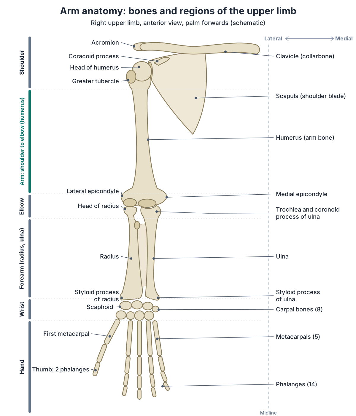
Bones of the upper limb
| Bone | Where | Number per limb | Key facts |
|---|---|---|---|
| Clavicle | Shoulder girdle, front | 1 (girdle, not in the 30) | Only bony link to the trunk, at the sternoclavicular joint; most commonly fractured bone |
| Scapula | Shoulder girdle, back | 1 (girdle, not in the 30) | Lies over ribs 2 to 7; its glenoid cavity forms the shoulder joint |
| Humerus | Arm | 1 | Single bone of the arm; radial groove on the back of the shaft |
| Ulna | Forearm, medial (little finger side) | 1 | Olecranon forms the point of the elbow |
| Radius | Forearm, lateral (thumb side) | 1 | Turns around the ulna in pronation and supination; styloid process at the wrist |
| Carpal bones | Wrist | 8 | Scaphoid, lunate, triquetrum, pisiform, trapezium, trapezoid, capitate, hamate |
| Metacarpal bones | Palm | 5 | Numbered 1 to 5 from the thumb |
| Phalanges | Thumb and fingers | 14 | Two in the thumb, three in each finger |
Sources: OpenStax Anatomy and Physiology 2e, 8.1 and 8.2; Kenhub.
Humerus: the upper arm bone
The humerus is the bone of the arm. At its upper end its head articulates with the glenoid cavity of the scapula to form the shoulder (glenohumeral) joint; at its lower end it articulates with the radius and ulna at the elbow. (Kenhub: Arm and shoulder anatomy.)
On the back of the shaft runs a shallow diagonal groove, the radial groove, which carries the radial nerve and the deep brachial (profunda brachii) artery. Just below the head, the surgical neck is a common fracture site, and the axillary nerve and posterior circumflex humeral artery wrap around it. (TeachMeAnatomy: The Humerus.)
Head, tubercles, radial groove, epicondyles and fractures of the arm bone. Humerus.
Radius and ulna: the forearm bones
The ulna is the medial bone of the forearm and the radius is the lateral one. The olecranon of the ulna forms the bony tip of the elbow, and the radius ends at the wrist in a pointed styloid process. A sheet of dense connective tissue, the interosseous membrane, unites their shafts. (OpenStax Anatomy and Physiology 2e, 8.2: Bones of the Upper Limb.)
In the anatomical position the forearm angles outwards from the line of the arm by 5 to 15 degrees. This is the carrying angle. (OpenStax Anatomy and Physiology 2e, 8.2: Bones of the Upper Limb.)
The radius and ulna meet at a proximal radioulnar joint, a pivot joint where the head of the radius turns inside the ring-shaped annular ligament. This turning lets the forearm pronate (palm down) and supinate (palm up). (OpenStax Anatomy and Physiology 2e, 9.6: Anatomy of Selected Synovial Joints.)
Head, tuberosity, distal end and fractures of the lateral forearm bone. Radius.
Clavicle and scapula: the shoulder girdle
The clavicle (collarbone) braces the scapula and shoulder joint, passes forces from the arm to the sternum and protects the nerves and vessels beneath it. OpenStax calls it the most commonly fractured bone in the body; breaks usually happen between its middle and lateral parts after a fall onto an outstretched arm or a blow to the shoulder. (OpenStax Anatomy and Physiology 2e, 8.1: The Pectoral Girdle.)
The scapula (shoulder blade) lies on the back of the chest over ribs two to seven. Its acromion, a lateral extension of the spine of the scapula, can be felt at the top of the shoulder and arches over the shoulder joint. (Kenhub: Scapula.)
Borders, angles, spine, acromion and glenoid cavity of the shoulder blade. Scapula.
Shoulder joint
The shoulder (glenohumeral) joint is a ball-and-socket joint between the head of the humerus and the glenoid cavity of the scapula. It has the largest range of motion of any joint in the body, but that mobility costs stability. Its own ligaments are weak, so the rotator cuff muscles hold it together and act as 'dynamic ligaments'. (OpenStax Anatomy and Physiology 2e, 9.6: Anatomy of Selected Synovial Joints.)
The humeral head is much larger than the glenoid fossa, and a rim of fibrocartilage, the glenoid labrum, deepens the socket. About 95% of shoulder dislocations are anterior. The axillary nerve runs close to the joint around the surgical neck of the humerus, so a dislocation can paralyse deltoid and numb the skin over the lower deltoid (the 'regimental badge' area). (TeachMeAnatomy: The Shoulder (Glenohumeral) Joint.)
Glenohumeral, acromioclavicular and sternoclavicular joints and ligaments. Shoulder anatomy.
Elbow joint
The elbow is a hinge-type synovial joint with two articulations: the trochlear notch of the ulna with the trochlea of the humerus, and the head of the radius with the capitulum of the humerus. The proximal radioulnar joint shares the same capsule. The radial collateral ligament runs from the lateral epicondyle and blends with the annular ligament, and the ulnar collateral ligament runs from the medial epicondyle to the coronoid process and olecranon. (TeachMeAnatomy: The Elbow Joint.)
Brachialis, biceps brachii and brachioradialis flex the elbow; triceps brachii and anconeus extend it. Overuse of the forearm muscle origins causes 'tennis elbow' at the lateral epicondyle (common extensor origin) and 'golfer's elbow' at the medial epicondyle (common flexor origin). (TeachMeAnatomy: The Elbow Joint.)
Humeroulnar, humeroradial and radioulnar joints, ligaments and bursae. Elbow anatomy.
Wrist joint
The wrist (radiocarpal) joint is a condyloid synovial joint between the lower end of the radius, with an articular disc, and the proximal row of carpal bones except the pisiform. The ulna does not touch the carpal bones because the articular disc separates them. The joint allows flexion, extension, adduction and abduction. (TeachMeAnatomy: The Wrist Joint.)
A fall on an outstretched hand can break the scaphoid, which causes tenderness in the anatomical snuffbox; because its blood enters at the far end, a fracture across its waist can starve the proximal piece (avascular necrosis). The same fall can cause a Colles' fracture, the most common wrist fracture, in which the end of the radius is pushed backwards into a 'dinner fork' shape. (TeachMeAnatomy: The Wrist Joint.)
Wrist bones, ligaments, carpal tunnel and anatomical snuffbox. Wrist anatomy.
The hand: carpals, metacarpals and phalanges
The hand skeleton is made of 8 carpal bones in the wrist, 5 metacarpal bones in the palm, numbered 1 to 5 from the thumb, and 14 phalanges. The thumb (pollex) has two phalanges, proximal and distal, and each finger has three: proximal, middle and distal. The pisiform is the bump you can feel at the medial base of the palm. (OpenStax Anatomy and Physiology 2e, 8.2: Bones of the Upper Limb.)
The small muscles of the hand form the thenar and hypothenar groups, the interossei and the lumbricals. Its joints are the wrist, intercarpal, carpometacarpal, metacarpophalangeal and interphalangeal joints, its blood comes through the superficial and deep palmar arches, and it is supplied by the radial, median and ulnar nerves. (Kenhub: Regions of the upper limb.)
Bones, joints, tendons, muscles and nerves of the hand, fingers and thumb. Hand anatomy.
Upper arm muscles: anterior and posterior compartments
The upper arm has two muscle compartments. The anterior compartment holds biceps brachii, coracobrachialis and brachialis, all supplied by the musculocutaneous nerve and mainly flexors. The posterior compartment holds triceps brachii, supplied by the radial nerve, which extends the elbow. Muscular branches of the brachial artery supply the front and the deep brachial artery supplies the back. (TeachMeAnatomy: Muscles of the Upper Arm.)
Brachialis arises from the medial and lateral surfaces of the humeral shaft and inserts on the ulnar tuberosity; it flexes the elbow and forms the floor of the cubital fossa. Coracobrachialis runs from the coracoid process to the medial humeral shaft, level with the deltoid tuberosity, and flexes and weakly adducts the arm at the shoulder. Triceps has three heads: the long head from the infraglenoid tubercle of the scapula and the lateral and medial heads from the humerus above and below the radial groove. All three insert on the olecranon, and the triceps reflex tests the C7 nerve root. (TeachMeAnatomy: Muscles of the Upper Arm.)
Biceps, brachialis, coracobrachialis, triceps and anconeus in detail. Arm muscles.
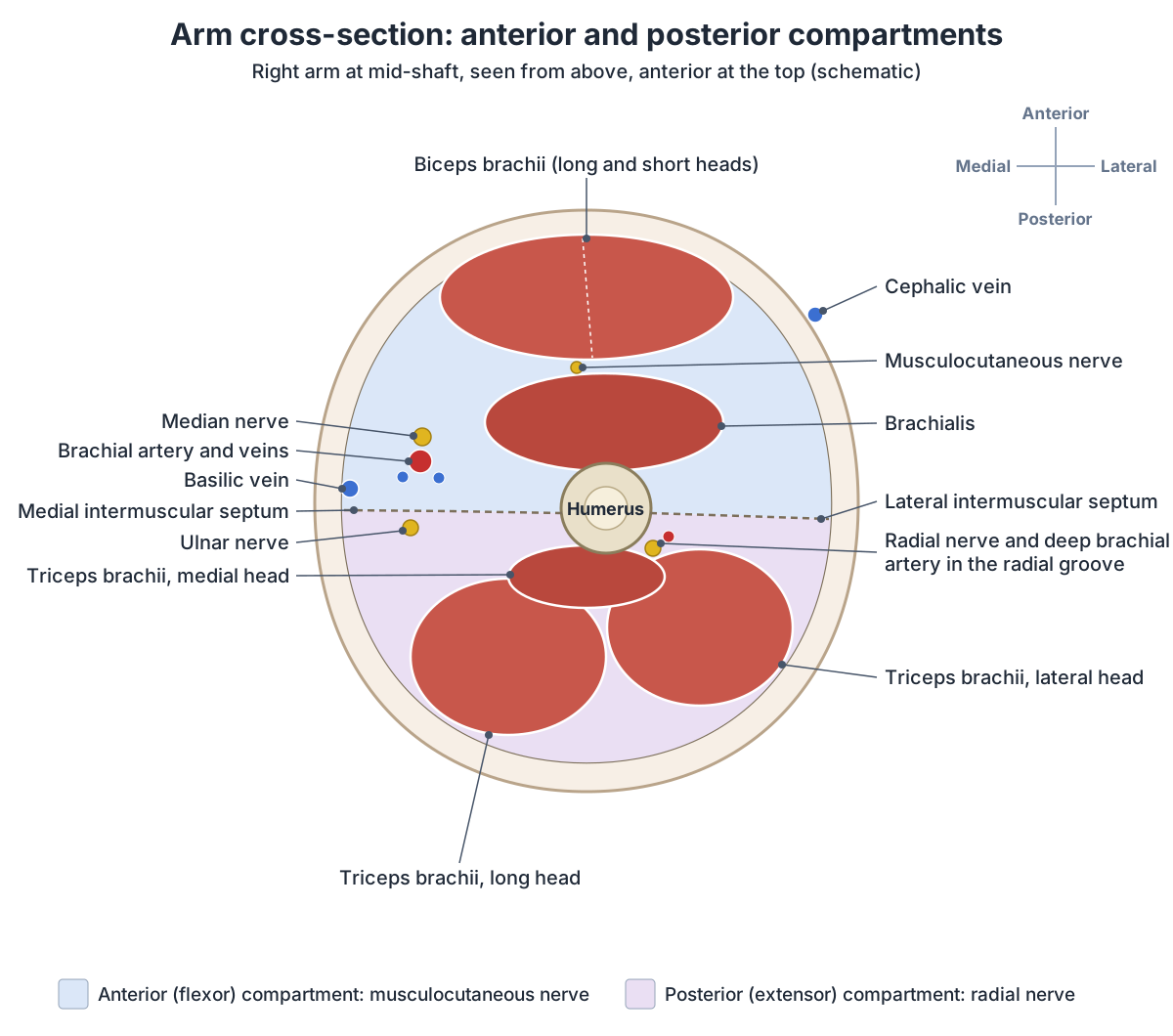
Muscles of the upper arm
| Muscle | Origin | Insertion | Nerve | Main action |
|---|---|---|---|---|
| Biceps brachii | Long head: supraglenoid tubercle; short head: coracoid process | Radial tuberosity; bicipital aponeurosis | Musculocutaneous | Supinates forearm; flexes elbow and shoulder |
| Brachialis | Medial and lateral surfaces of the humeral shaft | Ulnar tuberosity | Musculocutaneous (small radial contribution) | Flexes elbow |
| Coracobrachialis | Coracoid process | Medial humeral shaft, level with the deltoid tuberosity | Musculocutaneous | Flexes and weakly adducts the arm at the shoulder |
| Triceps brachii | Long head: infraglenoid tubercle; lateral and medial heads: humerus above and below the radial groove | Olecranon of the ulna | Radial | Extends elbow |
Source: TeachMeAnatomy, Muscles of the Upper Arm.
Biceps brachii
Biceps brachii has a long head from the supraglenoid tubercle of the scapula and a short head from the coracoid process. Both insert on the radial tuberosity and, through the bicipital aponeurosis, on the forearm fascia. Biceps supinates the forearm (turns the palm up) as well as flexing the elbow and shoulder, and the biceps reflex tests the C6 nerve root. (TeachMeAnatomy: Muscles of the Upper Arm.)
The tendon of the long head is one of the more commonly ruptured tendons, although complete ruptures are rare overall. A rupture leaves a bulge in the muscle belly when the elbow bends (the 'Popeye sign'), yet many people notice little weakness because brachialis and supinator take over. (TeachMeAnatomy: Muscles of the Upper Arm.)
Long and short heads, insertion, actions and biceps tendon injuries. Biceps anatomy.
Shoulder muscles that move the arm
Deltoid and trapezius give the shoulder its contour. Deeper lie the four rotator cuff muscles, remembered as SITS: supraspinatus, which also stabilises the humeral head, starts abduction of the arm up to about 15 degrees, infraspinatus and teres minor rotate the arm laterally, and subscapularis rotates it medially. The suprascapular nerve supplies supraspinatus and infraspinatus, the axillary nerve supplies teres minor, and the subscapular nerves supply subscapularis. (Kenhub: Arm and shoulder anatomy.)
After supraspinatus starts abduction, the middle fibres of deltoid continue it from about 15 to 90 degrees, and trapezius and serratus anterior then rotate the scapula to raise the arm higher. Pectoralis major, the anterior deltoid and coracobrachialis flex the shoulder; the posterior deltoid, latissimus dorsi and teres major extend it. (TeachMeAnatomy: The Shoulder (Glenohumeral) Joint.)
Supraspinatus, infraspinatus, teres minor and subscapularis. Rotator cuff.
Forearm muscles: flexors and extensors
The anterior compartment of the forearm has 8 muscles in three layers and mainly flexes the wrist and fingers and pronates the forearm. The superficial layer (flexor carpi ulnaris, palmaris longus, flexor carpi radialis and pronator teres) shares a common origin on the medial epicondyle. The median nerve supplies most of the compartment; the ulnar nerve supplies flexor carpi ulnaris and the medial half of flexor digitorum profundus. (TeachMeAnatomy: Muscles in the Anterior Compartment of the Forearm.)
The posterior compartment has 12 muscles in two layers: seven superficial, including brachioradialis and anconeus, and five deep. Four superficial extensors share a common origin on the lateral epicondyle. All are supplied by the radial nerve or its branches; they extend the wrist and fingers, and supinator turns the palm up. (TeachMeAnatomy: Muscles in the Posterior Compartment of the Forearm.)
All 20 forearm muscles by layer, with nerves and actions. Forearm muscles.
Nerves of the arm: the brachial plexus
All the main nerves of the upper limb come from the brachial plexus, formed by the anterior rami of C5, C6, C7, C8 and T1. The roots pass between the anterior and middle scalene muscles, join into three trunks in the neck, split into divisions and regroup in the axilla as lateral, posterior and medial cords, named by their position around the axillary artery. The cords give five major terminal branches. (TeachMeAnatomy: The Brachial Plexus.)
The musculocutaneous nerve pierces coracobrachialis and runs down the front of the arm between biceps brachii and brachialis, then continues as the lateral cutaneous nerve of the forearm. (TeachMeAnatomy: The Musculocutaneous Nerve.)
The median nerve runs down the arm beside the brachial artery, first lateral to it; halfway down it crosses the artery to lie on its medial side. It enters the forearm through the cubital fossa, passes between the two heads of pronator teres and reaches the hand through the carpal tunnel, where compression causes carpal tunnel syndrome. (TeachMeAnatomy: The Median Nerve.)
The ulnar nerve runs down the medial side of the arm, pierces the medial intermuscular septum at mid-arm to enter the posterior compartment, and passes behind the elbow through the ulnar tunnel between the medial epicondyle and the olecranon. (TeachMeAnatomy: The Ulnar Nerve.)
The radial nerve, from the posterior cord, winds around the back of the humerus in the radial groove, passes in front of the lateral epicondyle and divides into a deep motor branch and a superficial sensory branch. A fracture of the humeral shaft can injure it in the groove, causing wrist drop. (TeachMeAnatomy: The Radial Nerve.)
Brachial plexus roots, trunks, cords and every terminal branch. Nerves of the arm.
Terminal branches of the brachial plexus
| Nerve | Roots | Main muscles | Skin |
|---|---|---|---|
| Musculocutaneous | C5–C7 | Biceps brachii, brachialis, coracobrachialis | Lateral forearm |
| Axillary | C5–C6 | Deltoid, teres minor | Over the lower deltoid |
| Radial | C5–T1 | Triceps and the extensors of the forearm | Back of the arm and forearm, back of the hand on the thumb side |
| Median | C6–T1 (sometimes C5) | Most forearm flexors, thenar muscles, two lateral lumbricals | Palm and lateral fingers |
| Ulnar | C8–T1 | Most small hand muscles, flexor carpi ulnaris, medial half of flexor digitorum profundus | Medial fingers and palm |
Source: TeachMeAnatomy, The Brachial Plexus.
Arteries and veins of the arm
The right subclavian artery arises from the brachiocephalic trunk and the left directly from the aortic arch. Each becomes the axillary artery at the lateral border of the first rib and the brachial artery at the lower border of teres major. The brachial artery, the main artery of the arm, gives off the deep brachial artery, which runs with the radial nerve in the radial groove, and divides in the cubital fossa, under the bicipital aponeurosis, into the radial and ulnar arteries. (TeachMeAnatomy: Arterial Supply to the Upper Limb.)
In the hand, the ulnar artery forms the superficial palmar arch and the radial artery, after crossing the anatomical snuffbox, forms the deep palmar arch. The radial pulse is felt at the wrist just lateral to the tendon of flexor carpi radialis. (TeachMeAnatomy: Arterial Supply to the Upper Limb.)
Deep veins travel in pairs beside the arteries and share their names; the brachial veins are the largest. The two big superficial veins start from the dorsal venous network of the hand: the cephalic vein climbs the lateral side and runs in the groove between deltoid and pectoralis major to the axillary vein, and the basilic vein climbs the medial side. The median cubital vein links them in front of the elbow and is a common site for taking blood. (TeachMeAnatomy: Venous Drainage of the Upper Limb.)
The basilic vein pierces the deep fascia of the arm halfway between the elbow and the axilla, runs medial to the brachial artery and joins the brachial veins in the axilla to form the axillary vein. (Radiopaedia: Basilic vein.)
Subclavian to palmar arches, superficial and deep veins, pulses. Arteries and veins of the arm.
Armpit (axilla) and elbow pit (cubital fossa)
The axilla is bounded in front by pectoralis major, pectoralis minor and subclavius, behind by subscapularis, teres major and latissimus dorsi, medially by serratus anterior and the chest wall and laterally by the humerus, coracobrachialis and biceps. It contains the axillary artery and vein, the brachial plexus and the axillary lymph nodes, which receive about 75% of the lymph from the breast. (TeachMeAnatomy: The Axilla.)
The cubital fossa is bordered by brachioradialis laterally, pronator teres medially and a line between the humeral epicondyles above. From lateral to medial it contains the radial nerve, the biceps tendon, the brachial artery and the median nerve. The brachial pulse is felt just medial to the biceps tendon, and the median cubital vein crosses the roof of the fossa. (TeachMeAnatomy: The Cubital Fossa.)
Walls, apex, base and contents of the armpit. Axilla anatomy.
Common arm injuries and the nerves at risk
Each part of the humerus has a structure at risk. A surgical neck fracture endangers the axillary nerve; a mid-shaft fracture endangers the radial nerve in the radial groove, causing wrist drop; and a supracondylar fracture above the elbow can damage the brachial artery, whose loss of supply can lead to Volkmann's ischaemic contracture, a fixed bending of the wrist and fingers. (TeachMeAnatomy: The Humerus.)
The 'funny bone' is not a bone but the ulnar nerve, which runs just under the skin at the elbow. Bumping the elbow briefly pinches the nerve and causes a shock-like tingling. (Cleveland Clinic: Ulnar Nerve (Funny Bone).)
Injury to the upper roots of the brachial plexus (C5 and C6, Erb's palsy), often from stretching during a difficult birth, leaves the arm hanging in a 'waiter's tip' position. Injury to the lower roots (C8 and T1, Klumpke's palsy) causes a clawed hand. (TeachMeAnatomy: The Brachial Plexus.)
Olecranon, trochlear notch, shaft and distal head of the medial forearm bone. Ulna.
Exploring arm anatomy in the 3D model
The 3D view uses the BodyParts3D adult male data set. It highlights the clavicle, scapula, humerus, radius, ulna and the interosseous membrane of the forearm; the three parts of deltoid; biceps brachii, brachialis, coracobrachialis and the three heads of triceps; and the brachial artery with the cephalic, basilic and median cubital veins. The carpal bones and hand stay visible in grey. The model has no nerves of the brachial plexus, no ligaments of the shoulder or elbow and no lymph nodes, so the text and diagrams cover these.
Use landmarks you can feel on your own arm to orient the model: the medial epicondyle, the prominent bump on the inner side of the elbow; the olecranon at the point of the elbow; the styloid process of the radius on the thumb side of the wrist; and the pisiform at the medial base of the palm. (OpenStax Anatomy and Physiology 2e, 8.2: Bones of the Upper Limb.)
Arm anatomy in the 3D model
- Acromial part of deltoid
- The middle fibres of the deltoid, forming the rounded contour of the shoulder.
- Basilic vein
- The basilic vein is a large superficial vein on the medial side of the arm.
- Brachial artery
- The brachial artery is the main artery of the arm, continuing the axillary artery.
- Brachialis
- The main flexor of the elbow, lying deep to biceps brachii.
- Cephalic vein
- The cephalic vein is a large superficial vein on the lateral side of the upper limb.
- Clavicle
- The collarbone, an S-shaped long bone running between the sternum and the shoulder.
- Clavicular part of deltoid
- The anterior fibres of the deltoid.
- Coracobrachialis
- A slender muscle on the medial side of the upper arm.
- Humerus
- The long bone of the upper arm.
- Interosseous membrane of forearm
- A broad fibrous sheet joining the radius and ulna.
- Lateral head of triceps brachii
- The lateral head of the triceps.
- Long head of biceps brachii
- The long head of biceps, passing through the shoulder joint.
- Long head of triceps brachii
- The long head of triceps, crossing the shoulder joint.
- Medial head of triceps brachii
- The deep head of triceps, lying beneath the long and lateral heads.
- Median cubital vein
- A superficial vein across the front of the elbow linking the cephalic and basilic veins.
- Radius
- The lateral bone of the forearm.
- Scapula
- The shoulder blade, a flat triangular bone on the back of the thorax.
- Short head of biceps brachii
- The medial head of biceps brachii at the front of the arm.
- Spinal part of deltoid
- The posterior fibers of deltoid arising from the scapular spine.
- Ulna
- The medial bone of the forearm.
Frequently asked questions
What are the 3 bones of the arm?
People usually mean the humerus, radius and ulna. Strictly, the arm (shoulder to elbow) has only one bone, the humerus; the radius and ulna are the bones of the forearm, with the ulna on the little finger side and the radius on the thumb side. The clavicle and scapula form the shoulder girdle that attaches the limb to the trunk.
Is the forearm part of the arm?
Not in anatomy. The upper limb has three regions: the arm between the shoulder and the elbow, the forearm between the elbow and the wrist, and the hand. In everyday speech 'arm' means the whole upper limb, and 'upper arm' means what anatomists call the arm. Exams and medical notes use the strict terms.
How many bones are in the arm and hand?
Each upper limb has 30 bones: the humerus, radius and ulna, 8 carpal bones in the wrist, 5 metacarpals in the palm and 14 phalanges in the fingers and thumb. The thumb has two phalanges and each finger has three. The clavicle and scapula of the shoulder girdle are counted separately, which makes 32 bones per side including the girdle.
What are the main muscles of the upper arm?
The front (anterior) compartment holds biceps brachii, brachialis and coracobrachialis, supplied by the musculocutaneous nerve; they flex the elbow and shoulder, and biceps also turns the palm up. The back (posterior) compartment holds triceps brachii, supplied by the radial nerve, which straightens the elbow. Deltoid caps the shoulder and lifts the arm.
Which nerves supply the arm?
All come from the brachial plexus (C5 to T1). Its five main branches are the musculocutaneous nerve (front of the arm), the axillary nerve (deltoid and teres minor), the radial nerve (triceps and the forearm extensors), the median nerve (most forearm flexors and the thumb muscles) and the ulnar nerve (most small muscles of the hand).
What is the funny bone?
It is not a bone. The 'funny bone' is the ulnar nerve, which passes behind the elbow between the medial epicondyle of the humerus and the olecranon of the ulna. There it lies just under the skin, so a knock on that spot briefly pinches the nerve and causes a shock-like tingling.
What is the main artery of the arm?
The brachial artery. It continues the axillary artery from the lower border of teres major, runs down the inner side of the arm and divides in the cubital fossa, in front of the elbow, into the radial and ulnar arteries. Its pulse can be felt in the cubital fossa, just medial to the biceps tendon.
References
- OpenStax. Anatomy and Physiology 2e, §8.1 The Pectoral Girdle. Rice University, 2022.
- OpenStax. Anatomy and Physiology 2e, §8.2 Bones of the Upper Limb. Rice University, 2022.
- OpenStax. Anatomy and Physiology 2e, §9.6 Anatomy of Selected Synovial Joints. Rice University, 2022.
- Kenhub. Regions of the upper limb.
- Kenhub. Arm and shoulder anatomy.
- Kenhub. Scapula.
- TeachMeAnatomy. Muscles of the Upper Arm.
- TeachMeAnatomy. Muscles in the Anterior Compartment of the Forearm.
- TeachMeAnatomy. Muscles in the Posterior Compartment of the Forearm.
- TeachMeAnatomy. The Shoulder (Glenohumeral) Joint.
- TeachMeAnatomy. The Elbow Joint.
- TeachMeAnatomy. The Wrist Joint.
- TeachMeAnatomy. The Humerus.
- TeachMeAnatomy. The Brachial Plexus.
- TeachMeAnatomy. The Musculocutaneous Nerve.
- TeachMeAnatomy. The Median Nerve.
- TeachMeAnatomy. The Ulnar Nerve.
- TeachMeAnatomy. The Radial Nerve.
- TeachMeAnatomy. Arterial Supply to the Upper Limb.
- TeachMeAnatomy. Venous Drainage of the Upper Limb.
- TeachMeAnatomy. The Axilla.
- TeachMeAnatomy. The Cubital Fossa.
- Radiopaedia. Basilic vein.
- Cleveland Clinic. Ulnar Nerve (Funny Bone): What Is It, Branches & Function.
- Mitsuhashi N. et al. (2009). BodyParts3D: 3D structure database for anatomical concepts. Nucleic Acids Research 37, D782–D785. Data © The Database Center for Life Science, CC BY 4.0.
Related anatomy
- Arm musclesArm muscles labeled in 3D: biceps brachii, brachialis, coracobrachialis, triceps brachii, deltoid and anconeus, with…
- Forearm musclesMuscles of the forearm explained: every anterior flexor and posterior extensor, with origin, insertion, nerve and…
- HumerusHumerus bone labeled: head, anatomical and surgical neck, tubercles, deltoid tuberosity, radial groove, epicondyles…
- Elbow jointElbow anatomy with labelled diagrams and a 3D model: humerus, radius and ulna, the three joints, collateral and…
- Upper limb nervesBrachial plexus diagram with roots, trunks, cords and branches; axillary, musculocutaneous, median, ulnar and radial…
- Upper limb blood vesselsArteries and veins of the arm and hand: subclavian, axillary branches, brachial, radial and ulnar arteries, palmar…
Explore the whole body in the interactive 3D human anatomy model, or browse the A–Z list of human structures.