

Where is the heart located?
The heart lies in the mediastinum, the central space of the chest between the two lungs. Its back surface lies close to the bodies of the thoracic vertebrae, and its front surface lies deep to the sternum and the costal cartilages. (OpenStax Anatomy and Physiology 2e, 19.1: Heart Anatomy.)
It is not centred. About one-third of the heart lies to the right of the midline and two-thirds to the left, behind the body of the sternum. Its base points backwards and its apex points down and to the left. (Radiopaedia: Heart.)
The base of the heart sits at about the level of the third costal cartilage. The apex lies just left of the sternum, between the junctions of the fourth and fifth ribs with their costal cartilages. Because the apex points to the left, the left lung has a dent for it, the cardiac notch. (OpenStax Anatomy and Physiology 2e, 19.1: Heart Anatomy.)
Underneath, the heart rests on the diaphragm: its inferior (diaphragmatic) surface, formed by both ventricles, lies mainly on the central tendon of the diaphragm. (Radiopaedia: Heart.)
The chest wall, lungs and mediastinum around the heart are covered in Chest anatomy.
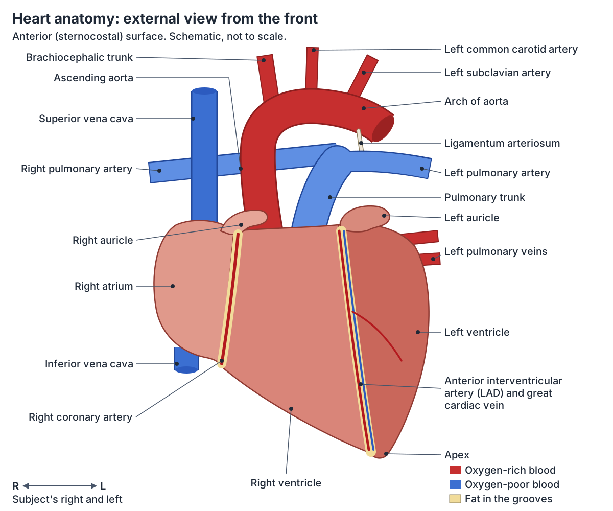
How big is the heart? Size, weight and shape of a real heart
An adult heart is about 12 cm (5 in) long, 8 cm (3.5 in) wide and 6 cm (2.5 in) thick. It weighs about 250 to 300 g in females and 300 to 350 g in males. Regular exercise can enlarge it, and so can disease such as hypertrophic cardiomyopathy. (OpenStax Anatomy and Physiology 2e, 19.1: Heart Anatomy.)
Radiopaedia gives similar figures: 12 × 8.5 × 6 cm, with an average weight of about 310 g in males and 255 g in females. (Radiopaedia: Heart.)
A real (anatomical) heart looks nothing like the ♥ symbol. It is shaped more like a pinecone: broad at the top, where the great arteries and veins attach, and tapering to the apex. OpenStax calls that broad upper end the base. (OpenStax Anatomy and Physiology 2e, 19.1: Heart Anatomy.)
Clinical anatomy books describe the heart as a pyramid that has fallen over. In that description the base is the back surface, formed by the left atrium, and the apex points forwards and downwards. (TeachMeAnatomy: The Surfaces and Borders of the Heart.)
Cleveland Clinic describes the heart as a fist-sized organ that sits in the front of the chest, behind the sternum and slightly left of centre, with the left lung a little smaller to make room for it. (Cleveland Clinic: Heart.)
Surfaces and borders of the heart
Each surface of the heart is formed mainly by one chamber. The anterior (sternocostal) surface is the right ventricle, the inferior (diaphragmatic) surface is both ventricles, the posterior surface (base) is the left atrium, the right pulmonary surface is the right atrium and the left pulmonary surface is the left ventricle. (TeachMeAnatomy: The Surfaces and Borders of the Heart.)
Seen from the front, the right border of the heart is made by the right atrium between the superior and inferior venae cavae. The left border is the left ventricle, with the left atrium, pulmonary trunk and arch of the aorta above it. The inferior border is the right ventricle, and the superior border is the two atria with the great vessels. (Radiopaedia: Heart.)
TeachMeAnatomy lists both ventricles along the inferior border, and the left ventricle with a small part of the left atrium along the left border, so textbooks differ slightly on how much of the lower edge belongs to the left ventricle. (TeachMeAnatomy: The Surfaces and Borders of the Heart.)
Fat-filled grooves (sulci) on the surface carry the coronary vessels. The deep coronary sulcus runs around the heart between the atria and the ventricles. The anterior and posterior interventricular sulci run down the front and back, marking the septum between the two ventricles. (OpenStax Anatomy and Physiology 2e, 19.1: Heart Anatomy.)
The coronary (atrioventricular) sulcus holds the right coronary artery, the circumflex artery, the small cardiac vein and the coronary sinus. The anterior interventricular sulcus holds the left anterior descending artery and the great cardiac vein, and the posterior one holds the posterior interventricular artery and the middle cardiac vein. (TeachMeAnatomy: The Surfaces and Borders of the Heart.)
Surfaces and borders of the heart
| Surface or border | Formed mainly by | Notes |
|---|---|---|
| Anterior (sternocostal) surface | Right ventricle | Lies behind the sternum and costal cartilages |
| Inferior (diaphragmatic) surface | Both ventricles | Rests mainly on the central tendon of the diaphragm |
| Posterior surface (base) | Left atrium | Receives the four pulmonary veins |
| Right (pulmonary) surface | Right atrium | Faces the right lung |
| Left (pulmonary) surface | Left ventricle | Sits in the cardiac notch of the left lung |
| Right border | Right atrium | Between the superior and inferior venae cavae |
| Left border | Left ventricle (and a little left atrium) | Pulmonary trunk and aortic arch lie above it |
| Inferior border | Right ventricle (both ventricles in some texts) | Ends at the apex |
| Apex | Left ventricle | Points down, forwards and to the left |
Compiled from TeachMeAnatomy, Radiopaedia and OpenStax (see references).
Pericardium: the sac around the heart
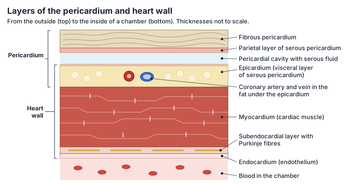
The heart sits inside a double-walled sac, the pericardium. Its tough outer layer, the fibrous pericardium, is dense connective tissue that protects the heart and holds it in place. Inside it, the serous pericardium has two layers: the parietal pericardium, fused to the fibrous layer, and the visceral pericardium (epicardium), fused to the heart. (OpenStax Anatomy and Physiology 2e, 19.1: Heart Anatomy.)
Between the two serous layers is the pericardial cavity. It holds a thin film of lubricating serous fluid, secreted by the mesothelial cells, so the heart can beat with little friction. (OpenStax Anatomy and Physiology 2e, 19.1: Heart Anatomy.)
The fibrous pericardium is continuous with the central tendon of the diaphragm, is attached to the sternum and blends with the outer coat (tunica adventitia) of the great vessels. It fixes the heart in the mediastinum, stops it filling too quickly and shields it from infection in neighbouring organs. Its sensory nerves are the phrenic nerves (C3 to C5), so pain from the pericardium can be felt in the shoulder. (TeachMeAnatomy: The Pericardium.)
Where the pericardium folds around the great vessels it leaves two passages. The transverse pericardial sinus lies behind the ascending aorta and pulmonary trunk and in front of the superior vena cava, separating the arteries from the veins; surgeons pass a clamp through it during coronary artery bypass surgery. The oblique pericardial sinus is a blind-ending pocket behind the heart. (TeachMeAnatomy: The Surfaces and Borders of the Heart.)
Fluid or blood that collects quickly in the pericardial cavity compresses the heart, a condition called cardiac tamponade. As little as 100 mL gathering rapidly after an injury can cause it, while a slowly growing effusion may reach more than a litre before it does. (OpenStax Anatomy and Physiology 2e, 19.1: Heart Anatomy.)
Layers of the heart wall: epicardium, myocardium and endocardium
The heart wall has three layers. From outside to inside they are the epicardium, which is the same layer as the visceral pericardium, the myocardium and the endocardium. (OpenStax Anatomy and Physiology 2e, 19.1: Heart Anatomy.)
The myocardium is the thickest layer. It is cardiac muscle supported by a framework of collagen fibres, and its muscle bundles spiral around the chambers in a figure-of-eight pattern that makes pumping more efficient. (OpenStax Anatomy and Physiology 2e, 19.1: Heart Anatomy.)
The endocardium lines the chambers and covers the valves. It is a simple squamous endothelium, continuous with the lining of the blood vessels. (OpenStax Anatomy and Physiology 2e, 19.1: Heart Anatomy.)
Two thin connective tissue layers sit between them. The subendocardial layer, under the endocardium, carries the Purkinje fibres of the conducting system. The subepicardial layer, under the epicardium, contains the coronary vessels and nerves. (TeachMeAnatomy: The Heart Wall.)
Each layer has its own inflammation. Endocarditis, usually a bacterial infection, mostly affects the valves and can damage them. Myocarditis often follows a viral infection such as coxsackie B and can weaken the heart muscle or cause arrhythmias. (TeachMeAnatomy: The Heart Wall.)
Cardiac muscle is compared with skeletal and smooth muscle and the other tissues of the body in Tissue types.
The four chambers of the heart
The heart has four chambers: two atria on top and two ventricles at the bottom. Cleveland Clinic notes that the left ventricle is slightly larger than the right. (Cleveland Clinic: Heart.)
The atria are receiving chambers: the right atrium takes blood from the venae cavae and the left atrium from the pulmonary veins. Each ventricle receives blood from the atrium above it and pumps it out of the heart, the right ventricle into the pulmonary trunk and the left into the aorta. (TeachMeAnatomy: The Chambers of the Heart.)
The chambers are separated by the interatrial and interventricular septa and, between atria and ventricles, by the atrioventricular septa. The right side handles oxygen-poor blood on its way to the lungs and the left side oxygen-rich blood on its way to the body. (Kenhub: Heart anatomy.)
Each atrium has an auricle (atrial appendage), a small ear-shaped pouch on its front that increases its capacity. The auricles are leaf-like and thin-walled. (TeachMeAnatomy: The Chambers of the Heart.)
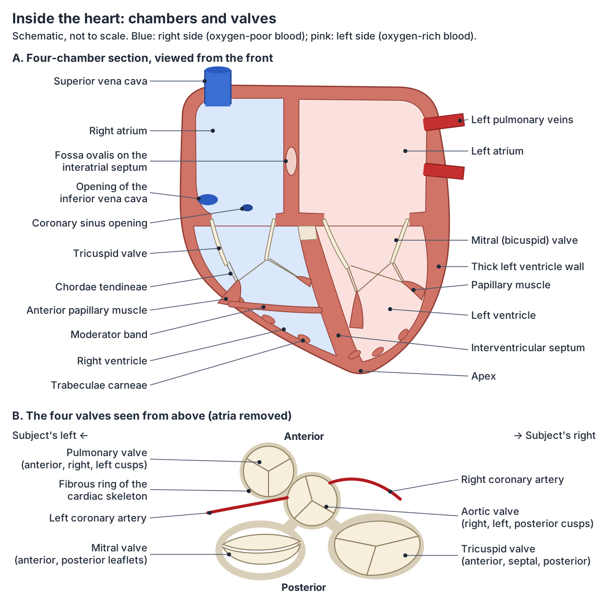
Right atrium
The right atrium receives oxygen-poor blood from the superior and inferior venae cavae and from the heart's own veins, and passes it through the tricuspid valve into the right ventricle. It forms the right border of the heart. (TeachMeAnatomy: The Chambers of the Heart.)
Inside, a muscular ridge called the crista terminalis divides it in two. Behind the ridge is the smooth-walled sinus venarum, which receives both venae cavae and develops from the embryonic sinus venosus. In front of it is the rough atrium proper, lined by pectinate muscles, which includes the right auricle. (TeachMeAnatomy: The Chambers of the Heart.)
The coronary sinus also opens into the right atrium. On its inner (septal) wall is the fossa ovalis, a shallow oval dip that marks the foramen ovale, the opening that let blood pass from the right to the left atrium and bypass the lungs before birth. (OpenStax Anatomy and Physiology 2e, 19.1: Heart Anatomy.)
If that opening does not seal after birth the result is an atrial septal defect, most often a patent foramen ovale. Left atrial pressure is normally higher, so blood shunts from left to right, and a large defect can overload the right ventricle. (TeachMeAnatomy: The Chambers of the Heart.)
Right ventricle
The right ventricle receives blood through the tricuspid valve and pumps it through the pulmonary valve into the pulmonary trunk. It is roughly triangular and forms most of the front (sternocostal) surface of the heart. (TeachMeAnatomy: The Chambers of the Heart.)
In cross-section it is crescent-shaped, wrapping around the front of the left ventricle. Its wall is thinner than that of the left ventricle because the pressures on the right side are lower. (Radiopaedia: Right ventricle.)
A muscular ridge, the supraventricular crest, separates the inflow part, whose walls are covered by irregular muscular ridges called trabeculae carneae, from the smooth outflow part, the conus arteriosus (infundibulum), which leads up to the pulmonary valve. (TeachMeAnatomy: The Chambers of the Heart.)
The moderator band (septomarginal trabecula) crosses the cavity from the lower interventricular septum to the anterior papillary muscle. It carries the right branch of the atrioventricular bundle, part of the conduction system. (Radiopaedia: Right ventricle.)
Left atrium
The left atrium receives oxygen-rich blood from the four pulmonary veins and pumps it through the mitral valve into the left ventricle. It forms most of the base, the back surface of the heart. (TeachMeAnatomy: The Chambers of the Heart.)
The part that receives the pulmonary veins has smooth walls. Pectinate muscles line only the front part and the left auricle, which curls forwards over the root of the pulmonary trunk. (TeachMeAnatomy: The Chambers of the Heart.)
Most blood flows from the atria into the ventricles before the atria contract. Atrial contraction then adds the last part, about 20 percent of ventricular filling. (OpenStax Anatomy and Physiology 2e, 19.1: Heart Anatomy.)
Left ventricle
The left ventricle receives blood from the left atrium through the mitral valve and pumps it through the aortic valve into the aorta. It forms the apex of the heart and most of its left and diaphragmatic borders. (TeachMeAnatomy: The Chambers of the Heart.)
Its wall is much thicker than the right ventricle's because it must generate enough pressure to drive blood through the whole systemic circuit, not just the lungs. (OpenStax Anatomy and Physiology 2e, 19.1: Heart Anatomy.)
Normal values from cardiac catheterisation show the difference: the left ventricle reaches a systolic pressure of about 90 to 140 mm Hg, while the pulmonary artery, fed by the right ventricle, has a systolic pressure of only 15 to 30 mm Hg. (Davis's Lab and Diagnostic Tests: Cardiac catheterization (normal pressures).)
The left ventricle is conical and longer than the right. Its wall is thickest at the base and thins to only 1 to 2 mm at the apex. Fine trabeculae carneae line it, mainly near the apex, while its inflow and outflow tracts are smooth. (Radiopaedia: Left ventricle.)
The smooth outflow part just below the aortic valve is called the aortic vestibule. (TeachMeAnatomy: The Chambers of the Heart.)
Chambers of the heart at a glance
| Chamber | Receives blood from | Sends blood through | Key internal features |
|---|---|---|---|
| Right atrium | Superior and inferior venae cavae, coronary sinus | Tricuspid valve to the right ventricle | Crista terminalis, pectinate muscles, right auricle, fossa ovalis |
| Right ventricle | Right atrium | Pulmonary valve to the pulmonary trunk | Trabeculae carneae, three papillary muscles, moderator band, conus arteriosus |
| Left atrium | Four pulmonary veins | Mitral valve to the left ventricle | Smooth walls, pectinate muscles only in the left auricle |
| Left ventricle | Left atrium | Aortic valve to the aorta | Thickest wall, two papillary muscles, fine trabeculae, aortic vestibule |
Septum of the heart and the fibrous skeleton
Walls called septa divide the heart. The interatrial septum separates the atria and carries the fossa ovalis. The interventricular septum separates the ventricles and is much thicker, because the ventricles generate far greater pressure than the atria. (OpenStax Anatomy and Physiology 2e, 19.1: Heart Anatomy.)
The interventricular septum has a small, thin membranous part at the top, which forms part of the fibrous skeleton, and a large muscular part that is as thick as the wall of the left ventricle. (TeachMeAnatomy: The Chambers of the Heart.)
The septum is concave towards the left ventricle and bulges into the right ventricle. (Radiopaedia: Left ventricle.)
Between the atria and the ventricles lies the atrioventricular septum. It contains the openings of the four valves and is reinforced by the cardiac (fibrous) skeleton, dense connective tissue that forms four rings to anchor the valves. The skeleton also forms an electrical boundary between the atria and the ventricles. (OpenStax Anatomy and Physiology 2e, 19.1: Heart Anatomy.)
Defects of the septa are among the commonest congenital heart conditions. In tetralogy of Fallot, a single developmental fault produces a ventricular septal defect, an aorta that overrides it, narrowing of the pulmonary valve and a thickened right ventricle, so oxygen-poor blood reaches the aorta. (TeachMeAnatomy: The Chambers of the Heart.)
Heart valves: tricuspid, pulmonary, mitral and aortic
Four valves keep blood moving one way. The atrioventricular valves lie between the atria and ventricles: the tricuspid valve on the right, with anterior, septal and posterior cusps, and the mitral (bicuspid) valve on the left, with anterior and posterior cusps. The semilunar valves guard the outflow arteries: the pulmonary valve (left, right and anterior cusps) and the aortic valve (right, left and posterior cusps). (TeachMeAnatomy: The Valves of the Heart.)
The atrioventricular valves are tied by chordae tendineae to papillary muscles. The semilunar valves have neither; each works as three pocket-like flaps. (OpenStax Anatomy and Physiology 2e, 19.1: Heart Anatomy.)
When the ventricles relax, blood falling back from the aorta and pulmonary trunk fills the small pockets (sinuses) behind the semilunar cusps and presses the cusps together. Each cusp has a thickened free edge, the lunule, which is widest at its midpoint, the nodule. The two coronary arteries open from the left and right aortic sinuses. (TeachMeAnatomy: The Valves of the Heart.)
The heart sounds come from valves closing. The first sound, S1 or 'lub', is the atrioventricular valves closing as the ventricles start to contract; the second, S2 or 'dub', is the semilunar valves closing as the ventricles relax. (OpenStax Anatomy and Physiology 2e, 19.3: Cardiac Cycle.)
Each valve is heard best at its own spot on the chest: the aortic valve at the right second intercostal space beside the sternum, the pulmonary valve at the left second intercostal space, the tricuspid valve at the lower left sternal edge, and the mitral valve at the left fifth intercostal space in the midclavicular line. (Kenhub: Heart valves.)
The tricuspid opening is supported by a fibrous ring about 10 to 11 cm around. The aortic and mitral valves are joined by fibrous tissue, while the pulmonary valve's skeleton is separate from the others. (Kenhub: Heart valves.)
Aortic stenosis is the valve disease most often met in anatomy teaching. Age-related calcification, a congenital bicuspid aortic valve or rheumatic fever narrows the valve, the left ventricle thickens, and severe disease causes breathlessness, fainting and angina. (TeachMeAnatomy: The Valves of the Heart.)
The four heart valves
| Valve | Between | Cusps | Stops backflow into | Best heard at |
|---|---|---|---|---|
| Tricuspid (right atrioventricular) | Right atrium and right ventricle | Anterior, septal, posterior | Right atrium | Lower left sternal edge, 5th intercostal space |
| Pulmonary (semilunar) | Right ventricle and pulmonary trunk | Left, right, anterior | Right ventricle | Left 2nd intercostal space beside the sternum |
| Mitral or bicuspid (left atrioventricular) | Left atrium and left ventricle | Anterior, posterior | Left atrium | Left 5th intercostal space, midclavicular line |
| Aortic (semilunar) | Left ventricle and aorta | Right, left, posterior (non-coronary) | Left ventricle | Right 2nd intercostal space beside the sternum |
Cusp names follow TeachMeAnatomy and Kenhub; auscultation points from Kenhub.
Papillary muscles and chordae tendineae
There are five papillary muscles: three in the right ventricle for the tricuspid valve and two in the left ventricle for the mitral valve. Chordae tendineae run from them to the free edges of the cusps. As the ventricles contract, the papillary muscles pull the chordae tight so the cusps cannot flip back (prolapse) into the atria. (TeachMeAnatomy: The Valves of the Heart.)
The chordae tendineae are strong cords made of about 80 percent collagen fibres. The right ventricle's papillary muscles are the anterior, posterior and septal; the left ventricle has an anterior and a posterior one. (OpenStax Anatomy and Physiology 2e, 19.1: Heart Anatomy.)
The anterior papillary muscle is the largest in the right ventricle. Radiopaedia names the posterior as the smallest. (Radiopaedia: Right ventricle.)
Kenhub instead names the septal papillary muscle as the smallest of the three, and calls the left ventricle's two muscles the superior (anterolateral) and inferior (posterior). (Kenhub: Heart valves.)
The blood supply of the left papillary muscles differs. The anterolateral muscle is usually fed by two arteries, the first diagonal branch of the left anterior descending artery and the first obtuse marginal branch of the circumflex. The posteromedial muscle is usually fed by the right coronary artery, and less often by the circumflex. (Radiopaedia: Left ventricle.)
Great vessels of the heart
The great vessels attach at the upper end of the heart. The superior and inferior venae cavae return blood from the body to the right atrium. The pulmonary trunk carries blood from the right ventricle and divides into the right and left pulmonary arteries. Four pulmonary veins bring oxygen-rich blood back to the left atrium, and the aorta carries blood from the left ventricle to the body, its first part giving off the coronary arteries. (OpenStax Anatomy and Physiology 2e, 19.1: Heart Anatomy.)
The superior vena cava is formed by the two brachiocephalic veins and enters the top of the right atrium at about the level of the third rib. The aortic arch gives three branches: the brachiocephalic trunk, the left common carotid artery and the left subclavian artery. The pulmonary veins, a superior and an inferior from each lung, enter the back of the left atrium. (TeachMeAnatomy: Great Vessels of the Heart.)
The inferior vena cava forms at about the level of L5 where the two common iliac veins join, runs up on the right of the aorta, passes through the diaphragm at the caval opening at T8 and drains into the lower part of the right atrium. (Radiopaedia: Inferior vena cava.)
The ascending aorta, the arch, the thoracic and abdominal aorta and their branches are described on The aorta.
Pulmonary artery anatomy: the pulmonary trunk and its branches
The pulmonary trunk continues straight up from the outflow tract of the right ventricle, separated from it by the pulmonary valve. It is about 50 mm long and 30 mm wide, and it climbs backwards and to the left of the ascending aorta. The two vessels share a sleeve of serous pericardium. (Radiopaedia: Pulmonary trunk.)
Kenhub gives the same size, about 5 cm long and 3 cm in diameter, and describes the trunk as short and stout. (Kenhub: Pulmonary arteries and veins.)
The trunk divides in the curve of the aortic arch, in front of the left main bronchus and to the left of the carina, into the right and left pulmonary arteries. On imaging its width is used when looking for pulmonary hypertension; some authors take 29 mm in males and 27 mm in females as the upper limit of normal. (Radiopaedia: Pulmonary trunk.)
TeachMeAnatomy puts the division at about the level of the fifth and sixth thoracic vertebrae (T5 to T6) and notes that the right pulmonary artery is thicker and longer than the left. (TeachMeAnatomy: Great Vessels of the Heart.)
The right pulmonary artery runs to the right lung root behind the arch of the aorta and the superior vena cava, with the right main bronchus and the oesophagus behind and above it. (GPnotebook: Right pulmonary artery (anatomy).)
The left pulmonary artery is shorter and narrower. It arches over the left main bronchus to lie on its upper side at the hilum of the left lung. The ligamentum arteriosum is attached to its upper surface just beyond its origin. (GPnotebook: Left pulmonary artery (anatomy).)
The ligamentum arteriosum is what remains of the ductus arteriosus, a fetal channel that carried blood from the pulmonary trunk to the aorta so that most of it bypassed the lungs. (OpenStax Anatomy and Physiology 2e, 20.6: Development of Blood Vessels and Fetal Circulation.)
Unlike other arteries, the pulmonary arteries carry oxygen-poor (deoxygenated) blood. Inside the lung they branch alongside the bronchial tree down to the capillaries around the alveoli, where gas exchange takes place. The bronchi themselves are fed by a separate bronchial circulation. (GPnotebook: Pulmonary arteries (anatomy).)
The lobes, hilum and branching of the pulmonary vessels inside the lungs are described in Lung anatomy.
Blood supply of the heart: coronary arteries and cardiac veins
The heart muscle has its own circulation. The right and left coronary arteries arise from the first part of the aorta. The left coronary artery supplies the left atrium, left ventricle and interventricular septum through its circumflex and left anterior descending (LAD) branches. The right coronary artery supplies the right atrium, parts of both ventricles and the conduction system through its marginal and posterior interventricular branches. (OpenStax Anatomy and Physiology 2e, 19.1: Heart Anatomy.)
Most cardiac veins, including the great, middle, small and posterior cardiac veins, drain into the coronary sinus, a large vein on the back of the heart that empties into the right atrium. The anterior cardiac veins open directly into the right atrium. (OpenStax Anatomy and Physiology 2e, 19.1: Heart Anatomy.)
Each coronary artery, its territory, coronary dominance and the cardiac veins are covered in detail on Coronary arteries.
Conduction system of the heart
The heartbeat starts in the heart itself. The sinoatrial (SA) node is a cluster of specialised cells in the upper back wall of the right atrium, near the opening of the superior vena cava. It depolarises faster than any other cells, so it is the heart's pacemaker; left to itself it would fire about 80 to 100 times a minute. (OpenStax Anatomy and Physiology 2e, 19.2: Cardiac Muscle and Electrical Activity.)
The impulse spreads across both atria, reaching the left atrium through Bachmann's bundle (the interatrial band), and runs along internodal pathways to the atrioventricular (AV) node in the lower right atrium, within the atrioventricular septum, in about 50 ms. (OpenStax Anatomy and Physiology 2e, 19.2: Cardiac Muscle and Electrical Activity.)
The AV node holds the impulse for about 100 ms so the atria can finish emptying into the ventricles. Because the fibrous skeleton blocks every other route, the AV node is the only way into the ventricles. (OpenStax Anatomy and Physiology 2e, 19.2: Cardiac Muscle and Electrical Activity.)
TeachMeAnatomy gives the AV nodal delay as about 120 ms and places the node near the opening of the coronary sinus. (TeachMeAnatomy: The Conducting System of the Heart.)
From the AV node the atrioventricular bundle (bundle of His) runs into the interventricular septum and splits into right and left bundle branches; the left branch has two fascicles. Purkinje fibres then spread the impulse through the ventricular walls in about 75 ms, so the ventricles contract from the apex towards the base. (OpenStax Anatomy and Physiology 2e, 19.2: Cardiac Muscle and Electrical Activity.)
Every part of the system can act as a backup pacemaker, but more slowly the further down it is: the AV node about 40 to 60 times a minute, the AV bundle 30 to 40, the bundle branches 20 to 30 and the Purkinje fibres 15 to 20. The whole journey from SA node to ventricular muscle takes about 225 ms. (OpenStax Anatomy and Physiology 2e, 19.2: Cardiac Muscle and Electrical Activity.)
The electrocardiogram (ECG) records these events from the skin: the P wave is atrial depolarisation, the QRS complex ventricular depolarisation and the T wave ventricular repolarisation. (OpenStax Anatomy and Physiology 2e, 19.2: Cardiac Muscle and Electrical Activity.)
When the system fails and the heart becomes too slow, an artificial pacemaker can take over. It is usually implanted under the left clavicle, with leads passed through the veins into the heart. (TeachMeAnatomy: The Conducting System of the Heart.)
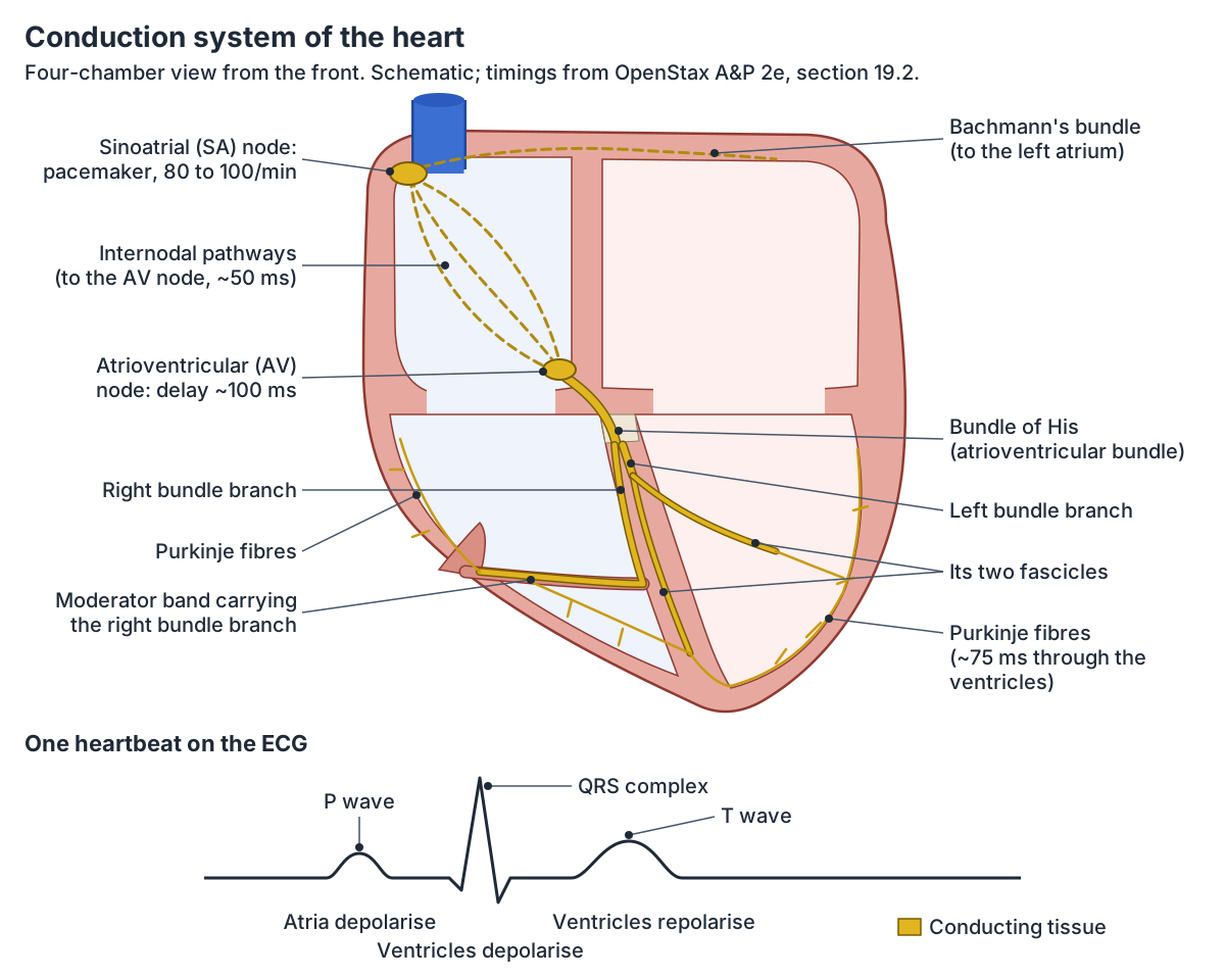
Conduction system of the heart
| Part | Where it is | What it does | Rate if it has to pace the heart |
|---|---|---|---|
| Sinoatrial (SA) node | Upper back wall of the right atrium, by the superior vena cava | Starts each beat (pacemaker) | 80 to 100 per minute |
| Internodal pathways and Bachmann's bundle | Walls of the atria | Carry the impulse to the AV node (about 50 ms) and to the left atrium | Not given |
| Atrioventricular (AV) node | Lower right atrium, in the atrioventricular septum | Delays the impulse about 100 ms | 40 to 60 per minute |
| AV bundle (bundle of His) | Top of the interventricular septum | Only electrical link through the fibrous skeleton | 30 to 40 per minute |
| Right and left bundle branches | Either side of the interventricular septum | Carry the impulse towards the apex | 20 to 30 per minute |
| Purkinje fibres | Under the endocardium of the ventricles | Spread the impulse through the ventricles (about 75 ms) | 15 to 20 per minute |
Timings and rates from OpenStax Anatomy and Physiology 2e, section 19.2.
Nerve supply of the heart: the cardiac plexus
Autonomic nerves reach the heart through the cardiac plexus around the aortic arch. Its superficial part lies below the arch, in front of the right pulmonary artery; its deep part lies behind the arch, in front of the tracheal bifurcation. (Radiopaedia: Cardiac plexus.)
Sympathetic fibres come from spinal cord segments T1 to T4, partly T5, through cardiac nerves from the cervical ganglia of the sympathetic trunk. Parasympathetic fibres come from cardiac branches of the vagus nerves and the recurrent laryngeal nerves. Both are most densely distributed to the SA and AV nodes. (Radiopaedia: Cardiac plexus.)
The nerves change the rate, they do not start the beat. Sympathetic stimulation speeds up the SA node and the heart rate; parasympathetic stimulation slows them. (TeachMeAnatomy: The Conducting System of the Heart.)
Lymph from the heart drains through a right collecting trunk to the anterior mediastinal nodes and through a left trunk to the tracheobronchial and then the paratracheal nodes. (Radiopaedia: Heart.)
The course of the vagus nerve and its cardiac branches are traced on the page about the Vagus nerve.
Blood flow through the heart in brief
Blood passes through the heart twice on each trip around the body. Oxygen-poor blood from the body enters the right atrium, passes to the right ventricle and is pumped through the pulmonary trunk to the lungs. Oxygen-rich blood returns through the pulmonary veins to the left atrium, passes to the left ventricle and is pumped into the aorta. (OpenStax Anatomy and Physiology 2e, 20.5: Circulatory Pathways.)
Each ventricle ejects about 70 mL of blood per beat in a resting adult. At 75 beats a minute that is about 5.25 litres a minute, and the heart beats about 108,000 times a day. (OpenStax Anatomy and Physiology 2e, 19.1: Heart Anatomy.)
Follow the path of blood step by step, valve by valve, with the cardiac cycle and an interactive 3D heart, on Blood flow through the heart.
How to draw and label a heart diagram
Start with the orientation. A labelled heart diagram is normally drawn in anterior view, so the subject's right is on your left. In that view the right ventricle forms most of the front, the right atrium makes the right border and the left ventricle, with a little of the left atrium, makes the left border. (TeachMeAnatomy: The Surfaces and Borders of the Heart.)
Draw the pulmonary trunk in front, rising from the right ventricle and slanting backwards to the left of the ascending aorta. The aorta arches over the point where the pulmonary trunk divides. (Radiopaedia: Pulmonary trunk.)
Two mnemonics help with the labels. 'Try Pulling My Aorta' gives the valves in the order blood meets them: tricuspid, pulmonary, mitral, aortic. 'LAB RAT' places the atrioventricular valves: left atrium, bicuspid; right atrium, tricuspid. (Kenhub: Heart anatomy.)
Most diagrams colour oxygen-poor blood blue and oxygen-rich blood red, so the pulmonary arteries are blue and the pulmonary veins red even though one is an artery and the other a vein. A full heart anatomy diagram should label the four chambers, four valves, the septum, the papillary muscles and chordae tendineae, and the great vessels: venae cavae, pulmonary trunk and arteries, pulmonary veins and aorta.
Heart anatomy in the 3D model
The 3D model on this page comes from BodyParts3D, a whole-body model of an adult human male built as an anatomical reference database and released under a CC BY licence. (BodyParts3D, Database Center for Life Science.)
The highlighted parts are the walls and cavities of the atria and ventricles, all nine valve leaflets and cusps (three tricuspid, two mitral, and the pulmonary and aortic cusps), the five papillary muscles, the ascending aorta and arch, the pulmonary trunk and arteries, the four pulmonary veins, the superior vena cava, the coronary artery trunks and the coronary sinus. Switch off the skeleton and rotate the heart to look at the valves from above, or click a leaflet to open its own page.
The model names the semilunar cusps by their older adult positions (anterior and posterior cusps) rather than the right, left, anterior and posterior names used above. It has no separate interventricular septum, pericardium, chordae tendineae, conduction tissue or fossa ovalis, so the diagrams on this page show those.
Heart in the 3D model
- Anterior cusp of aortic valve
- The anterior cusp is one of the three semilunar cusps of the aortic valve, lying toward the front of the aortic root.
- Anterior cusp of pulmonary valve
- Two of the three semilunar cusps of the pulmonary valve, named anterior in the older adult-position terminology used by this model: the right anterior cusp is the right cusp and the left anterior cusp is the anterior cusp of current terminology.
- Anterior leaflet of mitral valve
- The anterior leaflet is the larger leaflet of the mitral valve and lies between the left atrioventricular opening and the aortic outflow.
- Anterior leaflet of tricuspid valve
- The anterior leaflet is the largest of the three leaflets of the tricuspid valve.
- Anterior papillary muscle of ventricle
- The anterior papillary muscle is the largest papillary muscle of the right ventricle.
- Anterolateral head of lateral papillary muscle of ventricle
- This is the anterolateral papillary muscle (or its head) of the left ventricle, a muscular projection supporting the mitral valve.
- Arch of aorta
- The aortic arch is the curved part of the aorta between the ascending and descending aorta, giving the great vessels to the head and arms.
- Ascending aorta
- The ascending aorta is the first part of the aorta, rising from the left ventricle.
- Cavity of atrium
- The cavity of the atrium is the blood-filled chamber of the right or left atrium.
- Cavity of ventricle
- The cavity of the ventricle is the blood-filled chamber of the right or left ventricle.
- Coronary sinus
- The coronary sinus is the main vein of the heart, draining most of the blood from the heart wall into the right atrium.
- Inferior pulmonary vein
- The inferior pulmonary veins (one per lung) carry oxygenated blood from the lower lobes of the lungs to the left atrium.
- Lateral papillary muscle of ventricle
- The lateral (anterolateral) papillary muscle is one of the two papillary muscles of the left ventricle.
- Posterior cusp of aortic valve
- Two of the three semilunar cusps of the aortic valve, named posterior in the older adult-position terminology used by this model: the left posterior cusp is the left coronary cusp and the right posterior cusp is the non-coronary cusp.
- Posterior cusp of pulmonary valve
- One of the three semilunar cusps of the pulmonary valve; the cusp the model (following FMA) calls posterior is the left cusp of current Terminologia Anatomica.
- Posterior leaflet of mitral valve
- One of the two leaflets of the mitral valve. It attaches to about two thirds of the annulus but is shorter and smaller in area than the anterior leaflet.
- Posterior leaflet of tricuspid valve
- One of the three leaflets of the tricuspid valve.
- Posterior papillary muscle of ventricle
- The posterior (inferior) papillary muscle of the right ventricle, which anchors the tricuspid valve.
- Pulmonary artery
- An artery carrying deoxygenated blood from the heart to a lung.
- Pulmonary trunk
- The large artery arising from the right ventricle that carries blood to the lungs.
- Septal leaflet of tricuspid valve
- One of the three leaflets of the tricuspid valve, attached to the interventricular septum.
- Septal papillary muscle of ventricle
- A papillary muscle on the interventricular septum of the right ventricle.
- Superior pulmonary vein
- One of the pulmonary veins draining the upper lung lobes into the left atrium.
- Superior vena cava
- The large vein draining the upper body into the right atrium.
- Trunk of coronary artery
- The main stems of the right and left coronary arteries, from their openings in the aortic sinuses to their first divisions.
- Wall of atrium
- The muscular walls of the right and left atria, the thin-walled receiving chambers of the heart.
- Wall of ventricle
- The muscular wall of a ventricle of the heart.
Frequently asked questions
What are the four chambers of the heart?
The four chambers are the right atrium, right ventricle, left atrium and left ventricle. The atria are the upper receiving chambers: the right atrium takes blood from the body and the left atrium from the lungs. The ventricles are the lower pumping chambers: the right ventricle sends blood to the lungs and the left ventricle sends it to the rest of the body.
What are the four valves of the heart?
The tricuspid valve lies between the right atrium and right ventricle, and the pulmonary valve between the right ventricle and pulmonary trunk. On the left, the mitral (bicuspid) valve lies between the left atrium and left ventricle, and the aortic valve between the left ventricle and aorta. The mnemonic 'Try Pulling My Aorta' gives them in the order blood passes through.
Which side of the chest is the heart on?
The heart lies in the middle of the chest, in the mediastinum between the lungs, but it is tilted to the left. About two-thirds of it lies left of the midline and one-third right. Its apex points down and to the left and lies just left of the sternum, between the fourth and fifth ribs, and the left lung has a notch to make room for it.
What are the layers of the heart wall?
The heart wall has three layers. The outer epicardium is the visceral layer of the serous pericardium. The middle myocardium is the thick layer of cardiac muscle that does the pumping. The inner endocardium is a smooth endothelium that lines the chambers and covers the valves. The whole heart sits in the pericardial sac, with a thin layer of fluid between its layers.
Why is the left ventricle wall thicker than the right?
The left ventricle pumps blood around the whole body, which needs much higher pressure than the short trip to the lungs. Its systolic pressure is about 90 to 140 mm Hg, while the pulmonary artery, fed by the right ventricle, reaches only 15 to 30 mm Hg. Its wall is much thicker as a result, while the thinner right ventricle wraps around it like a crescent.
What do the papillary muscles and chordae tendineae do?
Papillary muscles are cone-shaped muscles on the inner walls of the ventricles: three in the right ventricle and two in the left. Chordae tendineae are collagen cords that join them to the free edges of the tricuspid and mitral valve cusps. When the ventricles contract, the papillary muscles tighten the cords so the cusps cannot be pushed back into the atria.
What does the pulmonary artery do?
The pulmonary trunk leaves the right ventricle through the pulmonary valve and divides under the aortic arch into the right and left pulmonary arteries, one for each lung. They carry oxygen-poor blood to the lungs to pick up oxygen and release carbon dioxide. Unlike most arteries they carry deoxygenated blood, and they work at low pressure, about 15 to 30 mm Hg systolic.
How big is the human heart?
An adult heart is about the size of a fist: roughly 12 cm long, 8 cm wide and 6 cm thick. It weighs about 250 to 300 g in women and 300 to 350 g in men, although regular exercise and some diseases can make it larger. At 75 beats a minute it beats about 108,000 times a day.
References
- OpenStax. Anatomy and Physiology 2e, §19.1 Heart Anatomy. Rice University, 2022.
- OpenStax. Anatomy and Physiology 2e, §19.2 Cardiac Muscle and Electrical Activity. Rice University, 2022.
- OpenStax. Anatomy and Physiology 2e, §19.3 Cardiac Cycle. Rice University, 2022.
- OpenStax. Anatomy and Physiology 2e, §20.5 Circulatory Pathways. Rice University, 2022.
- OpenStax. Anatomy and Physiology 2e, §20.6 Development of Blood Vessels and Fetal Circulation. Rice University, 2022.
- Jones O. The Surfaces and Borders of the Heart. TeachMeAnatomy.
- Jones O. The Chambers of the Heart. TeachMeAnatomy.
- Jones O. The Valves of the Heart. TeachMeAnatomy.
- Jones O. The Heart Wall. TeachMeAnatomy.
- Jones O. The Conducting System of the Heart. TeachMeAnatomy.
- Jones O. The Pericardium. TeachMeAnatomy.
- Jones O. Great Vessels of the Heart. TeachMeAnatomy.
- Heart valves. Kenhub anatomy library.
- Heart anatomy. Kenhub anatomy library.
- Pulmonary arteries and veins. Kenhub anatomy library.
- Heart. Radiopaedia reference article.
- Left ventricle. Radiopaedia reference article.
- Right ventricle. Radiopaedia reference article.
- Pulmonary trunk. Radiopaedia reference article.
- Cardiac plexus. Radiopaedia reference article.
- Inferior vena cava. Radiopaedia reference article.
- Pulmonary arteries (anatomy). GPnotebook.
- Right pulmonary artery (anatomy). GPnotebook.
- Left pulmonary artery (anatomy). GPnotebook.
- Cardiac Catheterization. In: Davis's Lab and Diagnostic Tests. F.A. Davis, via Nursing Central.
- Heart. Cleveland Clinic health library.
- BodyParts3D. Database Center for Life Science, Japan.
- Mitsuhashi N. et al. (2009). BodyParts3D: 3D structure database for anatomical concepts. Nucleic Acids Research 37, D782–D785. Data © The Database Center for Life Science, CC BY 4.0.
Related anatomy
- Coronary arteriesCoronary arteries explained: left main, LAD and diagonals, circumflex, RCA and posterior descending artery, dominance…
- Circulatory systemCardiovascular anatomy explained: heart, arteries, capillaries, vein anatomy, pulmonary vs systemic circulation and…
- AortaAorta anatomy explained: ascending aorta, aortic arch and its three branches, thoracic aorta, and every abdominal…
- LungsLung anatomy with labelled diagrams and a 3D model of all five lobes: fissures, hilum, segments, pleura, blood supply…
- Chest anatomyChest anatomy with labelled diagrams and a 3D model: chest wall layers, ribs and sternum, chest muscles, heart, lungs…
- Pulmonary trunkPulmonary trunk: The large artery arising from the right ventricle that carries blood to the lungs. See where it is in…
Explore the whole body in the interactive 3D human anatomy model, or browse the A–Z list of human structures.