

Where are the lungs located?
The lungs lie in the thoracic cavity, one on each side of the mediastinum, the central compartment of the chest that holds the heart. Each lung sits in its own pleural cavity and hangs from the mediastinum by its root. The left lung is slightly smaller than the right because the heart takes up room on the left. (TeachMeAnatomy: The Lungs.)
Each lung is roughly a cone with an apex, a base, three surfaces and three borders. The apex is the blunt upper end, which rises above the first rib into the root of the neck. The base rests on the diaphragm. (TeachMeAnatomy: The Lungs.)
Cleveland Clinic gives the length of an adult lung as a little over 9 inches (about 24 cm) during normal breathing and about 10.5 inches (27 cm) when fully expanded. The right lung is shorter and wider than the left. (Cleveland Clinic: Lungs.)
Radiopaedia gives the weight of each lung as about 1.1 kg and says the two lungs contain about 1500 miles of airways and 300 to 500 million alveoli, with a combined surface area of about 70 square metres, half a tennis court. About 10,000 litres of air and 7,000 litres of blood pass through the lungs of a resting adult each day. (Radiopaedia: Lungs.)
Kenhub gives lower weights, about 625 g for the right lung and 565 g for the left, and notes that men tend to have heavier lungs, though this depends on height. Published weights therefore range from about 0.5 to 1.1 kg per lung. Adult lungs look dark and mottled from inhaled carbon, while a newborn's lungs are light pink. (Kenhub: Lung anatomy.)
The ribs, sternum and thoracic vertebrae that surround and protect the lungs. Rib cage.
Lung lobes: how many lobes do the lungs have?
The lungs have five lobes in total. The right lung has three: the superior (upper), middle and inferior (lower) lobes. The left lung has two: the superior and inferior lobes. Fissures separate the lobes, and the visceral pleura that covers the lung dips into each fissure. (OpenStax Anatomy and Physiology 2e, 22.2: The Lungs.)
On the right, the upper part of the oblique fissure separates the upper lobe from the lower lobe, and its lower part separates the middle lobe from the lower lobe. On the left, the oblique fissure separates the upper and lower lobes. Normally only the right lung has a horizontal fissure, between the upper and middle lobes. (Radiopaedia: Oblique fissure.)
Why does the right lung have three lobes and the left only two? Kenhub explains that the left lung has one lobe and one fissure fewer than the right to make room for the heart, which lies more to the left. The left lung's front border also bends away around the heart, forming the cardiac notch. (Kenhub: Lung anatomy.)
In the 3D model each of the five lobes is a separate, see-through part. Click a lobe to read about it, or open its own page from the list below the model: the upper, middle and lower lobes of the right lung and the upper and lower lobes of the left lung.
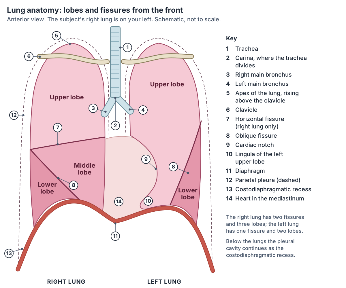
The five lobes of the lungs
| Lobe | Lung | Bounded by | Forms | Bronchopulmonary segments |
|---|---|---|---|---|
| Upper (superior) lobe | Right | Horizontal fissure below, upper part of the oblique fissure behind | Apex, upper costal and medial surfaces | 3: apical, posterior, anterior |
| Middle lobe | Right | Horizontal fissure above, oblique fissure below and behind | Lower front border and front of the base; the smallest lobe | 2: lateral, medial |
| Lower (inferior) lobe | Right | Oblique fissure in front and above | Back and lower costal and medial surfaces, most of the base; the largest lobe | 5: superior, medial basal, anterior basal, lateral basal, posterior basal |
| Upper (superior) lobe | Left | Oblique fissure behind and below | Apex, most of the costal and medial surfaces, whole front border, cardiac notch, lingula | Usually 4: apicoposterior, anterior, superior lingular, inferior lingular |
| Lower (inferior) lobe | Left | Oblique fissure in front and above | Most of the base and the back border; the larger left lobe | Usually 4: superior, anteromedial basal, lateral basal, posterior basal |
Lobe positions and relative sizes from Kenhub; segments from Radiopaedia. On the left, two pairs of segments share one bronchus, so counts of 8, 9 or 10 left segments are all in use.
Right lung lobes anatomy: upper, middle and lower lobes
The right lung has three lobes. The upper lobe lies above the horizontal fissure and contains the apex. The middle lobe, the smallest and wedge-shaped, lies below the horizontal fissure and in front of the oblique fissure, at the front and side of the lower chest. The lower lobe, the largest by volume, lies behind the oblique fissure and forms most of the base and the back of the lung. (Kenhub: Lung anatomy.)
Kenhub traces the right oblique fissure from the hilum across the base and onto the costal surface, about 7 cm from the front border, through the fifth intercostal space. Behind, it lies at or just below the spinous process of T4. The horizontal fissure is much shorter: it runs from the mid-axillary line in the fourth intercostal space forward to the front border at the fourth costochondral junction, then turns onto the medial surface to reach the hilum. (Kenhub: Lung anatomy.)
Radiopaedia notes a broad segmental symmetry between the two lungs, with a few differences. The right middle lobe and the lingula of the left upper lobe correspond to each other, and each has two segments. (Radiopaedia: Bronchopulmonary segmental anatomy.)
The right main bronchus is wider, shorter and more vertical than the left, so inhaled foreign bodies more often enter the right lung. The right upper lobe bronchus branches off the main bronchus before it enters the hilum. (TeachMeAnatomy: The Tracheobronchial Tree.)
The smallest lobe, between the horizontal and oblique fissures, with its own 3D page. Middle lobe of the right lung.
Left lung anatomy: upper lobe, lingula and cardiac notch
The left lung has an upper and a lower lobe. The larger lower lobe lies behind and below the oblique fissure and contains most of the base and the back border. The upper lobe lies in front of and above the fissure and contains the apex, most of the costal and medial surfaces, the whole front border, the cardiac notch and the lingula. The left oblique fissure is slightly more vertical than the right. (Kenhub: Lung anatomy.)
The front border of each lung is thin and sharp. On the left lung it carries a notch, the cardiac notch, made by the apex of the heart. (TeachMeAnatomy: The Lungs.)
Below the cardiac notch is the lingula, the part of the left upper lobe that corresponds to the right middle lobe. It has two bronchopulmonary segments, the superior and inferior lingular segments. (Radiopaedia: Bronchopulmonary segmental anatomy.)
The word lingula is the diminutive of the Latin lingua, tongue. Anatomists use it for several small, tongue-shaped parts, including ones on the mandible, the sphenoid bone and the cerebellum as well as the lung. (Radiopaedia: Lingula (disambiguation).)
The lobe with the cardiac notch and the lingula, with its own 3D page. Upper lobe of the left lung.
Fissures of the lung: oblique and horizontal
On the chest wall the oblique fissure is drawn as a curved line. On the right it begins at about the T4 spinous process, crosses the fifth intercostal space at the side and follows the sixth rib in front. On the left it begins a little higher, between the T3 and T4 spinous processes. The left oblique fissure is usually higher and more vertical than the right. (Radiopaedia: Oblique fissure.)
The horizontal fissure follows the fourth intercostal space from the sternum and ends where it meets the oblique fissure as that crosses the fifth rib in the mid-axillary line. On a frontal chest X-ray it appears as a thin horizontal line at about the level of the anterior fourth rib and is seen in about 65% of normal films. (Radiopaedia: Horizontal fissure.)
TeachMeAnatomy describes the same fissure as running horizontally from the sternum at the level of the 4th rib, so the two sources differ by about one intercostal space. (TeachMeAnatomy: The Lungs.)
A teaching guide from Loyola University Chicago gives the same pattern in bedside terms: the oblique fissure runs from the sixth rib in the midclavicular line to the fifth rib in the mid-axillary line and then, with the arms raised, along the medial border of the scapula to the third spinous process; the transverse (horizontal) fissure runs from the fifth rib in the mid-axillary line to the fourth rib in front. Sources therefore place the back end of the oblique fissure between T3 and T4. (Loyola University Chicago Stritch School of Medicine: Pulmonary anatomical principles.)
Fissures are often incomplete, which means lung tissue bridges two lobes near the hilum. Radiopaedia reports incomplete fissures in 48% of right and 43% of left oblique fissures; a missing oblique fissure is rare, under 1%. (Radiopaedia: Oblique fissure.)
The horizontal fissure varies more. Post-mortem studies find it incomplete in 8 to 35% of right lungs and absent in 3 to 50%, and a minority of people also have a horizontal fissure in the left lung. (Radiopaedia: Horizontal fissure.)
The large lobe behind the oblique fissure, with its own 3D page. Lower lobe of the right lung.
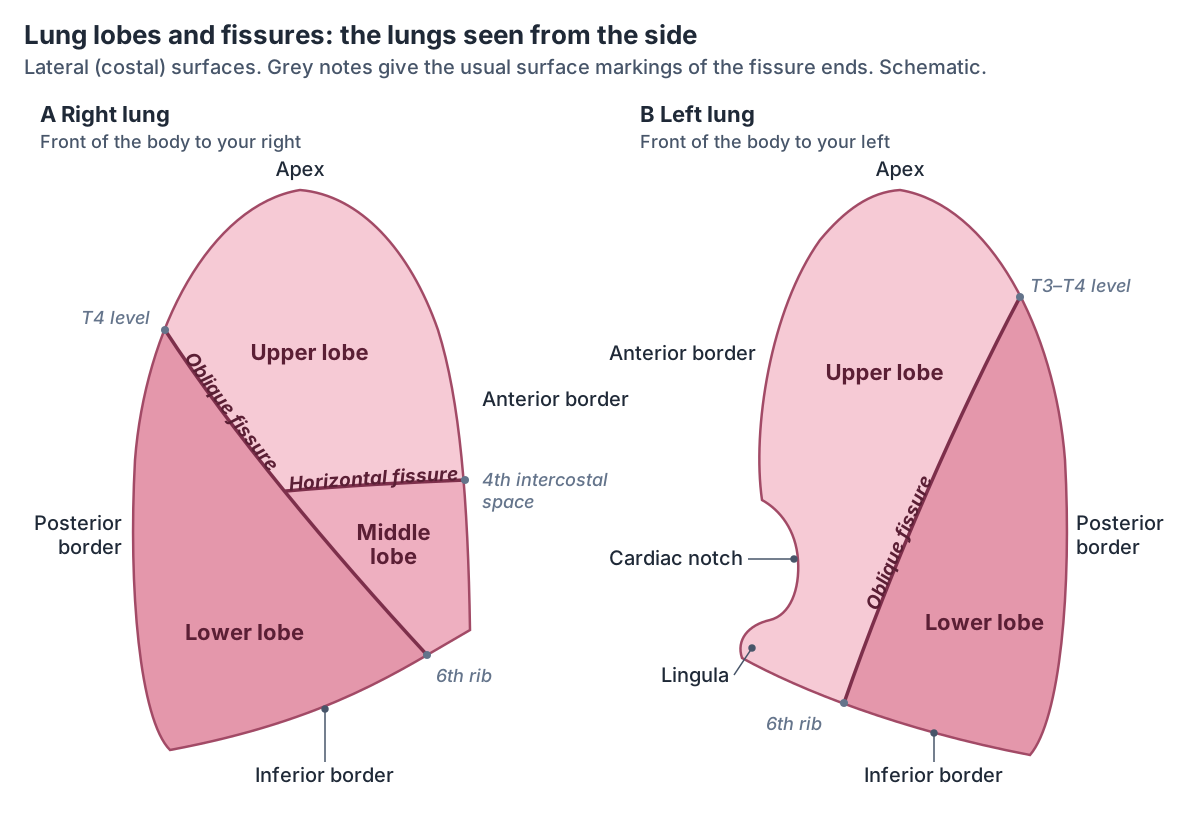
Surfaces and borders of the lung
Each lung has three surfaces. The costal surface is smooth and convex and faces the ribs, separated from them and from the innermost intercostal muscles by the costal pleura. The mediastinal surface faces the middle of the chest and contains the hilum. The diaphragmatic surface, or base, is concave, and deeper on the right because the right dome of the diaphragm sits higher over the liver. (TeachMeAnatomy: The Lungs.)
Each lung also has three borders: a thin, sharp anterior border, a sharp inferior border and a smooth, rounded posterior border. (TeachMeAnatomy: The Lungs.)
Kenhub divides the medial surface into a front mediastinal part, which carries the cardiac impression, and a back vertebral part that lies against the thoracic vertebrae and discs. Below the diaphragm, the base of the left lung lies over the spleen and stomach and the base of the right lung over the liver. (Kenhub: Lung anatomy.)
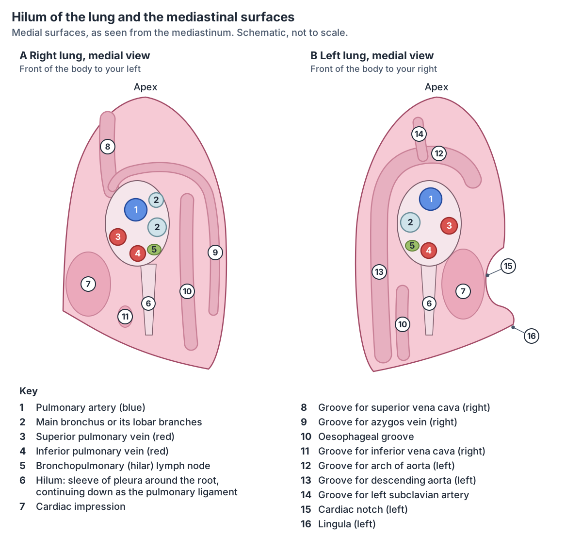
The organs of the mediastinum lie against the medial surfaces. The left lung is related to the heart, the arch of the aorta, the thoracic aorta and the oesophagus. The right lung is related to the oesophagus, the heart, the inferior and superior venae cavae and the azygos vein. The hilum diagram shows where each of these lies. (TeachMeAnatomy: The Lungs.)
The apex projects above the first costal cartilage and the medial third of the clavicle. The subclavian artery separates it from scalenus anterior. On the left it is related to the left brachiocephalic vein and the left subclavian artery; on the right, to the trachea, the brachiocephalic trunk and the right brachiocephalic vein. (Kenhub: Lung anatomy.)
The heart that makes the cardiac impression and the cardiac notch. Heart anatomy.
Hilum and root of the lung
The hilum is a wedge-shaped area on the mediastinal surface of each lung where structures enter and leave it. The root is the bundle of those structures, which suspends the lung from the mediastinum: a main bronchus, a pulmonary artery, two pulmonary veins, bronchial vessels, the pulmonary plexus of nerves and lymphatic vessels. The visceral and parietal pleura are continuous around the root. (TeachMeAnatomy: The Lungs.)
The roots lie at the level of the fifth to seventh thoracic vertebrae. In the left hilum the pulmonary artery is the highest structure, the main bronchus lies below and behind it, and the lower pulmonary vein is the lowest; the upper pulmonary vein lies in front of and below the artery. In the right hilum the main bronchus lies behind the pulmonary artery, and the upper pulmonary vein lies in front of and below the artery and above the lower vein. (Kenhub: Lung anatomy.)
Radiopaedia also places the roots between T5 and T7 and describes the hilum as enclosed in a sleeve of pleura that continues downwards as the pulmonary ligament. The left hilum lies below the aortic arch and in front of the descending aorta, with the bronchus at its back and the veins always in front of and below the artery. The right hilum lies below the arch of the azygos vein and behind the superior vena cava and right atrium. The hilar point, where the upper lobe vein crosses the pulmonary artery, usually forms an angle of about 120°. (Radiopaedia: Lung hilum.)
In front of each root run the phrenic nerve, the pericardiacophrenic vessels and the anterior pulmonary plexus. Behind it lie the vagus nerve and the posterior pulmonary plexus, and below it hangs the pulmonary ligament. (Kenhub: Lung anatomy.)
The main bronchi from the carina to the hilum, and how they branch inside the lungs. Trachea and bronchial tree.
Bronchopulmonary segments of the lung
A bronchopulmonary segment is a division of a lobe that receives air from its own tertiary (segmental) bronchus and blood from its own artery. Some lung diseases stay within one segment, and a diseased segment can sometimes be removed surgically with little effect on its neighbours. (OpenStax Anatomy and Physiology 2e, 22.2: The Lungs.)
The right lung has 10 segments: three in the upper lobe, two in the middle lobe and five in the lower lobe. The left lung is usually given 8, because two pairs of segments share a single bronchus: the apicoposterior segment of the upper lobe and the anteromedial basal segment of the lower lobe. (Radiopaedia: Bronchopulmonary segmental anatomy.)
Counts differ by convention. Duke University's histology notes give 10 segments on the right and 8 or 10 on the left depending on the classification, and stress that segmental anatomy matters for surgical resection. Beyond the segment, the acinus, all the lung beyond one terminal bronchiole, is the main unit of gas exchange and measures 6 to 10 mm across; a secondary lobule of 3 to 5 acini measures 10 to 20 mm. (Duke University Microscopic Anatomy: The Respiratory System (course notes, 2015).)
Kenhub gives a third figure for the left lung, 9 to 10 segments, while agreeing on 10 for the right. (Kenhub: Lung anatomy.)
Each segment is divided into pulmonary lobules, each supplied by its own large bronchiole, and the lobules are separated by connective tissue walls called interlobular septa. (OpenStax Anatomy and Physiology 2e, 22.2: The Lungs.)
In the 3D model each coloured branch inside the lobes is a segmental bronchial tree from BodyParts3D, named after the segment it supplies, such as the apical, superior lingular or posterior basal segmental bronchial tree.
Bronchopulmonary segments of each lung
| Lobe | Right lung (10 segments) | Left lung (usually 8 segments) |
|---|---|---|
| Upper lobe | Apical (S1), posterior (S2), anterior (S3) | Apicoposterior (S1+2), anterior (S3) |
| Middle lobe or lingula | Middle lobe: lateral (S4), medial (S5) | Lingula of the upper lobe: superior lingular (S4), inferior lingular (S5) |
| Lower lobe | Superior (S6), medial basal (S7), anterior basal (S8), lateral basal (S9), posterior basal (S10) | Superior (S6), anteromedial basal (S7+8), lateral basal (S9), posterior basal (S10) |
From Radiopaedia. Each segment has its own segmental (tertiary) bronchus and artery. The left S1 and S2, and S7 and S8, usually share a bronchus.
Pleura of the lungs and the pleural recesses
The pleura is a thin serous membrane around each lung, made of flat cells (mesothelium) on connective tissue. Its visceral layer covers the lung and dips into the fissures. Its parietal layer lines the chest wall and is thicker. The two layers are continuous at the hilum, and between them lies the pleural cavity, a potential space that holds a small amount of serous fluid. (TeachMeAnatomy: The Pleurae.)
The parietal pleura has four parts: cervical (the dome over the apex in the root of the neck), costal (over the ribs, costal cartilages and intercostal muscles), mediastinal and diaphragmatic. The pleural fluid lets the layers slide, and its surface tension holds them together, so the lung expands when the chest expands. (TeachMeAnatomy: The Pleurae.)
Where the lungs do not fill the pleural cavity, two layers of parietal pleura meet to form recesses. The costomediastinal recess lies behind the sternum, between the costal and mediastinal pleura. The costodiaphragmatic recess lies between the costal and diaphragmatic pleura around the base of the lung. Fluid in the chest, as in a pleural effusion, collects in these recesses. The right and left pleural cavities do not connect. (Kenhub: Pleura.)
Radiopaedia lists two smaller recesses as well, the phrenicomediastinal and vertebromediastinal recesses. On the surface, the lower edge of the parietal pleura lies about two ribs below the lung: it crosses the midclavicular line at the 8th rib and the mid-axillary line at the 10th rib and reaches the 12th rib at the back. Above, the pleura rises about 1 inch (2.5 cm) above the medial third of the clavicle. (Radiopaedia: Pleura.)
The parietal pleura is sensitive to pain, temperature and pressure and is supplied by the intercostal and phrenic nerves, so pain from it is sharp and well localised. The visceral pleura senses only stretch and receives autonomic fibres from the pulmonary plexus. The parietal pleura takes its blood from the intercostal arteries and the visceral pleura from the bronchial arteries. (TeachMeAnatomy: The Pleurae.)
The dome-shaped muscle under the lungs, covered on top by the diaphragmatic pleura. Diaphragm.
Blood supply of the lungs: pulmonary and bronchial circulations
The lungs have two blood supplies. The paired pulmonary arteries bring deoxygenated blood from the heart to be oxygenated, and four pulmonary veins, two from each lung, return oxygenated blood. The bronchial arteries, from the descending aorta, bring oxygen-rich blood to nourish the bronchi, the lung roots, the visceral pleura and the supporting tissue. (TeachMeAnatomy: The Lungs.)
The pulmonary trunk leaves the right ventricle and divides into the right and left pulmonary arteries, and each of these divides into superior and inferior branches. The left pulmonary artery passes under the aortic arch into the oblique fissure and gives a lingular branch. On the right the superior and middle lobar veins usually join, so each lung returns two pulmonary veins to the left atrium. (Kenhub: Lung anatomy.)
Inside the lung the pulmonary arteries branch with the airways, each branch narrower than the last. One arteriole and one venule serve each pulmonary lobule, and the pulmonary capillaries, whose very thin walls have no smooth muscle, meet the alveolar walls to form the respiratory membrane. (OpenStax Anatomy and Physiology 2e, 22.2: The Lungs.)
The bronchial arteries usually arise from the descending aorta between T5 and T6 (about 70% of people). The commonest pattern, also in about 70%, is two left and one right bronchial artery; the right one often shares a trunk with a posterior intercostal artery, the intercostobronchial trunk (27%). They are small, about 1.5 mm across at their origin, and carry only about 1% of the blood that flows through the lungs. (Radiopaedia: Bronchial arteries.)
Bronchial veins drain the hila. Superficial bronchial veins drain to the azygos vein on the right and to the accessory hemiazygos, left superior intercostal or brachiocephalic vein on the left. Deep bronchial veins drain into a pulmonary vein or straight into the left atrium. (Kenhub: Lung anatomy.)
How blood travels from the right ventricle to the lungs and back to the left atrium. Blood flow through the heart.
Lymphatic drainage and nerves of the lungs
Lymph from the lung tissue drains through a superficial (subpleural) plexus and from the root structures through a deep plexus. Both drain to the tracheobronchial lymph nodes around the bifurcations of the trachea and main bronchi, and from there to the right and left bronchomediastinal trunks. (TeachMeAnatomy: The Lungs.)
Radiopaedia gives the chain as bronchopulmonary (hilar) nodes, then tracheobronchial and paratracheal nodes, then the bronchomediastinal trunks. It places the sympathetic supply in the T1 to T4 sympathetic ganglia, reaching the lung through the pulmonary plexus, with parasympathetic fibres from the vagus nerve. (Radiopaedia: Lungs.)
Kenhub adds that the lower lobes drain to the inferior tracheobronchial nodes and the upper lobes to the superior tracheobronchial nodes, and that superficial and deep channels communicate, so lymph can take another route when disease blocks a channel. (Kenhub: Lung anatomy.)
The lungs are supplied by the pulmonary plexuses. Parasympathetic fibres from the vagus nerve make the bronchial glands secrete, constrict the bronchial smooth muscle and widen the pulmonary vessels. Sympathetic fibres from the sympathetic trunks relax the bronchial smooth muscle and narrow the vessels. Visceral sensory fibres carry pain to the sensory ganglion of the vagus nerve. (TeachMeAnatomy: The Lungs.)
OpenStax adds that sensory fibres reach the lungs from the vagus nerve and from the second to fifth thoracic ganglia, and that the pulmonary plexus forms at the hilum and follows the bronchi to the muscle fibres, glands and blood vessels. Autonomic control of the airways also takes part in reflexes such as coughing. (OpenStax Anatomy and Physiology 2e, 22.2: The Lungs.)
Surface anatomy: where are the lungs under the ribs and in the back?
In quiet breathing the lower border of the lung lies at about the 6th rib in the midclavicular line, the 8th rib in the mid-axillary line and the 10th rib at the back. The right lung starts about 3 cm above the clavicle, and the front border of the left lung follows the edge of the heart down to the 6th rib. (Loyola University Chicago Stritch School of Medicine: Pulmonary anatomical principles.)
Older anatomical surveys collected in Anatomy Atlases show how much these levels vary. Authors place the apex 1 to 3.8 cm above the clavicle and the lower border at the back anywhere from the 9th to the 11th rib. The lowest point of the right lung lay on average at the middle of the eleventh thoracic vertebra, with a range from the ninth thoracic disc to the twelfth vertebra. Some authors found the right apex 0.5 to 1 cm higher than the left; others found no difference. (Bergman RA, Afifi AK, Jew JJ, Reimann PC. Topography of the lungs. Anatomy Atlases.)
The apex lies just under the skin above the clavicle, where it can be felt, and it is a typical site for Pancoast tumours and tuberculosis. Because the pleural gutter is deep at the back, fluid in the pleural cavity tends to collect behind the lung. (Loyola University Chicago Stritch School of Medicine: Pulmonary anatomical principles.)
The ribs, scapula and muscles that lie over the back of the lungs. Back anatomy.
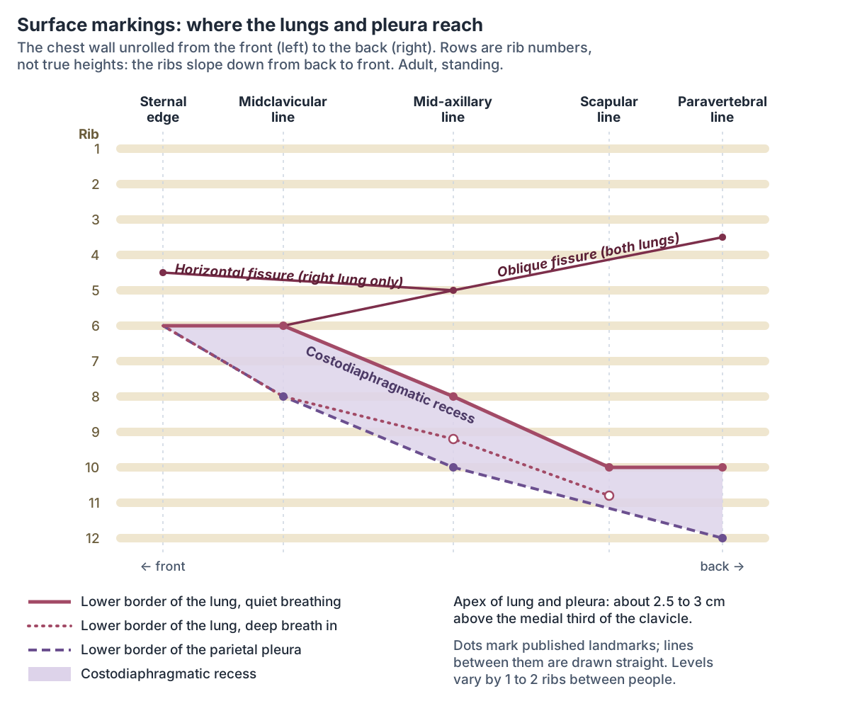
Surface markings of the lungs and pleura
| Landmark | Lung, quiet breathing | Lung, deep breath in | Parietal pleura |
|---|---|---|---|
| Apex | About 3 cm above the clavicle | Not given | About 2.5 cm (1 inch) above the medial third of the clavicle |
| Midclavicular line | 6th rib | 8th rib | 8th rib |
| Mid-axillary line | 8th rib | Lower border of the 9th rib | 10th rib |
| Back (scapular or paravertebral line) | 10th rib | Upper border of the 11th rib (scapular line) | 12th rib |
Quiet-breathing levels from Loyola University Chicago and Corning (Anatomy Atlases); deep-breath levels from Gerlach (Anatomy Atlases); pleural levels from Radiopaedia. The pleura reaches about two ribs lower than the lung.
How do the lungs move when you breathe?
The lungs follow the chest wall. When the diaphragm and external intercostal muscles contract, the chest gets bigger, the pressure inside the alveoli falls below atmospheric pressure and air flows in. In quiet breathing, breathing out is passive: the muscles relax and the elastic recoil of the lungs pushes air out. (OpenStax Anatomy and Physiology 2e, 22.3: The Process of Breathing.)
The lungs stay stretched against the chest wall because the pressure in the pleural cavity stays below the pressure in the alveoli, at about –4 mm Hg throughout the breathing cycle. The outward pull of the chest wall and pleural fluid is greater than the inward pull of the lungs' elastic tissue and surface tension. Surfactant made by type II alveolar cells keeps the alveoli from collapsing when you breathe out. (OpenStax Anatomy and Physiology 2e, 22.3: The Process of Breathing.)
As the diaphragm descends, the lower borders of the lungs slide down into the costodiaphragmatic recess. In a deep breath in, Gerlach's figures in Anatomy Atlases put the lower border at the 8th rib in the midclavicular line, the lower border of the 9th rib in the mid-axillary line and the upper border of the 11th rib in the scapular line. Other authors measured the border moving 2 to 3 cm in the midclavicular line and 3 to 4 cm in the axillary lines, and found the lungs 1 to 2 cm lower when lying down. (Bergman RA, Afifi AK, Jew JJ, Reimann PC. Topography of the lungs. Anatomy Atlases.)
A quiet breath (the tidal volume) moves about 0.5 L. TeachMePhysiology gives average adult values of about 4.5 L for the vital capacity, the most you can breathe out after a full breath in, and about 6 L for the total lung capacity. (TeachMePhysiology: Lung Volumes and Capacities.)
OpenStax gives matching figures: a tidal volume of about 500 mL, a vital capacity of 4000 to 5000 mL, and a total lung capacity of about 6000 mL in men and about 4200 mL in women. (OpenStax Anatomy and Physiology 2e, 22.3: The Process of Breathing.)
The airway, the muscles of breathing and gas exchange in the alveoli. Respiratory system.
Left vs right lung: what is the difference?
The right lung is shorter, wider and larger overall, with three lobes, two fissures and 10 segments. The left lung has one lobe and one fissure fewer, a cardiac notch and a lingula, and 9 to 10 segments in Kenhub's count. The right hemidiaphragm sits higher, which is why the right lung is shorter. (Kenhub: Lung anatomy.)
The two lungs drain differently too. The right bronchial vein drains to the azygos vein and the left to the accessory hemiazygos vein, and the medial surface of each lung is shaped by different neighbours: the venae cavae and azygos vein on the right, the aorta on the left. (TeachMeAnatomy: The Lungs.)
Why inhaled objects usually end up in the right lung. Right main bronchus.
Left vs right lung
| Feature | Right lung | Left lung |
|---|---|---|
| Lobes | 3: upper, middle, lower | 2: upper (with the lingula), lower |
| Fissures | Oblique and horizontal | Oblique only |
| Bronchopulmonary segments | 10 | 8 to 10, depending on how they are counted |
| Shape | Shorter and wider; base more concave over the higher right dome of the diaphragm | Longer and narrower; cardiac notch in the front border |
| Weight (Kenhub) | About 625 g | About 565 g |
| Main bronchus | Wider, shorter, more vertical; inhaled objects go here more often | Narrower, longer, runs under the aortic arch |
| Pulmonary veins | Two (the upper and middle lobe veins usually join) | Two |
| Medial relations | Heart, superior and inferior venae cavae, azygos vein, oesophagus | Heart, arch of the aorta, thoracic aorta, oesophagus |
| Bronchial arteries | Usually one, often from the intercostobronchial trunk | Usually two, from the thoracic aorta |
| Bronchial vein drains to | Azygos vein | Accessory hemiazygos vein |
| Oblique fissure begins behind | About the T4 spinous process | Between the T3 and T4 spinous processes |
Compiled from TeachMeAnatomy, Kenhub and Radiopaedia.
Lung development and common variants
The lungs begin as a lung bud from the foregut in weeks 4 to 7 of development. The bronchial buds branch until all the segmental bronchi have formed by about week 16. Respiratory bronchioles and the first alveolar ducts form between weeks 16 and 24, and by about week 28 enough alveoli have matured for a premature baby usually to breathe on its own. Surfactant is not generally adequate until about the eighth month, and a full set of functional alveoli is reached at around 8 years of age. (OpenStax Anatomy and Physiology 2e, 22.7: Embryonic Development of the Respiratory System.)
Kenhub names the stages pseudoglandular (about weeks 6 to 16), canalicular (16 to 26), terminal sac (about week 26 to birth) and alveolar (week 32 to about 8 years), and notes that most alveoli form after birth. Its stage boundaries differ from OpenStax's by a couple of weeks. (Kenhub: Lung anatomy.)
The best-known variant is the azygos lobe, found in about 1% of anatomical specimens and about twice as often in males. It forms when the developing azygos vein fails to slide over the apex of the right lung and cuts into it instead, pulling a deep fold of pleura into the upper lobe. It is not a true lobe because it has no bronchus of its own. (Radiopaedia: Azygos fissure (azygos lobe).)
Other variants listed by Radiopaedia include accessory fissures, a tracheal bronchus, horseshoe lung and, rarely, pulmonary aplasia, in which a lung does not develop. (Radiopaedia: Lungs.)
The vein that arches over the right lung root and can create an azygos lobe. Azygos vein.
How the lungs in the 3D model were made
The BodyParts3D dataset behind the model is an adult male body that includes the trachea, the main, lobar and segmental bronchial trees and the pulmonary arteries and veins, but no lung tissue. For this site each lung was built as the space its pleural cavity fills inside the BodyParts3D chest: inside the ribs, costal cartilages, sternum and intercostal muscles, above the diaphragm and outside the spine and mediastinum. The apex rises above the first rib, the lower border follows the quiet-breathing surface markings (6th rib in the midclavicular line, 8th rib in the mid-axillary line, 10th rib at the back), and the left lung has a cardiac notch.
The fissures were first placed from surface anatomy. The oblique fissure runs from the T4 spinous level on the right, or between T3 and T4 on the left, across the side of the chest to the sixth rib in front, and on the right the horizontal fissure runs from the sternum at the level of the 4th rib to meet it at the fifth rib in the mid-axillary line. (Radiopaedia: Oblique fissure.)
Each lobe's share of its lung was then checked against CT measurements in healthy adults, which range from full inspiration to the end of a quiet breath out: the right middle lobe takes about 8 to 11% of both lungs, the left upper lobe 21 to 25% and the right lower lobe 24 to 27%. (Silva M, Nemec SF, Heidinger BH, et al. Lung volume and heterogeneity: CT quantification of lobar contribution. RSNA 2014 abstract.)
A second CT study of 32 adults gave similar shares lying down and standing (right middle lobe 9.3% and 8.5%, left upper lobe 23.1% and 22.4%). Where a lobe of the model fell outside these ranges, its fissure was moved parallel to itself just far enough to bring it in: by about 1 cm for the left oblique fissure and for the horizontal fissure. (Yamada Y, Yamada M, Yokoyama Y, et al. Differences in lung and lobe volumes between supine and standing positions scanned with conventional and newly developed 320-detector-row upright CT. Respiration 2020.)
The result is a reconstruction, not a scan of real lung tissue. The pleura is not modelled, and the lobes are drawn see-through so that the bronchial tree and pulmonary vessels inside stay visible. Use the Focus selected switch to isolate one lobe, Hover labels to name parts under the pointer, and the Skin and Muscles sliders to clear the chest wall.
Clinical anatomy: pneumothorax, pleural effusion and pulmonary embolism
In a pneumothorax, air enters the pleural cavity and breaks the surface-tension seal between the pleural layers, so the lung no longer expands with the chest. It causes chest pain, shortness of breath and uneven chest movement. It may be spontaneous or follow injury such as a rib fracture; small primary ones often need little treatment, while larger or traumatic ones may need a chest drain. (TeachMeAnatomy: The Pleurae.)
A pleural effusion is fluid in the pleural cavity. Radiopaedia gives the normal amount of pleural fluid as about 5 to 15 mL (0.1 to 0.2 mL per kg). An upright chest X-ray shows an effusion only once about 250 to 600 mL has collected, while ultrasound can detect as little as 3 to 5 mL. (Radiopaedia: Pleural effusion.)
A pulmonary embolism is a blockage of a pulmonary artery by material carried from elsewhere, most often a blood clot from a distant vein, but also fat after a fracture or air. It reduces blood flow through the lung and oxygenation and makes blood back up in the right ventricle, causing breathlessness, chest pain, cough, coughing up blood and fast breathing. (TeachMeAnatomy: The Lungs.)
Radiopaedia lists haemoptysis (coughing up blood), enlargement of the bronchial arteries and bronchial artery aneurysm as the main conditions involving the bronchial arteries. (Radiopaedia: Bronchial arteries.)
Other common lung conditions include asthma, chronic obstructive pulmonary disease (COPD), bronchitis, bronchiectasis, cystic fibrosis, interstitial lung disease, pneumonia, infections such as tuberculosis, and lung cancer. (Cleveland Clinic: Lungs.)
The artery that carries blood to the lungs and is blocked in a pulmonary embolism. Pulmonary artery.
Lungs in the 3D model
- Anterior basal segmental bronchial tree
- The anterior basal bronchial tree is the segmental bronchus and its branches supplying a bronchopulmonary segment.
- Anterior segmental bronchial tree
- The anterior bronchial tree is the segmental bronchus and its branches supplying a bronchopulmonary segment.
- Apical segmental bronchial tree
- The apical bronchial tree is the segmental bronchus and its branches supplying a bronchopulmonary segment.
- Bronchial artery
- The bronchial arteries are small arteries that supply the bronchial tree and supporting tissue of the lungs.
- Inferior lingular bronchial tree
- The inferior lingular bronchial tree is the segmental bronchus and its branches supplying a bronchopulmonary segment.
- Inferior pulmonary vein
- The inferior pulmonary veins (one per lung) carry oxygenated blood from the lower lobes of the lungs to the left atrium.
- Lateral basal segmental bronchial tree
- The lateral basal bronchial tree is the segmental bronchus and its branches supplying a bronchopulmonary segment.
- Lateral segmental bronchial tree
- The lateral bronchial tree is the segmental bronchus and its branches supplying a bronchopulmonary segment.
- Lower lobe of left lung
- The lower lobe of the left lung lies below and behind the oblique fissure. It forms the back and the base of the left lung, resting on the left dome of the diaphragm.
- Lower lobe of right lung
- The lower lobe of the right lung lies below and behind the oblique fissure. It forms the back and the base of the right lung, resting on the right dome of the diaphragm above the liver.
- Main bronchus
- The left main (primary) bronchus, one of the two airways formed by the division of the trachea.
- Main bronchus proper
- The right main (primary) bronchus, the extrapulmonary airway from the carina to the hilum of the right lung.
- Medial basal segmental bronchial tree
- The medial basal bronchial tree is the segmental bronchus and its branches supplying a bronchopulmonary segment.
- Medial segmental bronchial tree
- The medial bronchial tree is the segmental bronchus and its branches supplying a bronchopulmonary segment.
- Middle lobe of right lung
- The middle lobe is the wedge of the right lung that lies between the horizontal and oblique fissures, at the front and side of the lower chest. Only the right lung has one.
- Posterior basal segmental bronchial tree
- The posterior basal bronchial tree is the segmental bronchus and its branches supplying a bronchopulmonary segment.
- Posterior segmental bronchial tree
- The posterior bronchial tree is the segmental bronchus and its branches supplying a bronchopulmonary segment.
- Pulmonary artery
- An artery carrying deoxygenated blood from the heart to a lung.
- Pulmonary trunk
- The large artery arising from the right ventricle that carries blood to the lungs.
- Superior lingular bronchial tree
- The superior lingular bronchial tree is the segmental bronchus and its branches supplying a bronchopulmonary segment.
- Superior pulmonary vein
- One of the pulmonary veins draining the upper lung lobes into the left atrium.
- Superior segmental bronchial tree
- The superior bronchial tree is the segmental bronchus and its branches supplying a bronchopulmonary segment.
- Trachea
- The trachea (windpipe) is a flexible air tube extending from the larynx to the bronchi.
- Upper lobe of left lung
- The upper lobe of the left lung is one of the left lung's two lobes. It lies above and in front of the oblique fissure, forms the apex of the lung and includes the lingula.
- Upper lobe of right lung
- The upper lobe of the right lung is the top of the right lung's three lobes. It lies above the horizontal fissure and above the upper part of the oblique fissure, and forms the apex of the lung.
Frequently asked questions
How many lobes do the lungs have?
Five altogether. The right lung has three lobes (upper, middle and lower), separated by the oblique and horizontal fissures. The left lung has two lobes (upper and lower), separated by the oblique fissure. The lingula of the left upper lobe sits where the right middle lobe would be, but it is part of the upper lobe, not a separate lobe.
Why is the left lung smaller than the right?
Because the heart lies more to the left. The left lung has one lobe and one fissure fewer than the right, and the apex of the heart makes a cardiac notch in its front border. The right lung is shorter, because the right dome of the diaphragm sits higher over the liver, but it is wider and larger overall.
Where are your lungs in your back?
At the back the lungs reach down to about the 10th rib in quiet breathing, and the pleura that surrounds them reaches the 12th rib. In a deep breath the lung border moves down to about the 11th rib. At the top, each lung rises above the first rib and about 3 cm above the clavicle into the root of the neck.
What is the hilum of the lung?
The hilum is the wedge-shaped area on the inner (mediastinal) surface of each lung where the root enters it. The root contains the main bronchus, the pulmonary artery, two pulmonary veins, bronchial vessels, nerves and lymph vessels, wrapped in a sleeve of pleura that continues below as the pulmonary ligament. The roots lie at about T5 to T7.
What is the lingula of the lung?
The lingula is the tongue-shaped lower part of the left upper lobe, just below the cardiac notch. It has two bronchopulmonary segments, the superior and inferior lingular segments, and corresponds to the middle lobe of the right lung. Its name is Latin for little tongue.
How many bronchopulmonary segments are in each lung?
The right lung has 10 segments: three in the upper lobe, two in the middle lobe and five in the lower lobe. The left lung is usually given 8, because two pairs of segments share a bronchus, but some sources count 9 or 10. Each segment has its own segmental bronchus and artery, so a surgeon can remove one segment alone.
How big are the lungs?
Cleveland Clinic gives an adult lung length of about 24 cm in normal breathing and 27 cm when fully expanded. Total lung capacity is about 6 litres in men and 4.2 litres in women, and a quiet breath moves about 0.5 litres. Weights quoted for one lung range from about 0.5 kg to 1.1 kg depending on the source.
What is the pleura?
The pleura is a thin double membrane around each lung. The visceral layer covers the lung and dips into its fissures; the parietal layer lines the chest wall, mediastinum and diaphragm. Between them is a thin film of fluid, normally only about 5 to 15 mL, that lets the lung slide and holds it against the chest wall.
References
- Betts JG, et al. Anatomy and Physiology 2e, §22.2 The Lungs. OpenStax, Rice University, 2022.
- Betts JG, et al. Anatomy and Physiology 2e, §22.3 The Process of Breathing. OpenStax, Rice University, 2022.
- Betts JG, et al. Anatomy and Physiology 2e, §22.7 Embryonic Development of the Respiratory System. OpenStax, Rice University, 2022.
- TeachMeAnatomy. The Lungs: Position, Structure, Vasculature.
- Silva M, Nemec SF, Heidinger BH, Occhipinti M, Dufresne V, Molinari F, Bankier AA. Lung volume and heterogeneity: CT quantification of lobar contribution. RSNA Annual Meeting 2014 (abstract).
- Yamada Y, Yamada M, Yokoyama Y, et al. Differences in lung and lobe volumes between supine and standing positions scanned with conventional and newly developed 320-detector-row upright CT. Respiration 2020.
- TeachMeAnatomy. The Pleurae: Parietal, Visceral, Nerve Supply.
- TeachMeAnatomy. The Tracheobronchial Tree.
- Kenhub. Lung: Anatomy, blood supply, innervation, functions.
- Kenhub. Pleura: Anatomy, parts and recesses.
- Radiopaedia. Lungs (reference article).
- Radiopaedia. Oblique fissure (reference article).
- Radiopaedia. Horizontal fissure (reference article).
- Radiopaedia. Lung hilum (reference article).
- Radiopaedia. Bronchopulmonary segmental anatomy (reference article).
- Radiopaedia. Pleura (reference article).
- Radiopaedia. Bronchial arteries (reference article).
- Radiopaedia. Azygos fissure (reference article).
- Radiopaedia. Pleural effusion (reference article).
- Radiopaedia. Lingula (disambiguation).
- Cleveland Clinic. Lungs: Function, Location and Anatomy.
- Loyola University Chicago Stritch School of Medicine. Pulmonary anatomical principles: surface projections of the lungs and pleura.
- Bergman RA, Afifi AK, Jew JJ, Reimann PC. Atlas of Human Anatomy in Cross Section, appendix: topography of the lungs. Anatomy Atlases.
- Duke University. Microscopic Anatomy: The Respiratory System (course notes), 2015.
- TeachMePhysiology. Lung Volumes and Capacities.
- Mitsuhashi N. et al. (2009). BodyParts3D: 3D structure database for anatomical concepts. Nucleic Acids Research 37, D782–D785. Data © The Database Center for Life Science, CC BY 4.0.
- Radiopaedia. Oblique fissure (surface anatomy used to place the oblique fissures in the 3D lungs).
- Radiopaedia. Horizontal fissure (surface anatomy used to place the horizontal fissure in the 3D right lung).
Related anatomy
- Respiratory systemAirway and respiratory system anatomy with labelled diagrams and a 3D model: nose, pharynx, larynx, trachea, bronchi…
- TracheaTrachea anatomy explained: where the windpipe runs, C-shaped rings, the carina, right vs left main bronchi and the…
- HeartHeart anatomy with labelled diagrams and a 3D model: location, pericardium, wall layers, four chambers, valves, great…
- Rib cageRib cage anatomy: 12 pairs of ribs, true, false and floating ribs, parts of a rib, sternum and jugular notch, costal…
- DiaphragmDiaphragm muscle anatomy with a labelled diagram and 3D model: attachments, crura, central tendon, openings at T8, T10…
- Middle lobe of right lungMiddle lobe of right lung: The middle lobe is the wedge of the right lung that lies between the horizontal and oblique…
Explore the whole body in the interactive 3D human anatomy model, or browse the A–Z list of human structures.