

What is the ankle joint?
The ankle joint (talocrural joint) is a synovial joint formed by the tibia and fibula of the leg and the talus of the foot. Functionally it is a hinge joint, so its main movements are dorsiflexion and plantarflexion. The calcaneus (heel bone) is not part of the ankle joint itself. (TeachMeAnatomy: The Ankle Joint.)
OpenStax describes the talocrural joint as a uniaxial hinge that allows only dorsiflexion and plantar flexion, with the sides of the talus held firmly by the malleoli. The subtalar joint, between the talus and calcaneus, is the most important joint for inversion and eversion of the foot. (OpenStax Anatomy and Physiology 2e, 9.6: Anatomy of Selected Synovial Joints.)
In biomechanics, "the ankle" often means an ankle joint complex: the talocrural (tibiotalar) joint, the subtalar (talocalcaneal) joint and the transverse tarsal joint. The distal tibiofibular joint is not a synovial joint; it is held by an interosseous membrane and ligaments and acts as a stabiliser. (Brockett CL, Chapman GJ. Biomechanics of the ankle. Orthop Trauma. 2016.)
Some textbooks call the talocrural joint the upper ankle joint, and the subtalar and talocalcaneonavicular joints together the lower ankle joint. (Kenhub: Ankle and foot anatomy.)
Ankle bones: tibia, fibula and talus
The ankle is made of three bones. The smooth lower end of the tibia (shinbone) rests on the talus, and the large bump on the inner side of the tibia is the medial malleolus ("little hammer"), the inner ankle bone. The lower end of the fibula forms the lateral malleolus, the outer ankle bone, whose inner surface also articulates with the talus. (OpenStax Anatomy and Physiology 2e, 8.4: Bones of the Lower Limb.)
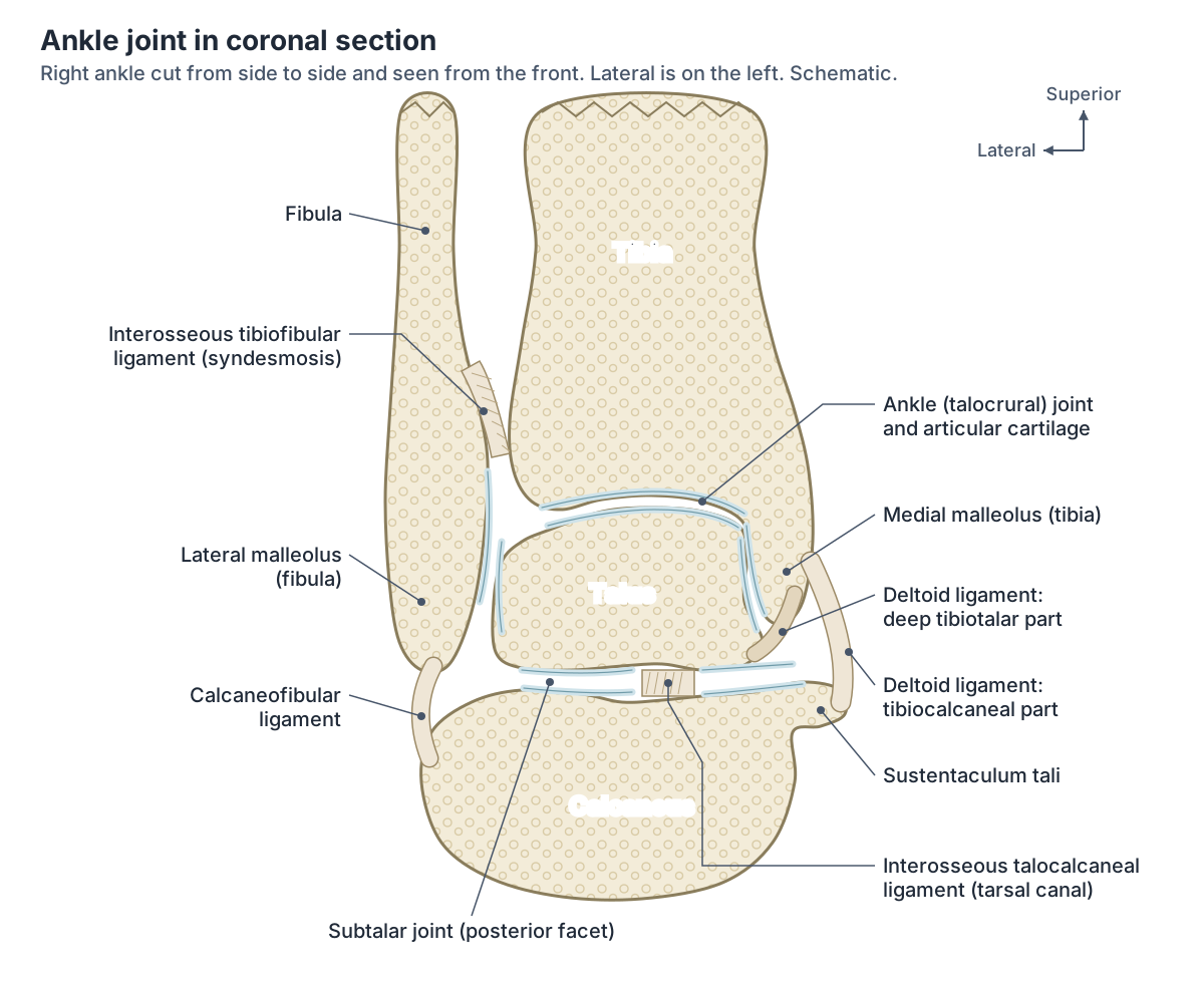
Bound together by strong tibiofibular ligaments, the tibia and fibula form a bracket-shaped socket called the mortise, lined with hyaline cartilage, into which the body of the talus fits. The articular part of the talus is wedge-shaped, broad in front and narrow behind. In dorsiflexion the broad front part sits in the mortise and the joint is more stable; in plantarflexion the narrow back part sits in it and the joint is less stable. (TeachMeAnatomy: The Ankle Joint.)
Kenhub calls this a tenon and mortise joint: the two malleoli and the inferior transverse tibiofibular ligament make a rectangular socket for the talar trochlea, the pulley-shaped top of the talus. The lateral malleolus sits lower and further back than the medial malleolus, which you can feel on your own ankle. (Kenhub: Ankle joint.)
The talus has a head, a neck and a body. The curved dome of the body, the trochlea, forms the ankle joint, and the head joins the navicular bone in front. Unlike most bones, the talus has no muscles attached to it. (Cleveland Clinic: Talus Bone.)
The joint capsule is thin and relatively weak. It attaches to the margins of the malleoli above and of the talar trochlea below, and it is reinforced on each side by the collateral ligaments. Its synovial lining extends a short way up into the distal tibiofibular joint. (Kenhub: Ankle joint.)
The shinbone and its medial malleolus, in 3D. Tibia.
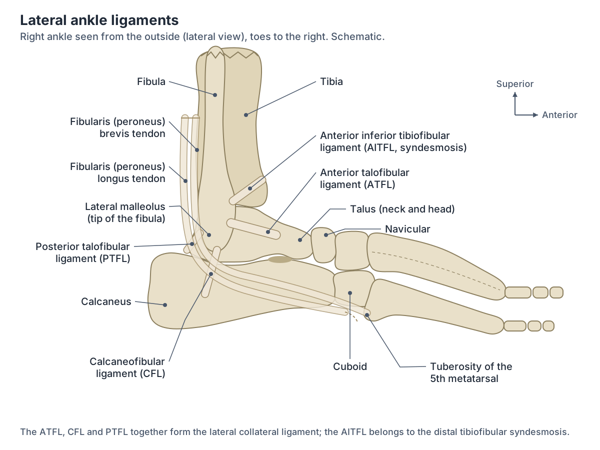
Lateral ankle anatomy: ATFL, CFL and PTFL
The lateral ligament of the ankle starts on the lateral malleolus and resists over-inversion, rolling the sole too far inwards. It has three separate parts: the anterior talofibular ligament (ATFL) to the side of the talus, the posterior talofibular ligament (PTFL) to the back of the talus, and the calcaneofibular ligament (CFL) to the calcaneus. (TeachMeAnatomy: The Ankle Joint.)
Kenhub describes the ATFL as a weak, flat band from the lateral malleolus to the side of the talar neck; the PTFL as a strong band from the lateral malleolar fossa to the lateral tubercle of the talus; and the CFL as a long band from the tip of the lateral malleolus to a tubercle on the outer surface of the calcaneus. (Kenhub: Ankle joint.)
In a cadaver study of 15 ankles, the ATFL had a single band in 8 and two bands (superior and inferior) in 7. Single-band ATFLs measured on average 14 to 16 mm long and 7.6 mm wide, the CFL 18 to 21 mm long and the PTFL 18 to 21 mm long. In 10 of the 15 ankles the CFL was joined to the ATFL and PTFL by connecting fibres. (Ahmed A et al. Lateral Ankle Ligaments: An Insight Into Their Functional Anatomy, Variations, and Surgical Importance. Cureus. 2024.)
The CFL is the only ligament that directly links the tibiotalar (ankle) joint and the subtalar joint. (Brockett CL, Chapman GJ. Biomechanics of the ankle. Orthop Trauma. 2016.)
Two tendons wrap around the back of the lateral malleolus: fibularis (peroneus) brevis and fibularis longus. Fibularis brevis glides between the malleolus and the longus tendon, then runs forward in a canal bounded by the calcaneus and the inferior fibular retinaculum to the tuberosity of the fifth metatarsal. Repeated shear in this groove can lead to tears and tendinitis. (Kenhub: Fibularis brevis muscle.)
The small saphenous vein also passes behind the lateral malleolus, carrying blood from the outer end of the dorsal venous arch of the foot up the back of the leg. (TeachMeAnatomy: Venous Drainage of the Lower Limb.)
The lateral malleolus and the tibiofibular joints. Fibula.
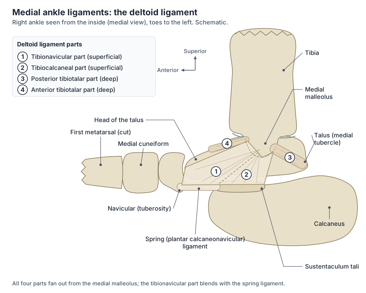
Medial ankle ligament: the deltoid ligament
The medial ligament, called the deltoid ligament, attaches to the medial malleolus and fans out to the talus, calcaneus and navicular. Its main job is to resist over-eversion of the foot. The lateral ligament is weaker than the deltoid ligament, which is one reason lateral sprains are far more common. (TeachMeAnatomy: The Ankle Joint.)
Kenhub describes the deltoid ligament as a strong triangular band with three to six parts, usually grouped as three: the tibionavicular part (superficial fibres to the navicular tuberosity, blending with the spring ligament), the tibiocalcaneal part (nearly vertical fibres to the sustentaculum tali of the calcaneus) and the deep tibiotalar part, whose anterior and posterior portions attach to the talus. (Kenhub: Ankle joint.)
Many guides list four named parts instead: the anterior tibiotalar, posterior tibiotalar, tibiocalcaneal and tibionavicular ligaments, all running from the medial malleolus to the talus, calcaneus and navicular. (Cleveland Clinic: Ankle Ligaments.)
Radiopaedia divides the deltoid ligament into two layers: a superficial triangular part and a deep tibiotalar part made of the deep posterior tibiotalar and anterior tibiotalar ligaments. (Radiopaedia: Ankle joint.)
Just in front of the deltoid ligament is the spring (plantar calcaneonavicular) ligament, which runs from the sustentaculum tali to the navicular and forms a sling under the head of the talus. With the tibialis posterior tendon it is a main stabiliser of the medial arch; when these fail, the hindfoot drifts outwards and the arch flattens (pes planus). (Radiopaedia: Spring ligament complex.)
Ligaments of the ankle
| Group | Ligament | From | To | Main role |
|---|---|---|---|---|
| Lateral | Anterior talofibular (ATFL) | Front of the lateral malleolus | Neck of the talus | Limits inversion and plantar flexion; the most often sprained |
| Lateral | Calcaneofibular (CFL) | Tip of the lateral malleolus | Lateral surface of the calcaneus | Limits inversion and dorsiflexion; crosses both the ankle and subtalar joints |
| Lateral | Posterior talofibular (PTFL) | Lateral malleolar fossa | Lateral tubercle of the talus | Strong band; rarely torn |
| Medial (deltoid) | Tibionavicular | Medial malleolus | Navicular tuberosity and spring ligament | Resists eversion |
| Medial (deltoid) | Tibiocalcaneal | Medial malleolus | Sustentaculum tali | Resists eversion |
| Medial (deltoid) | Anterior and posterior tibiotalar (deep) | Medial malleolus | Talus | Anterior part limits plantar flexion; posterior part limits dorsiflexion |
| Syndesmosis | AITFL, PITFL, transverse and interosseous ligaments | Distal tibia | Distal fibula | Hold the fibula against the tibia; injured in high ankle sprains |
Attachments from Kenhub and TeachMeAnatomy; syndesmosis from Hermans et al. 2010.
The syndesmosis: high ankle ligaments
Just above the ankle joint, the lower ends of the tibia and fibula are joined by a fibrous joint, the distal tibiofibular syndesmosis. It is made of two bones and four ligaments: the anterior inferior tibiofibular ligament (AITFL), the posterior inferior tibiofibular ligament (PITFL), the transverse ligament and the interosseous ligament, which is the thickened lower end of the interosseous membrane. (Hermans JJ et al. Anatomy of the distal tibiofibular syndesmosis in adults: a pictorial essay with a multimodality approach. J Anat. 2010.)
The AITFL runs obliquely from the anterior tubercle of the tibia down to the anterior tubercle of the fibula. It is the weakest of the four and the first to fail when the fibula is forced into external rotation. (Hermans JJ et al. Anatomy of the distal tibiofibular syndesmosis in adults: a pictorial essay with a multimodality approach. J Anat. 2010.)
Counts differ between sources: Cleveland Clinic lists five syndesmotic ligaments because it names the inferior transverse ligament and the transverse tibiofibular ligament separately. (Cleveland Clinic: Ankle Ligaments.)
An injury to these ligaments is called a high ankle sprain. It is thought to follow forceful external rotation of the foot with internal rotation of the leg. Radiopaedia estimates that about 10% of ankle injuries involve the syndesmosis, with a reported range of 1 to 20%. (Radiopaedia: Distal tibiofibular syndesmosis injury (high ankle sprain).)
Radiopaedia's anatomy article gives a lower figure of about 5% (range 1 to 11%) and notes that syndesmotic injury occurs in about half of Weber B ankle fractures and in all Weber C fractures. (Radiopaedia: Distal tibiofibular syndesmosis.)
Subtalar joint: the joint below the ankle
The subtalar (talocalcaneal) joint lies directly under the ankle joint, between the posterior articular surface on the underside of the talus and the posterior facet of the calcaneus. It is a plane synovial joint held mainly by the strong interosseous talocalcaneal ligament, which sits in the sinus tarsi, a small tunnel between the two bones. (TeachMeAnatomy: The Subtalar Joint.)
Because its axis is oblique, the subtalar joint is the main site of inversion and eversion of the foot and plays no part in dorsiflexion or plantarflexion. Eversion is produced by the muscles of the lateral compartment of the leg and inversion by tibialis anterior. (TeachMeAnatomy: The Subtalar Joint.)
In normal walking the subtalar joint moves through about 15 degrees of inversion and eversion. Across the whole ankle complex, total side-to-side motion is about 35 degrees: 23 degrees of inversion and 12 degrees of eversion. (Brockett CL, Chapman GJ. Biomechanics of the ankle. Orthop Trauma. 2016.)
The talus, calcaneus and the other 24 bones of the foot. Foot bones.
Tendons, vessels and nerves that cross the ankle
Every tendon from the leg to the foot crosses the ankle. In front, under the extensor retinaculum, lie the tibialis anterior, extensor hallucis longus and extensor digitorum longus tendons with the dorsalis pedis artery and the deep peroneal (fibular) nerve. Behind the medial malleolus, from front to back, are tibialis posterior, flexor digitorum longus, the posterior tibial artery, the tibial nerve and flexor hallucis longus. On the lateral side are fibularis longus and brevis, the sural nerve and the end of the peroneal artery. (Radiopaedia: Ankle joint.)
The medial group passes through the tarsal tunnel, whose roof is the flexor retinaculum, stretched from the medial malleolus to the medial tubercle of the calcaneus. The mnemonic "Tom, Dick and a Very Nervous Harry" gives the order: tibialis posterior, flexor digitorum longus, posterior tibial artery and vein, tibial nerve, flexor hallucis longus. Compression of the tibial nerve here is tarsal tunnel syndrome. (TeachMeAnatomy: The Tarsal Tunnel.)
At the front of the ankle the extensor hallucis longus tendon emerges between tibialis anterior and extensor digitorum longus. All three dorsiflex the foot and are supplied by the deep fibular nerve. (TeachMeAnatomy: Muscles in the Anterior Compartment of the Leg.)
The dorsalis pedis pulse is felt on the top of the foot just lateral to the extensor hallucis longus tendon, and the posterior tibial pulse just below and behind the medial malleolus. (TeachMeAnatomy: Arterial Supply to the Lower Limb.)
Behind the ankle, gastrocnemius and soleus merge into the calcaneal (Achilles) tendon, which inserts on the calcaneus. The thin plantaris tendon blends into it; plantaris is absent in about 10% of people. (TeachMeAnatomy: Muscles in the Posterior Compartment of the Leg.)
The great saphenous vein forms from the dorsal venous arch and the dorsal vein of the big toe and passes in front of the medial malleolus on its way up the inner side of the leg. (TeachMeAnatomy: Venous Drainage of the Lower Limb.)
The calf and shin muscles whose tendons cross the ankle. Leg muscles.
Structures that pass around the ankle
| Position | Structures | Held by |
|---|---|---|
| Front | Tibialis anterior, extensor hallucis longus and extensor digitorum longus tendons; dorsalis pedis artery; deep fibular nerve | Extensor retinacula |
| Behind the medial malleolus | Tibialis posterior, flexor digitorum longus, posterior tibial artery and vein, tibial nerve, flexor hallucis longus (front to back) | Flexor retinaculum (tarsal tunnel) |
| Behind the lateral malleolus | Fibularis brevis and longus tendons; sural nerve and small saphenous vein more superficially | Inferior fibular retinaculum below the malleolus |
| Behind the ankle | Calcaneal (Achilles) tendon and plantaris | Inserts on the calcaneus |
| Medial side, superficial | Great saphenous vein (in front of the medial malleolus) and saphenous nerve | Lie superficially |
Sources: Radiopaedia, TeachMeAnatomy (tarsal tunnel, venous drainage), Kenhub (fibularis brevis).
Ankle movements: dorsiflexion and plantarflexion
The ankle moves about a side-to-side axis. Kenhub gives about 20 degrees of dorsiflexion and 30 to 50 degrees of plantar flexion. In standing, the foot and leg form a 90-degree angle; maximum dorsiflexion is the close-packed position, in which the joint is most stable. (Kenhub: Ankle joint.)
Measured ranges vary. A biomechanics review gives 10 to 20 degrees of dorsiflexion and 40 to 55 degrees of plantarflexion, and reports that walking uses about 30 degrees of this range, climbing stairs 37 degrees and going down stairs 56 degrees. (Brockett CL, Chapman GJ. Biomechanics of the ankle. Orthop Trauma. 2016.)
In a study of 96 healthy men aged 30 to 40, passive dorsiflexion averaged 15 degrees (range 5 to 40) and plantar flexion about 40 degrees (range 10 to 55), with no difference between right and left ankles. (Roaas A, Andersson GB. Normal range of motion of the hip, knee and ankle joints in male subjects, 30-40 years of age. Acta Orthop Scand. 1982.)
Plantar flexion is produced mainly by gastrocnemius and soleus, helped by tibialis posterior, flexor digitorum longus, flexor hallucis longus and fibularis longus, all of which pass behind the malleoli. Dorsiflexion is produced by tibialis anterior, extensor digitorum longus, extensor hallucis longus and fibularis tertius. (Kenhub: Ankle joint.)
The ankle carries large loads: about five times body weight in the stance phase of walking and up to 13 times body weight in running. About 83% of the load passes through the tibiotalar joint and about 17% through the fibula. (Brockett CL, Chapman GJ. Biomechanics of the ankle. Orthop Trauma. 2016.)
Movements at the ankle and the joints and muscles involved
| Movement | Main joint | Typical range | Main muscles |
|---|---|---|---|
| Dorsiflexion (toes up) | Ankle (talocrural) | About 10 to 20 degrees | Tibialis anterior, extensor digitorum longus, extensor hallucis longus, fibularis tertius |
| Plantarflexion (toes down) | Ankle (talocrural) | About 30 to 55 degrees | Gastrocnemius, soleus; helped by tibialis posterior, long toe flexors, fibularis longus |
| Inversion (sole inwards) | Subtalar and transverse tarsal joints | About 23 degrees | Tibialis anterior, tibialis posterior |
| Eversion (sole outwards) | Subtalar and transverse tarsal joints | About 12 degrees | Fibularis longus, brevis and tertius |
Ranges vary between studies and methods: Kenhub, Brockett and Chapman 2016, and Roaas and Andersson 1982 give overlapping values.
Blood supply and nerve supply of the ankle
The anterior tibial, posterior tibial and fibular (peroneal) arteries supply the ankle. Their malleolar branches form an anastomosis around the malleoli, and the veins follow the arteries. (Kenhub: Ankle joint.)
Kenhub gives the nerve supply as roots L4 to S2, carried by the deep fibular (peroneal), tibial and sural nerves. (Kenhub: Ankle joint.)
TeachMeAnatomy lists the tibial, superficial fibular and deep fibular nerves instead of the sural nerve, so textbooks differ on the smaller branches. (TeachMeAnatomy: The Ankle Joint.)
Radiopaedia notes that the joint receives articular branches from several nerves, in keeping with Hilton's law, including the tibial and deep peroneal nerves. (Radiopaedia: Ankle joint.)
The tibial, fibular and dorsalis pedis arteries and their pulses. Arteries of the leg.
Ankle sprain anatomy: which ligament tears?
The ankle is the most frequently injured joint in the body, and the most common injury is an inversion sprain, in which the sole is forced inwards. The ATFL is injured most often, followed by the CFL. Eversion sprains, which stretch the deltoid ligament, are less common. (OpenStax Anatomy and Physiology 2e, 9.6: Anatomy of Selected Synovial Joints.)
A sprain is a partial or complete tear of ankle ligaments. The usual mechanism is excessive inversion of a plantarflexed, weight-bearing foot, the position in which the narrow back of the talus sits in the mortise. The ATFL is the lateral ligament most at risk of lasting damage. (TeachMeAnatomy: The Ankle Joint.)
Radiopaedia estimates that the ATFL is involved in about 73% of ankle sprains. It grades sprains as grade 1 (ligament stretched, no visible tear or instability), grade 2 (partial rupture) and grade 3 (complete rupture). X-rays look for fractures, and MRI is used when pain persists. (Radiopaedia: Ankle sprain.)
A 2024 review attributes 66 to 80% of ankle sprains to the ATFL and reports combined ATFL and CFL ruptures in 20 to 30% of lateral ankle injuries. About 40% of patients still report instability six months after a sprain. (Ahmed A et al. Lateral Ankle Ligaments: An Insight Into Their Functional Anatomy, Variations, and Surgical Importance. Cureus. 2024.)
Ankle fractures: Pott's fracture and the Weber classification
Seen from the front, the ankle forms a ring: the tibia and fibula above, the talus and calcaneus (the subtalar joint) below, and the medial and lateral ligaments at the sides. Like a polo mint, a ring usually breaks in two places, so a fracture on one side may come with a ligament tear on the other that does not show on an X-ray. (TeachMeAnatomy: The Ankle Joint.)
Pott's fracture-dislocation is a bimalleolar or trimalleolar fracture caused by forced eversion. The medial ligament first pulls off the medial malleolus, the talus then moves outwards and breaks the lateral malleolus, and finally the back of the lower tibia shears off against the talus. (TeachMeAnatomy: The Ankle Joint.)
Fractures of the lateral malleolus are graded by their level relative to the syndesmosis. Weber A lies below it and leaves it intact; Weber B starts at its level and has variable stability; Weber C lies above it, disrupts it and is unstable, usually needing surgical fixation. (Radiopaedia: Weber classification of ankle fractures.)
The ankle is less prone to primary osteoarthritis than the hip or knee, but osteoarthritis after injury (post-traumatic osteoarthritis) is more common at the ankle. (Brockett CL, Chapman GJ. Biomechanics of the ankle. Orthop Trauma. 2016.)
Ankle and foot anatomy: how they work together
The talus links the leg to the rest of the foot's 26 bones: 7 tarsals, 5 metatarsals and 14 phalanges. The foot's own joints are grouped as intertarsal, tarsometatarsal, metatarsophalangeal and interphalangeal joints. (Kenhub: Ankle and foot anatomy.)
The ankle also acts as a shock absorber at heel strike, while some pronation and supination of the foot take place through the subtalar and midtarsal joints below it. (Kenhub: Ankle joint.)
Bones, arches, muscles, tendons and the sole of the foot. Foot anatomy.
Ankle joint in the 3D model
- Calcaneal tendon
- The thick tendon joining gastrocnemius and soleus to the heel bone.
- Calcaneus
- The largest tarsal bone, forming the heel.
- Fibula
- The slender lateral bone of the leg.
- Navicular bone of foot
- A boat-shaped tarsal bone on the medial side of the foot.
- Talus
- A tarsal bone that sits between the tibia and fibula above and the calcaneus below.
- Tibia
- The shinbone, the medial and main weight-bearing bone of the leg.
Frequently asked questions
What are the three bones of the ankle?
The ankle joint is formed by the tibia, the fibula and the talus. The lower ends of the tibia and fibula, including the medial and lateral malleoli, form a socket called the mortise, and the body of the talus fits into it. The calcaneus (heel bone) lies below the talus and is not part of the ankle joint itself.
What are the main ligaments of the ankle?
On the outside are the anterior talofibular, calcaneofibular and posterior talofibular ligaments, together called the lateral ligament. On the inside is the fan-shaped deltoid ligament, with tibionavicular, tibiocalcaneal and tibiotalar parts. Above the joint, the syndesmosis ligaments (AITFL, PITFL, transverse and interosseous ligaments) hold the tibia and fibula together.
Which ankle ligament is most commonly injured?
The anterior talofibular ligament (ATFL). It is a weak, flat band on the outside of the ankle, and it is tight when the foot points down, the position in which most inversion sprains happen. Reported figures range from about 66 to 80% of sprains. The calcaneofibular ligament is the next most often torn.
What are the bumps on the sides of the ankle called?
They are the malleoli. The inner bump is the medial malleolus, the lower end of the tibia, and the outer bump is the lateral malleolus, the lower end of the fibula. The lateral malleolus sits lower and further back than the medial one. Together they grip the sides of the talus.
What tendons run behind the ankle bones?
Behind the medial malleolus run tibialis posterior, flexor digitorum longus and flexor hallucis longus, with the posterior tibial artery and tibial nerve between them, in the tarsal tunnel. Behind the lateral malleolus run the fibularis (peroneus) brevis and longus tendons. The Achilles tendon runs down the back of the ankle to the heel.
What is the difference between the ankle joint and the subtalar joint?
The ankle (talocrural) joint lies between the tibia, fibula and talus and moves the foot up and down. The subtalar joint lies just below it, between the talus and calcaneus, and is the main joint for tilting the sole inwards (inversion) and outwards (eversion). It plays no part in dorsiflexion or plantarflexion.
What is a high ankle sprain?
A high ankle sprain is an injury to the distal tibiofibular syndesmosis, the ligaments that hold the lower tibia and fibula together just above the ankle joint. It is thought to follow forceful outward rotation of the foot. It is less common than an ordinary lateral sprain; estimates range from about 1 to 20% of ankle injuries.
References
- TeachMeAnatomy. The Ankle Joint.
- Kenhub. Ankle joint.
- Kenhub. Ankle and foot anatomy.
- OpenStax. Anatomy and Physiology 2e, §9.6 Anatomy of Selected Synovial Joints. Rice University, 2022.
- OpenStax. Anatomy and Physiology 2e, §8.4 Bones of the Lower Limb. Rice University, 2022.
- Cleveland Clinic. Talus Bone: Anatomy, Function & Common Conditions.
- Cleveland Clinic. Ankle Ligaments: What They Are, Anatomy & Function.
- Radiopaedia. Ankle joint.
- Radiopaedia. Ankle sprain.
- Radiopaedia. Weber classification of ankle fractures.
- Radiopaedia. Distal tibiofibular syndesmosis.
- Radiopaedia. Distal tibiofibular syndesmosis injury (high ankle sprain).
- Radiopaedia. Spring ligament complex.
- Hermans JJ, Beumer A, de Jong TAW, Kleinrensink GJ. Anatomy of the distal tibiofibular syndesmosis in adults: a pictorial essay with a multimodality approach. J Anat. 2010;217(6):633-645.
- Ahmed A, Mishra P, Patra B, Ravi PK. Lateral Ankle Ligaments: An Insight Into Their Functional Anatomy, Variations, and Surgical Importance. Cureus. 2024;16(2):e53826.
- Brockett CL, Chapman GJ. Biomechanics of the ankle. Orthop Trauma. 2016;30(3):232-238.
- Roaas A, Andersson GB. Normal range of motion of the hip, knee and ankle joints in male subjects, 30-40 years of age. Acta Orthop Scand. 1982;53(2):205-208.
- TeachMeAnatomy. The Subtalar Joint.
- TeachMeAnatomy. The Tarsal Tunnel.
- TeachMeAnatomy. Muscles in the Anterior Compartment of the Leg.
- TeachMeAnatomy. Muscles in the Posterior Compartment of the Leg.
- TeachMeAnatomy. Arterial Supply to the Lower Limb.
- TeachMeAnatomy. Venous Drainage of the Lower Limb.
- Kenhub. Fibularis brevis muscle.
- Mitsuhashi N. et al. (2009). BodyParts3D: 3D structure database for anatomical concepts. Nucleic Acids Research 37, D782–D785. Data © The Database Center for Life Science, CC BY 4.0.
Related anatomy
- Foot anatomyFoot anatomy with labelled diagrams and a 3D model: the 26 bones, joints, arches, muscles, tendons, nerves, vessels…
- Foot bonesFoot bone anatomy with a labeled diagram and 3D model: all 26 bones of the foot, the 7 tarsals, 5 metatarsals and 14…
- Leg musclesLeg muscle anatomy with a 3D model and labeled diagrams: all leg muscle groups, the calf muscles, tibialis anterior…
- TibiaTibia anatomy explained: where the shinbone is, the proximal tibia, tibial plateau, shaft and medial malleolus, its…
- FibulaFibula anatomy: the head of fibula, shaft and lateral malleolus, the proximal and distal tibiofibular joints and…
- Lower limb nervesLower limb nerves explained: lumbar and sacral plexus diagram, femoral, saphenous, sciatic, tibial, common peroneal…
Explore the whole body in the interactive 3D human anatomy model, or browse the A–Z list of human structures.