

Where is the biceps brachii?
The biceps brachii lies in the anterior (front) compartment of the arm, the segment between the shoulder and the elbow. It is the most superficial muscle there: it forms the front of the arm and covers brachialis and coracobrachialis, while the deltoid covers its two origin tendons at the shoulder. (Kenhub: Biceps brachii muscle.)
The anterior compartment of the arm contains three muscles, biceps brachii, coracobrachialis and brachialis, and all three are supplied by the musculocutaneous nerve. The posterior compartment holds triceps brachii, supplied by the radial nerve. (TeachMeAnatomy: Muscles of the Upper Arm.)
Because it crosses both the shoulder and the elbow, OpenStax describes biceps brachii as a two-headed muscle that crosses the shoulder and elbow joints to flex the forearm, the movement of a biceps curl. (OpenStax Anatomy and Physiology 2e, 11.5: Muscles of the Pectoral Girdle and Upper Limbs.)
Muscles are often named for their number of origins: the prefix bi- means the muscle has two origins, and OpenStax uses biceps brachii as its example. So 'biceps' is already singular, and 'bicep' is an everyday back-formation. The full name, biceps brachii, tells it apart from biceps femoris, a hamstring muscle of the thigh. (OpenStax Anatomy and Physiology 2e, 11.2: Naming Skeletal Muscles.)
All the muscles of the upper arm together, including triceps, brachialis and coracobrachialis. Arm muscles.
Long head and short head of the biceps
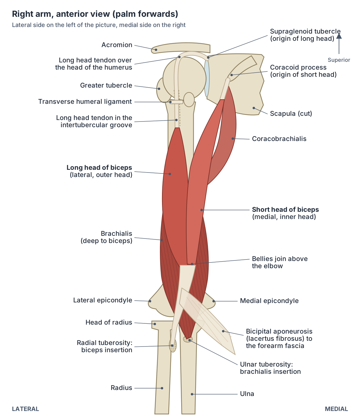
The biceps has two heads, both arising from the scapula. The short head arises from the tip (apex) of the coracoid process. The long head arises from the supraglenoid tubercle, a small bump just above the glenoid cavity of the shoulder joint. (University of Washington Musculoskeletal Atlas: Biceps brachii.)
In the arm the long head lies laterally (the outer head) and the short head medially (the inner head). The short head travels vertically through the axilla into the arm before its fibres unite with the long head. (Elsevier Complete Anatomy: Short head of biceps brachii.)
The long head starts as a long cylindrical tendon inside the shoulder joint capsule, continuous with the glenoid labrum. It runs over the head of the humerus inside a synovial sheath and then descends in the intertubercular (bicipital) groove, where the transverse humeral ligament holds it in place. (Elsevier Complete Anatomy: Long head of biceps brachii.)
Kenhub describes the long head tendon as intracapsular but extrasynovial. It makes a sharp turn over the humeral head, held by capsular ligaments known as the biceps pulley, before entering the groove. Radiopaedia adds that it passes through the rotator cuff interval on the way. (Kenhub: Biceps brachii muscle.)
A 2025 review describes the long head as arising from both the supraglenoid tubercle and the superior labrum, and notes that the transverse humeral and superior glenohumeral ligaments help keep the tendon in its groove; when they fail, the tendon can slip medially out of the groove. (Elgendy M et al. Biceps tendon rupture: contemporary evidence, evolving techniques, and a mechanics-to-management clinical framework. Cureus. 2025.)
Textbooks describe the short head arising by a short flat tendon, partly blended with the origin of coracobrachialis. A dissection study of five cadaver shoulders challenged this: it found muscle fibres attaching directly to the tip of the coracoid, covered only by a thin aponeurosis, and could not separate the short head from coracobrachialis at their attachment. (Crichton JCI, Funk L. The anatomy of the short head of biceps: not a tendon. Int J Shoulder Surg. 2009.)
The two bellies stay separate for most of the arm and merge in the distal arm above the elbow. (Radiopaedia: Biceps brachii.)
The glenohumeral joint, labrum and capsule that the long head tendon runs through. Shoulder anatomy.
Where does the biceps insert?
The biceps inserts on the radius. Its two bellies converge on a single tendon that attaches to the radial tuberosity, and this tendon gives off a flat expansion, the bicipital aponeurosis, which fans out into the deep fascia of the forearm. (Elsevier Complete Anatomy: Short head of biceps brachii.)
Radiopaedia describes the distal tendon as bifid, reaching the posterior margin of the radial tuberosity, with a small bursa there that eases pronation and supination. The bicipital aponeurosis (lacertus fibrosus) arises from the medial side of the tendon and crosses over the brachial artery and median nerve before blending with the fascia over the forearm flexors. (Radiopaedia: Biceps brachii.)
The two parts of the distal tendon behave differently. In a 2021 review, the part from the long head is described as the chief supinator and the part from the short head as the principal elbow flexor, and both attach on the back of the radial tuberosity. Surgeons aim to restore this footprint behind the apex of the tuberosity when they repair a torn tendon. (Vishwanathan K, Soni K. Distal biceps rupture: evaluation and management. J Clin Orthop Trauma. 2021.)
The aponeurosis forms the roof of the cubital fossa at the front of the elbow, and the biceps tendon itself can be felt in the crook of the arm. (Kenhub: Biceps brachii muscle.)
The radius, the radial tuberosity and the cubital fossa where the biceps tendon ends. Elbow anatomy.
Biceps brachii: origin, insertion, nerve, action and blood supply
| Part | Origin | Insertion | Nerve (roots) | Main actions | Blood supply |
|---|---|---|---|---|---|
| Long head | Supraglenoid tubercle of the scapula (and superior glenoid labrum) | Radial tuberosity (posterior part); bicipital aponeurosis into forearm fascia | Musculocutaneous nerve (C5, C6) | Supinates forearm; flexes elbow; abducts arm at the shoulder | Muscular branches of the brachial artery |
| Short head | Tip (apex) of the coracoid process of the scapula | Radial tuberosity with the long head; bicipital aponeurosis | Musculocutaneous nerve (C5, C6) | Flexes elbow; supinates forearm; adducts and flexes arm at the shoulder | Muscular branches of the brachial artery |
| Whole muscle | Two heads on the scapula | One distal tendon to the radius | Musculocutaneous nerve, a branch of the lateral cord (C5, C6) | Supination (strongest with the elbow bent), elbow flexion (strongest with the palm up), weak shoulder flexion | Up to eight branches of the brachial artery (Kenhub) |
Compiled from the University of Washington atlas, Kenhub, Radiopaedia, Elsevier Complete Anatomy and Elgendy et al. 2025.
Nerve and blood supply of the biceps
The biceps is supplied by the musculocutaneous nerve, from spinal roots C5 and C6. Radiopaedia notes that the nerve sends a separate branch to each belly. (Radiopaedia: Biceps brachii.)
The musculocutaneous nerve is the continuation of the lateral cord of the brachial plexus. (TeachMeAnatomy: The Brachial Plexus.)
The biceps tendon reflex is used to test the nerve roots. TeachMeAnatomy links it to spinal cord segment C6, while Kenhub describes the muscle as a reference muscle for both C5 and C6. (TeachMeAnatomy: Muscles of the Upper Arm.)
The blood supply comes from muscular branches of the brachial artery. Kenhub gives the number as up to eight vessels arising from the brachial artery in the middle third of the arm. (Kenhub: Biceps brachii muscle.)
The brachial plexus, the lateral cord and the full course of the musculocutaneous nerve. Nerves of the arm.
What does the biceps do?
The biceps has three jobs: it supinates the forearm (turns the palm up), flexes the elbow and helps flex the shoulder. The University of Washington atlas sums it up as supinating the forearm and, when the forearm is supine, flexing it. (University of Washington Musculoskeletal Atlas: Biceps brachii.)
Supination is strongest when the elbow is already bent. At the shoulder the biceps is only a weak flexor. It also helps hold the head of the humerus in its socket, and its antagonist at the elbow is triceps brachii. (Kenhub: Biceps brachii muscle.)
The two heads do slightly different things at the shoulder. Radiopaedia lists abduction of the arm for the long head and adduction and flexion for the short head. It adds that with the elbow extended the biceps produces only elbow flexion, and that it can help start shoulder abduction when the deltoid is weak or paralysed. (Radiopaedia: Biceps brachii.)
Kenhub agrees that the long head abducts the arm and adds a role in inward (medial) rotation, while the short head adducts it; when both heads contract together the arm flexes at the shoulder. (Kenhub: Biceps brachii muscle.)
Forearm position matters. Elsevier's Complete Anatomy notes that biceps flexes the elbow most strongly when the forearm is supinated, and that the muscle is tested by flexing the elbow against resistance with the palm up, when it can be felt contracting. (Elsevier Complete Anatomy: Long head of biceps brachii.)
Biceps vs brachialis
Brachialis lies deep to the biceps on the front of the humerus. It arises from the anterior surface of the distal half of the humerus and inserts on the ulnar tuberosity and the coronoid process of the ulna, so unlike the biceps it pulls on the ulna, not the radius. (Kenhub: Brachialis muscle.)
Kenhub calls brachialis the workhorse of the elbow and the only pure flexor of the elbow joint. It flexes the elbow in every forearm position, is not affected by pronation or supination, and produces most of the force during elbow flexion. (Kenhub: Brachialis muscle.)
OpenStax puts the same idea simply: deep to the biceps brachii, the brachialis provides additional power in flexing the forearm. (OpenStax Anatomy and Physiology 2e, 11.5: Muscles of the Pectoral Girdle and Upper Limbs.)
Brachialis is supplied mainly by the musculocutaneous nerve, like the biceps, but TeachMeAnatomy and Kenhub both note a contribution from the radial nerve, which Kenhub places in a small lateral part of the muscle (C7). (TeachMeAnatomy: Muscles of the Upper Arm.)
The deep elbow flexor in the 3D model, with its own structure page. Brachialis.
Biceps brachii vs brachialis
| Feature | Biceps brachii | Brachialis |
|---|---|---|
| Position | Superficial, front of the arm | Deep to biceps, on the front of the humerus |
| Origin | Scapula (two heads) | Anterior surface of the distal half of the humerus |
| Insertion | Radial tuberosity and forearm fascia | Ulnar tuberosity and coronoid process of the ulna |
| Joints crossed | Shoulder and elbow | Elbow only |
| Nerve | Musculocutaneous (C5, C6) | Musculocutaneous (C5, C6), small lateral part from the radial nerve (C7) |
| Elbow flexion | Strongest with the forearm supinated | Equal in every forearm position; produces most flexion force |
| Supination | Main supinator when the elbow is bent | None |
Based on Kenhub (biceps brachii and brachialis), TeachMeAnatomy and Elsevier Complete Anatomy.
Can the biceps have three heads?
Yes. An extra (accessory) head is one of the commonest muscle variations in the arm. A 2022 meta-analysis of 78 studies covering 10,603 upper limbs found accessory heads of the biceps in 9.6% of limbs (95% confidence interval 8 to 11%). (Benes M et al. Accessory heads of the biceps brachii muscle: a systematic review and meta-analysis. J Anat. 2022.)
The commonest form is a humeral head arising from the front of the humerus near the insertion of coracobrachialis and the origin of brachialis, found in 6.8% of limbs. The musculocutaneous nerve supplied the extra head in 94.4% of cases and the median nerve in 5.6%. Accessory heads were more often one-sided than on both arms (4.4% versus 2.6%) and somewhat more frequent in males than females (10.2% versus 7.3%). (Benes M et al. Accessory heads of the biceps brachii muscle: a systematic review and meta-analysis. J Anat. 2022.)
Extra heads are usually harmless. Reports suggest that some compress the median nerve or the brachial artery near the elbow, and they can be mistaken for soft-tissue masses on imaging. (Benes M et al. Accessory heads of the biceps brachii muscle: a systematic review and meta-analysis. J Anat. 2022.)
Radiopaedia lists other variants seen on scans: a duplicated long head tendon, which can mimic a longitudinal tear, an accessory slip of the long head tendon, and a bifid insertion, where the distal tendons of the two heads stay separate. (Radiopaedia: Biceps brachii.)
Biceps tendon tears: shoulder and elbow
The biceps has two tendons at the shoulder and one at the elbow, and each can tear. At the shoulder, the long head tendon is the one at risk; the short head rarely tears. Most long head tears come from gradual fraying with age rather than a single injury. (OrthoInfo (AAOS): Biceps Tendon Tear at the Shoulder.)
When the long head tears, the muscle belly slides down and bunches into a bulge above the elbow, the 'Popeye' sign. Most people can still use the biceps afterwards, because the short head remains attached. (OrthoInfo (AAOS): Biceps Tendon Tear at the Shoulder.)
TeachMeAnatomy notes that weakness after a long head rupture is often minimal because brachialis and supinator compensate. (TeachMeAnatomy: Muscles of the Upper Arm.)
A 2025 review likewise describes proximal rupture as mainly changing the contour of the arm with minimal loss of strength, and calls the deformity after a distal rupture a 'reverse Popeye': the muscle retracts towards the shoulder and the front of the elbow looks flatter. The same review quotes 2 to 5 distal ruptures per 100,000 person-years. (Elgendy M et al. Biceps tendon rupture: contemporary evidence, evolving techniques, and a mechanics-to-management clinical framework. Cureus. 2025.)
Tears at the elbow (distal biceps tears) are less common. OrthoInfo gives a rate of 3 to 5 people per 100,000 each year, rarely in women, most often in men over 30, usually when the bent elbow is forced straight against resistance. A pop, bruising at the elbow and forearm and a bunched-up muscle are typical. (OrthoInfo (AAOS): Biceps Tendon Tear at the Elbow.)
Published rates differ. A 2021 review gives about 5.4 cases per 100,000 people per year, with peaks in young athletes and in people in their 40s to 60s. Typical complaints are pain at the front of the elbow and weak bending and twisting, such as difficulty with a screwdriver or a jar lid. (Vishwanathan K, Soni K. Distal biceps rupture: evaluation and management. J Clin Orthop Trauma. 2021.)
An intact bicipital aponeurosis can stop a torn distal tendon retracting far. Without repair, studies summarised in the 2021 review found about 40 to 50% loss of supination strength and about 30% loss of elbow flexion strength. The hook test, which checks for the taut, cord-like tendon at the front of the elbow, is reported to have 100% specificity and 80 to 100% sensitivity for complete tears. (Vishwanathan K, Soni K. Distal biceps rupture: evaluation and management. J Clin Orthop Trauma. 2021.)
OrthoInfo gives a similar estimate of about 30 to 40% loss of strength after an unrepaired distal tear, mainly in turning the palm up. (OrthoInfo (AAOS): Biceps Tendon Tear at the Elbow.)
The bones, compartments, nerves and vessels of the whole upper limb. Arm anatomy.
The biceps in the 3D model
In the front view the biceps is the first arm muscle you see, because it lies superficial to brachialis and coracobrachialis; only its two origin tendons are hidden, under the deltoid. (Kenhub: Biceps brachii muscle.)
The model highlights the long head and the short head of the biceps as two separate parts, with the skeleton ghosted around them. Rotate to the front of the shoulder to see the short head leaving the coracoid process, then follow the long head up between the tubercles of the humerus towards the top of the glenoid cavity. Click either head to open its own page. The model does not include tendons as separate parts, nerves or the bicipital aponeurosis, so the diagram above shows those. The model is BodyParts3D, built from one adult male.
Biceps brachii in the 3D model
- Long head of biceps brachii
- The long head of biceps, passing through the shoulder joint.
- Short head of biceps brachii
- The medial head of biceps brachii at the front of the arm.
Frequently asked questions
What are the two heads of the biceps?
The biceps has a long head and a short head. The long head starts on the supraglenoid tubercle just above the shoulder socket and runs through the shoulder joint and down the intertubercular groove of the humerus; it lies on the outer side of the arm. The short head starts on the coracoid process of the scapula and lies on the inner side. Both join one tendon that inserts on the radius.
Where does the biceps attach at the elbow?
The biceps tendon attaches to the radial tuberosity, a roughened bump on the inner side of the upper radius, just below the elbow. A flat sheet called the bicipital aponeurosis (lacertus fibrosus) also leaves the tendon and blends into the fascia over the forearm flexor muscles. Because it pulls on the radius, the biceps can rotate the forearm as well as bend the elbow.
What nerve supplies the biceps?
The musculocutaneous nerve supplies the biceps, carrying fibres from spinal roots C5 and C6. It is the continuation of the lateral cord of the brachial plexus and sends a separate branch to each head. The same nerve supplies brachialis and coracobrachialis. Doctors test the C5 and C6 roots, and especially C6, with the biceps tendon reflex.
What is the main function of the biceps?
The biceps bends (flexes) the elbow and turns the palm up (supination). Supination is strongest when the elbow is already bent, as when using a screwdriver, and flexion is strongest when the palm faces up. Because both heads start on the scapula, the biceps also helps flex the arm at the shoulder, although only weakly.
Is the brachialis stronger than the biceps?
For bending the elbow, brachialis does much of the work. It lies under the biceps, pulls on the ulna and flexes the elbow equally whatever the position of the forearm, so Kenhub calls it the workhorse of the elbow and credits it with most of the force in elbow flexion. The biceps adds flexion power, especially with the palm up, and is the main supinator.
Can you still use your arm after a biceps tendon tear?
After a tear of the long head at the shoulder, most people keep good function because the short head is still attached, though a Popeye-shaped bulge appears. A complete tear at the elbow is different: without repair, studies report roughly 30 to 50% loss of supination strength and about 30% loss of elbow flexion strength. Treatment decisions are made by a doctor.
How common is a third head of the biceps?
Fairly common for a muscle variation. A 2022 meta-analysis of more than 10,000 upper limbs found an accessory head in about 9.6% of limbs. The usual form arises from the front of the humerus near the brachialis origin and is supplied by the musculocutaneous nerve. Extra heads rarely cause symptoms, although some can press on the median nerve or brachial artery.
References
- OpenStax. Anatomy and Physiology 2e, §11.5 Muscles of the Pectoral Girdle and Upper Limbs. Rice University, 2022.
- OpenStax. Anatomy and Physiology 2e, §11.2 Naming Skeletal Muscles. Rice University, 2022.
- Kenhub. Biceps brachii muscle: origin, insertion, innervation and function.
- Kenhub. Brachialis muscle.
- Radiopaedia. Biceps brachii.
- TeachMeAnatomy. Muscles of the Upper Arm: biceps brachii, coracobrachialis, brachialis and triceps.
- TeachMeAnatomy. The Brachial Plexus.
- University of Washington Department of Radiology. Musculoskeletal Atlas: Biceps brachii.
- Elsevier Complete Anatomy. Long head of biceps brachii.
- Elsevier Complete Anatomy. Short head of biceps brachii.
- Crichton JCI, Funk L. The anatomy of the short head of biceps: not a tendon. Int J Shoulder Surg. 2009;3(4):75-79.
- Benes M, Kachlik D, Lev D, Kunc V. Accessory heads of the biceps brachii muscle: a systematic review and meta-analysis. J Anat. 2022;241(2):461-477.
- Vishwanathan K, Soni K. Distal biceps rupture: evaluation and management. J Clin Orthop Trauma. 2021;19:132-138.
- Elgendy M, Mersal M, Embaby O, Elsaidy S, Alsonbaty M. Biceps tendon rupture: contemporary evidence, evolving techniques, and a mechanics-to-management clinical framework. Cureus. 2025;17(11):e97718.
- American Academy of Orthopaedic Surgeons, OrthoInfo. Biceps Tendon Tear at the Shoulder.
- American Academy of Orthopaedic Surgeons, OrthoInfo. Biceps Tendon Tear at the Elbow.
- Mitsuhashi N. et al. (2009). BodyParts3D: 3D structure database for anatomical concepts. Nucleic Acids Research 37, D782–D785. Data © The Database Center for Life Science, CC BY 4.0.
Related anatomy
- Arm musclesArm muscles labeled in 3D: biceps brachii, brachialis, coracobrachialis, triceps brachii, deltoid and anconeus, with…
- Arm anatomyArm anatomy explained: upper limb regions, the 30 arm bones, shoulder, elbow and wrist joints, muscle compartments…
- Shoulder jointShoulder anatomy with labelled diagrams and a 3D model: clavicle, scapula and humerus, the glenohumeral, AC and SC…
- Elbow jointElbow anatomy with labelled diagrams and a 3D model: humerus, radius and ulna, the three joints, collateral and…
- BrachialisBrachialis: The main flexor of the elbow, lying deep to biceps brachii. See where it is in an interactive 3D human…
- Upper limb nervesBrachial plexus diagram with roots, trunks, cords and branches; axillary, musculocutaneous, median, ulnar and radial…
Explore the whole body in the interactive 3D human anatomy model, or browse the A–Z list of human structures.