

What is the elbow joint?
The elbow joint is where the distal humerus meets the proximal radius and ulna. It is a uniaxial hinge formed mainly by the humeroulnar joint, with two associated joints, the humeroradial and proximal radioulnar joints. All three are enclosed in a single articular capsule. (OpenStax Anatomy and Physiology 2e, 9.6: Anatomy of Selected Synovial Joints.)
TeachMeAnatomy describes the elbow as a hinge-type synovial joint that permits flexion and extension of the forearm. The proximal radioulnar joint lies in the same capsule but is usually described separately, because it is the joint that allows pronation and supination. (TeachMeAnatomy: The Elbow Joint.)
Radiopaedia calls the elbow a trochoginglymoid joint, meaning a combined hinge (ginglymus) and pivot (trochoid). (Radiopaedia: Elbow joint.)
Kenhub classes it as a compound synovial joint, because it contains more than one articulation, and functionally as a uniaxial hinge. (Kenhub: Elbow joint.)
In anatomy the arm is only the segment between the shoulder and elbow joints, with the humerus as its single bone. The forearm lies between the elbow and wrist joints and contains the ulna (medially) and the radius (laterally), so the elbow is the joint between arm and forearm. (OpenStax Anatomy and Physiology 2e, 8.2: Bones of the Upper Limb.)
The whole upper limb from shoulder to hand: bones, muscle compartments, nerves and vessels. Arm anatomy.
Elbow bones: humerus, radius and ulna
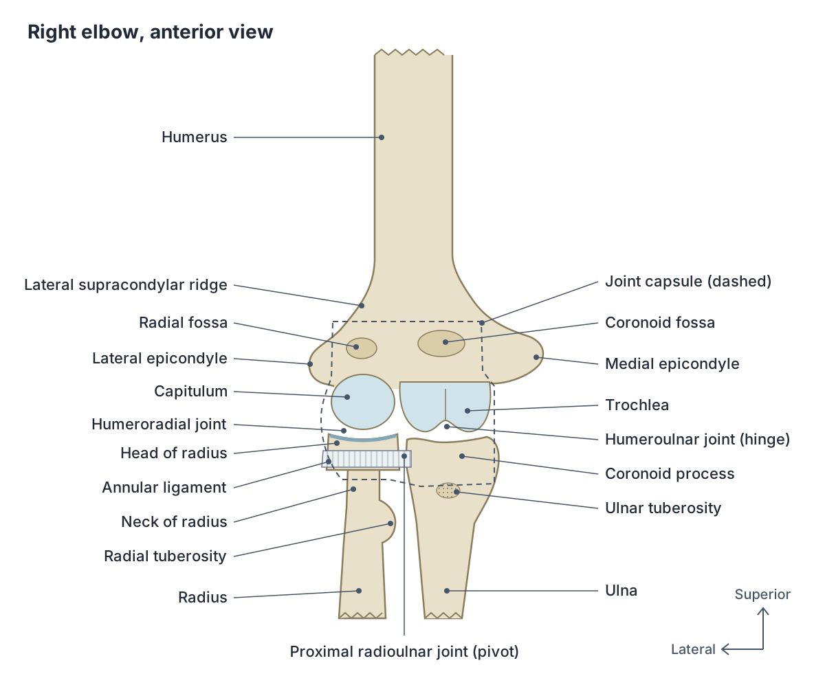
The distal end of the humerus carries two articular surfaces. The pulley-shaped trochlea lies medially and articulates with the ulna; the knob-like capitulum lies just lateral to it and articulates with the head of the radius. Above them stand the large medial epicondyle and the much smaller lateral epicondyle, with the lateral supracondylar ridge rising above the lateral epicondyle. (OpenStax Anatomy and Physiology 2e, 8.2: Bones of the Upper Limb.)
Three hollows on the distal humerus receive the forearm bones. The coronoid fossa above the trochlea receives the coronoid process of the ulna, and the radial fossa above the capitulum receives the head of the radius when the elbow is flexed. The larger olecranon fossa on the back of the humerus receives the olecranon when the forearm is fully extended. (OpenStax Anatomy and Physiology 2e, 8.2: Bones of the Upper Limb.)
The proximal ulna looks like a crescent wrench. Its C-shaped trochlear notch grips the trochlea. The posterior and superior part of the bone is the olecranon, the point of the elbow, and the lower lip of the notch is the coronoid process, with the roughened ulnar tuberosity just below it. A small smooth area below the trochlear notch, the radial notch, is where the head of the radius articulates with the ulna. (OpenStax Anatomy and Physiology 2e, 8.2: Bones of the Upper Limb.)
The proximal radius has a disc-shaped head, a narrowed neck and, below that on the medial side, the radial tuberosity. The head articulates with the capitulum above and with the radial notch of the ulna at its side. (OpenStax Anatomy and Physiology 2e, 8.2: Bones of the Upper Limb.)
Every landmark of the upper arm bone, from the head at the shoulder to the trochlea and capitulum at the elbow. Humerus.
Humeroulnar, humeroradial and proximal radioulnar joints
The elbow capsule holds two articulations between the humerus and the forearm. At the humeroulnar joint the trochlear notch of the ulna wraps around the trochlea of the humerus, and at the humeroradial joint the head of the radius sits against the capitulum. (TeachMeAnatomy: The Elbow Joint.)
Proximal radioulnar joint: the head of the radius turns inside a fibro-osseous ring made by the radial notch of the ulna and the annular ligament. Kenhub describes it as functionally a pivot joint, and TeachMeAnatomy notes that it lies immediately distal to the elbow joint inside the same capsule. (Kenhub: Elbow joint.)
Flexion and extension take place at the humeroulnar joint, while pronation and supination take place at the humeroradial and proximal radioulnar joints, where the radial head spins on the capitulum and inside the annular ligament. (Radiopaedia: Elbow joint.)
The two forearm bones share the load unevenly. Orthobullets gives the split for an axial load through the straight elbow as about 60% through the radiohumeral joint and 40% through the ulnohumeral joint. (Orthobullets: Elbow Anatomy & Biomechanics.)
The olecranon, trochlear notch, coronoid process and radial notch that make the elbow's hinge. Ulna.
The three joints inside the elbow capsule
| Joint | Bones and surfaces | Type | Movement |
|---|---|---|---|
| Humeroulnar | Trochlea of humerus with trochlear notch of ulna | Synovial hinge | Flexion and extension |
| Humeroradial | Capitulum of humerus with head of radius | Synovial; moves with the hinge | Follows flexion and extension; radial head spins on the capitulum in pronation and supination |
| Proximal radioulnar | Head of radius in the radial notch of ulna, held by the annular ligament | Synovial pivot | Pronation and supination |
All three share one fibrous capsule. Based on OpenStax §9.6, TeachMeAnatomy and Radiopaedia.
Joint capsule, synovial membrane and fat pads
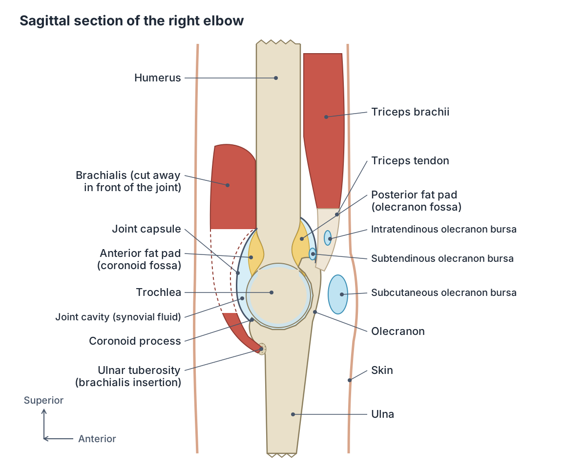
The articular surfaces are covered with hyaline cartilage and lubricated by viscous synovial fluid. A fibrous capsule lined on its inner surface by a synovial membrane encloses the whole joint. (Kenhub: Elbow joint.)
Above, the capsule is attached around the radial, coronoid and olecranon fossae; below, it is attached to the annular ligament and the coronoid process of the ulna. Radiopaedia gives its volume as 24 to 30 mL. (Radiopaedia: Elbow joint.)
The capsule is thin in front and behind, and thickened at its sides by the collateral ligaments, which limit side-to-side movement. (OpenStax Anatomy and Physiology 2e, 9.6: Anatomy of Selected Synovial Joints.)
Between the layers of the capsule lie three fat pads, outside the synovial membrane: the anterior pads in the coronoid and radial fossae and a posterior pad in the olecranon fossa. When the joint fills with fluid these pads are pushed out of their fossae, which is a sign of an elbow effusion on an X-ray. A thin anterior fat pad is normal, but an elevated one suggests a fracture such as a supracondylar fracture. (Radiopaedia: Elbow joint.)
Ligaments of the elbow
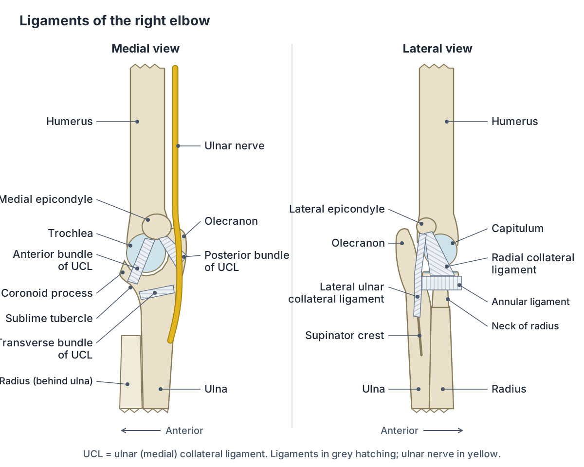
The ulnar (medial) collateral ligament is triangular. It runs from the medial epicondyle of the humerus to the proximal ulna. Its anterior part is the strongest and resists hyperextension. Frequent, forceful extension of the forearm can injure it, as in baseball pitchers, and its surgical reconstruction is known as Tommy John surgery. (OpenStax Anatomy and Physiology 2e, 9.6: Anatomy of Selected Synovial Joints.)
Radiopaedia divides the medial collateral complex into three bundles. The cord-like anterior bundle runs from the lower medial epicondyle to the sublime tubercle on the medial side of the coronoid process. The posterior bundle, a thickening of the capsule, runs to the medial olecranon. The transverse (oblique) bundle, also called Cooper's ligament, joins the lower ends of the other two and does almost nothing for stability. The complex as a whole resists valgus stress. (Radiopaedia: Medial collateral ligament complex of the elbow.)
The lateral collateral complex is Y-shaped. The radial collateral ligament runs from the lateral epicondyle to the annular ligament and supinator fascia. The lateral ulnar collateral ligament runs from the lateral epicondyle to the supinator crest of the ulna. The annular ligament forms a sling around the radial head, attached to the ulna at the radial notch and the supinator crest. An accessory collateral ligament is present in up to 33% of people. (Radiopaedia: Lateral collateral ligament complex of the elbow.)
The lateral complex is a major lateral stabiliser and resists varus stress. Injury to it can cause posterolateral rotatory instability of the elbow. (Radiopaedia: Lateral collateral ligament complex of the elbow.)
Two smaller ligaments complete the picture. The quadrate ligament is a thickening of the capsule from just below the radial notch to the medial neck of the radius. The oblique cord, from the ulnar tuberosity to just below the radial tuberosity, is inconstant and of little functional importance. (Radiopaedia: Elbow joint.)
Ligaments of the elbow
| Ligament | From | To | Main job |
|---|---|---|---|
| Ulnar (medial) collateral: anterior bundle | Lower medial epicondyle | Sublime tubercle of the coronoid process | Strongest part; resists valgus stress and hyperextension |
| Ulnar collateral: posterior bundle | Medial epicondyle | Medial olecranon | Capsular thickening on the medial side |
| Ulnar collateral: transverse bundle | Coronoid process | Olecranon | Little role in stability |
| Radial (lateral) collateral | Lateral epicondyle | Annular ligament and supinator fascia | Resists varus stress |
| Lateral ulnar collateral | Lateral epicondyle | Supinator crest of ulna | Prevents posterolateral rotatory instability |
| Annular | Radial notch of the ulna | Supinator crest of the ulna (encircles the radial head) | Holds the radial head against the ulna |
| Quadrate | Just below the radial notch | Medial neck of the radius | Stays tense in pronation and supination (Kenhub) |
Attachments from Radiopaedia; functions from Radiopaedia, Kenhub and OpenStax §9.6.
Bursae of the elbow
A bursa is a small sac containing a little synovial fluid that reduces friction between moving structures. Three bursae sit around the olecranon: an intratendinous bursa within the triceps tendon, a subtendinous bursa between the olecranon and the triceps tendon, and a subcutaneous bursa between the olecranon and the skin. (TeachMeAnatomy: The Elbow Joint.)
Radiopaedia adds a fourth, the bicipitoradial bursa, named for its position between the biceps tendon and the radius at the front of the elbow. (Radiopaedia: Elbow joint.)
Inflammation of the subcutaneous olecranon bursa (olecranon bursitis) is the most common elbow bursitis. It follows repeated friction or pressure, and the bursa can become infected after minor trauma or a superficial cut over the point of the elbow. (TeachMeAnatomy: The Elbow Joint.)
Elbow movements and the muscles that cross the elbow
The elbow flexors are biceps brachii, brachialis and brachioradialis; the extensors are triceps brachii and anconeus. Pronator teres and pronator quadratus turn the palm to face backwards (pronation), and supinator, helped by biceps brachii, turns it forwards (supination). (OpenStax Anatomy and Physiology 2e, 11.5: Muscles of the Pectoral Girdle and Upper Limbs.)
Pronation is produced by rotation of the radius at the proximal radioulnar joint. Supination rotates the radius back so the radius and ulna lie parallel again. (OpenStax Anatomy and Physiology 2e, 9.5: Types of Body Movements.)
Brachialis runs from the shaft of the humerus to the ulnar tuberosity, just below the elbow, and flexes the elbow. Biceps brachii inserts on the radial tuberosity and, through the bicipital aponeurosis, into the forearm fascia; it supinates the forearm and flexes both the elbow and the shoulder. (TeachMeAnatomy: Muscles of the Upper Arm.)
Many forearm muscles also start at the elbow. The superficial muscles of the anterior forearm share a common flexor tendon that arises from the medial epicondyle of the humerus. (TeachMeAnatomy: Muscles of the Anterior Forearm.)
On the lateral side, extensor carpi radialis brevis, extensor digitorum, extensor digiti minimi and extensor carpi ulnaris share a common extensor origin on the lateral epicondyle. (TeachMeAnatomy: Muscles of the Posterior Forearm.)
How far does the elbow bend? One review gives the normal arc of motion as 0 to 145 degrees of flexion and, citing Morrey's biomechanical study, notes that an arc between 30 and 130 degrees is enough for about 90% of everyday activities. (Blonna D et al. Arthroscopic Treatment of Stiff Elbow. ISRN Surgery. 2011.)
Other sources give similar figures. Vieira and colleagues cite the American Academy of Orthopaedic Surgeons definition of normal elbow flexion as 0 to 146 degrees, and the same 30 to 130 degree functional arc. (Vieira LAG et al. Arthroscopic treatment of elbow stiffness. Rev Bras Ortop. 2015.)
Orthobullets quotes a total flexion-extension range of 0 to 150 degrees and a functional range of 30 to 130 degrees, and lists anconeus, brachialis, triceps and biceps as dynamic stabilisers that press the joint surfaces together. (Orthobullets: Elbow Anatomy & Biomechanics.)
The flexors and extensors that start at the epicondyles, plus the pronators and supinator, with a full attachment table. Forearm muscles.
Muscles that move the elbow and forearm
| Muscle | Origin | Insertion | Nerve | Main action |
|---|---|---|---|---|
| Biceps brachii | Long head: supraglenoid tubercle; short head: coracoid process | Radial tuberosity and bicipital aponeurosis | Musculocutaneous | Flexes elbow, supinates forearm |
| Brachialis | Anterior shaft of the humerus (distal half) | Ulnar tuberosity | Musculocutaneous (small radial nerve contribution) | Flexes elbow in any forearm position |
| Brachioradialis | Lateral supracondylar ridge of the humerus | Distal radius just above the styloid process | Radial | Flexes elbow |
| Triceps brachii | Long head: infraglenoid tubercle; lateral and medial heads: posterior humerus | Olecranon | Radial | Extends elbow |
| Anconeus | Lateral epicondyle | Lateral olecranon and upper posterior ulna | Radial | Assists extension, stabilises the elbow |
| Pronator teres | Medial epicondyle and coronoid process | Middle of the lateral radius | Median | Pronates forearm |
| Supinator | Lateral epicondyle and posterior ulna | Proximal shaft of the radius | Deep branch of radial (posterior interosseous) | Supinates forearm |
| Pronator quadratus | Distal anterior ulna | Distal anterior radius | Anterior interosseous (median) | Pronates forearm (does not cross the elbow) |
Based on TeachMeAnatomy (upper arm, anterior and posterior forearm), Kenhub and the University of Washington musculoskeletal atlas.
What is the carrying angle of the elbow?
The carrying angle is the slight outward (valgus) angle between the long axis of the humerus and the long axis of the forearm, seen when the elbow is straight. It helps the forearms swing clear of the hips when walking, and the two axes come into line when the elbow is fully flexed. (Radiopaedia: Carrying angle.)
Radiopaedia gives average adult values of 11 degrees in males and 14 degrees in females. Orthobullets quotes ranges of 5 to 10 degrees in males and 10 to 15 degrees in females and notes that the angle decreases as the elbow flexes. (Orthobullets: Elbow Anatomy & Biomechanics.)
Measured values vary between populations. In a 2025 study of 300 right-handed adults in central India, the mean carrying angle of the dominant arm was 10.2 degrees in men and 13.4 degrees in women. (Thakur TS et al. Elbow Joint Anthropometry in Central Indian Population. J Pharm Bioallied Sci. 2025.)
A reduced carrying angle makes the forearm point towards the body. This is cubitus varus, also called gunstock deformity. (Radiopaedia: Carrying angle.)
Cubital fossa: the front of the elbow
The cubital fossa is the triangular hollow at the front of the elbow. Its lateral border is brachioradialis, its medial border is pronator teres and its upper border is an imaginary line between the two epicondyles. Brachialis and supinator form its floor, and the bicipital aponeurosis, fascia and skin form its roof. (TeachMeAnatomy: The Cubital Fossa.)
From lateral to medial it contains the radial nerve, the biceps tendon, the brachial artery and the median nerve. The brachial artery divides here into the radial and ulnar arteries, and its pulse can be felt just medial to the biceps tendon. (TeachMeAnatomy: The Cubital Fossa.)
In the roof, the median cubital vein joins the cephalic and basilic veins. It is a common site for taking blood (venepuncture). (TeachMeAnatomy: The Cubital Fossa.)
The brachial artery, its division at the elbow and the superficial veins used for blood tests. Arteries and veins of the arm.
Nerves and blood supply of the elbow (and the funny bone)
The so-called funny bone is the ulnar nerve. At the elbow it runs through the cubital tunnel under the bony bump of the medial epicondyle, so bumping the elbow against a hard surface can briefly pinch it. The ulnar nerve carries feeling from the little finger and the half of the ring finger next to it. (Cleveland Clinic: Ulnar Nerve (Funny Bone).)
TeachMeAnatomy describes the ulnar nerve passing behind the elbow in the space between the medial epicondyle and the olecranon, giving an articular branch to the joint and then passing between the two heads of flexor carpi ulnaris into the forearm. (TeachMeAnatomy: The Ulnar Nerve.)
The median nerve enters the forearm through the cubital fossa and passes between the two heads of pronator teres. (TeachMeAnatomy: The Median Nerve.)
The radial nerve crosses in front of the lateral epicondyle and divides into a superficial sensory branch and a deep motor branch, which is called the posterior interosseous nerve after it pierces supinator. (TeachMeAnatomy: The Radial Nerve.)
The joint itself receives articular branches from the musculocutaneous, radial, ulnar and median nerves, the same nerves that supply the muscles crossing it (Hilton's law). (Radiopaedia: Elbow joint.)
The blood supply comes from a ring of anastomosing arteries. From above, the brachial artery gives the superior and inferior ulnar collateral arteries and the profunda brachii gives the middle and radial collateral arteries. From below, the radial recurrent artery and the anterior and posterior ulnar recurrent arteries climb back up to meet them. Kenhub calls this a periarticular anastomotic loop. (Kenhub: Elbow joint.)
The anconeus muscle is supplied by two of these vessels, the middle collateral branch of the deep brachial artery and the recurrent interosseous artery. All of the anastomosis arteries named here are highlighted in the 3D model. (University of Washington Musculoskeletal Atlas: Anconeus.)
The brachial plexus and the course of the median, ulnar and radial nerves past the elbow. Nerves of the upper limb.
Elbow growth plates in children (CRITOE)
The ossification centres of a child's elbow appear in a fixed order, remembered as CRITOE: capitulum, radial head, internal (medial) epicondyle, trochlea, olecranon and external (lateral) epicondyle. (Radiopaedia: CRITOE.)
Kenhub warns that these secondary ossification centres can be mistaken for fractures, and gives approximate ages of 1 year for the capitulum, 5 years for the radial head and medial epicondyle, 11 years for the trochlea and 12 years for the olecranon. Ages vary between children and between references. (Kenhub: Elbow joint.)
Common elbow injuries and conditions
Supracondylar fracture: a transverse break of the distal humerus between the epicondyles, most often in children after a fall on an outstretched hand. It can damage the brachial artery, which if untreated can lead to Volkmann's ischaemic contracture, and the median, ulnar or radial nerves, so the circulation and nerves are checked carefully. (TeachMeAnatomy: The Elbow Joint.)
Elbow dislocation: most dislocations are posterior. The ulnar collateral ligament is usually torn and the ulnar nerve can be injured. (TeachMeAnatomy: The Elbow Joint.)
Pulled elbow (nursemaid's elbow): the radial head slips partly out of the annular ligament when a young child, typically aged 6 months to 3 years, is pulled or lifted by the arm. X-rays are usually normal, and supinating the forearm with the elbow flexed almost always reduces it. (Radiopaedia: Pulled elbow.)
Tennis elbow (lateral epicondylitis) is an overuse injury of the common extensor origin, and golfer's elbow (medial epicondylitis) an overuse injury of the common flexor origin. (TeachMeAnatomy: The Elbow Joint.)
Radial head fractures often follow a fall on an outstretched hand and can cause loss of full extension. (Kenhub: Elbow joint.)
The radial head and neck, the radial tuberosity and the fractures of the thumb-side forearm bone. Radius.
Elbow joint in the 3D model
- Anconeus
- A small triangular muscle on the posterior elbow.
- Anterior ulnar recurrent artery
- The anterior ulnar recurrent artery is a small branch of the ulnar artery that supplies the elbow.
- Brachialis
- The main flexor of the elbow, lying deep to biceps brachii.
- Brachioradialis
- An elbow flexor that forms the lateral border of the cubital fossa; unusually for a flexor, it is supplied by the radial nerve.
- Humeral head of pronator teres
- The larger, superficial head of pronator teres.
- Humerus
- The long bone of the upper arm.
- Inferior ulnar collateral artery
- The inferior ulnar collateral artery is a branch of the brachial artery that supplies the elbow.
- Median cubital vein
- A superficial vein across the front of the elbow linking the cephalic and basilic veins.
- Middle collateral branch of deep brachial artery
- A terminal branch of the deep brachial artery (profunda brachii) in the posterior arm.
- Posterior ulnar recurrent artery
- A branch of the ulnar artery that ascends behind the medial epicondyle.
- Radial collateral branch of deep brachial artery
- A terminal branch of the deep brachial artery that runs with the radial nerve.
- Radial recurrent artery
- A branch of the radial artery that ascends toward the elbow.
- Radius
- The lateral bone of the forearm.
- Recurrent interosseous artery
- A branch of the posterior interosseous artery running back toward the elbow.
- Superior ulnar collateral artery
- A branch of the brachial artery in the arm.
- Supinator
- A deep forearm muscle wrapping around the upper radius.
- Ulna
- The medial bone of the forearm.
- Ulnar head of pronator teres
- The smaller ulnar head of pronator teres.
Frequently asked questions
What are the three bones of the elbow?
The elbow is formed by the humerus of the upper arm and the radius and ulna of the forearm. The trochlea of the humerus fits into the trochlear notch of the ulna, the capitulum meets the head of the radius, and the radial head also turns against the radial notch of the ulna. The olecranon of the ulna is the bony point you lean on.
What type of joint is the elbow?
The elbow is a synovial hinge joint, so its main movements are flexion and extension. It is often called a compound or hinge-and-pivot joint because the proximal radioulnar joint, a pivot joint that allows pronation and supination, shares the same capsule with the humeroulnar and humeroradial joints.
What are the main ligaments of the elbow?
The ulnar (medial) collateral ligament runs from the medial epicondyle to the coronoid process and olecranon and resists valgus stress; its anterior bundle is the strongest. The lateral collateral complex includes the radial collateral ligament, the lateral ulnar collateral ligament and the annular ligament, which wraps around the radial head. The lateral complex resists varus stress.
Why does hitting your funny bone tingle?
The funny bone is not a bone but the ulnar nerve. It passes behind the elbow through the cubital tunnel, under the bony bump of the medial epicondyle, where it lies close to the surface. Bumping the elbow briefly pinches the nerve and causes a shock-like tingle. The same nerve carries feeling from the little finger and the neighbouring half of the ring finger.
What is the carrying angle and why is it bigger in women?
The carrying angle is the slight outward angle of the forearm relative to the upper arm when the elbow is straight. Radiopaedia gives average values of about 11 degrees in men and 14 degrees in women, and studies in several populations find it larger in females. Exact values vary between populations and measuring methods, and the angle disappears when the elbow is fully bent.
Which muscles bend and straighten the elbow?
Biceps brachii, brachialis and brachioradialis bend (flex) the elbow, which Kenhub remembers as the three Bs. Brachialis pulls on the ulna, while biceps pulls on the radius and also turns the palm up (supination). Triceps brachii straightens (extends) the elbow, assisted by the small anconeus muscle on the back of the joint.
What is nursemaid's elbow?
Nursemaid's elbow, or pulled elbow, is a partial slip of the radial head out of the annular ligament in a young child, usually between 6 months and 3 years old. It typically happens when a child is lifted or pulled by the hand. X-rays are normally unremarkable, and a doctor can usually reduce it quickly by supinating the forearm with the elbow bent.
References
- OpenStax. Anatomy and Physiology 2e, §9.6 Anatomy of Selected Synovial Joints. Rice University, 2022.
- OpenStax. Anatomy and Physiology 2e, §8.2 Bones of the Upper Limb. Rice University, 2022.
- OpenStax. Anatomy and Physiology 2e, §11.5 Muscles of the Pectoral Girdle and Upper Limbs. Rice University, 2022.
- OpenStax. Anatomy and Physiology 2e, §9.5 Types of Body Movements. Rice University, 2022.
- TeachMeAnatomy. The Elbow Joint: structure, movements and injuries.
- TeachMeAnatomy. The Radioulnar Joints.
- TeachMeAnatomy. The Cubital Fossa: borders and contents.
- TeachMeAnatomy. The Ulnar Nerve.
- TeachMeAnatomy. The Median Nerve.
- TeachMeAnatomy. The Radial Nerve.
- TeachMeAnatomy. Arteries of the Upper Limb.
- TeachMeAnatomy. Muscles of the Upper Arm.
- TeachMeAnatomy. Muscles of the Posterior Forearm.
- TeachMeAnatomy. Muscles of the Anterior Forearm.
- Kenhub. Elbow joint: bones, ligaments, movements and clinical notes.
- Radiopaedia. Elbow joint.
- Radiopaedia. Medial collateral ligament complex of the elbow.
- Radiopaedia. Lateral collateral ligament complex of the elbow.
- Radiopaedia. Carrying angle.
- Radiopaedia. Pulled elbow.
- Radiopaedia. CRITOE (mnemonic for elbow ossification centres).
- Orthobullets. Elbow Anatomy & Biomechanics.
- Vieira LAG, Dal Molin FF, Visco A, et al. Arthroscopic treatment of elbow stiffness. Rev Bras Ortop. 2015;46(4):398-402.
- Blonna D, Bellato E, Marini E, Scelsi M, Castoldi F. Arthroscopic Treatment of Stiff Elbow. ISRN Surg. 2011;2011:378135.
- Thakur TS, Singh MP, Sharma MK, et al. Elbow Joint Anthropometry in Central Indian Population: A Cross-Sectional Study. J Pharm Bioallied Sci. 2025.
- Cleveland Clinic. Ulnar Nerve (Funny Bone): What Is It, Branches & Function.
- University of Washington Department of Radiology. Musculoskeletal Atlas: Anconeus.
- Mitsuhashi N. et al. (2009). BodyParts3D: 3D structure database for anatomical concepts. Nucleic Acids Research 37, D782–D785. Data © The Database Center for Life Science, CC BY 4.0.
Related anatomy
- HumerusHumerus bone labeled: head, anatomical and surgical neck, tubercles, deltoid tuberosity, radial groove, epicondyles…
- RadiusRadius labeled: head of radius, neck, radial tuberosity, shaft, Lister's tubercle, ulnar notch and styloid process…
- UlnaUlna labeled: olecranon, trochlear notch, coronoid process, radial notch, shaft, head and styloid process, how the…
- Forearm musclesMuscles of the forearm explained: every anterior flexor and posterior extensor, with origin, insertion, nerve and…
- Arm anatomyArm anatomy explained: upper limb regions, the 30 arm bones, shoulder, elbow and wrist joints, muscle compartments…
- Upper limb nervesBrachial plexus diagram with roots, trunks, cords and branches; axillary, musculocutaneous, median, ulnar and radial…
Explore the whole body in the interactive 3D human anatomy model, or browse the A–Z list of human structures.