

Where is the breast? Female chest anatomy
The breasts are paired structures on the front of the chest wall, present in both sexes but more prominent in females after puberty. Each breast lies on pectoralis major and serratus anterior and extends vertically from the 2nd to the 6th costal cartilage and horizontally from the lateral border of the sternum to the midaxillary line. It has a rounded body and an axillary tail that runs along the lower outer edge of pectoralis major towards the armpit. (TeachMeAnatomy: The Breasts.)
Radiopaedia gives the same extent, from the 2nd to the 6th rib, with the clavicle above, the sternum medially and the midaxillary line laterally, and names the extension into the armpit the axillary tail (of Spence). (Radiopaedia: Breast.)
GPnotebook gives a slightly lower range, ribs 3 to 7 between the parasternal and anterior axillary lines, and places the nipple at about the fourth intercostal space, just lateral to the midclavicular line, a position that changes with age. (GPnotebook: Breast anatomy.)
Behind the breast lie the deep fascia and the pectoral muscles. The breast is separated from them by the retromammary space, a layer of loose connective tissue. (Kenhub: Female breast anatomy.)
The chest wall, muscles and organs under the breast, male and female. Chest anatomy.
Parts of the breast
In females the breast is an accessory organ of the reproductive system that supplies milk to an infant. Milk is made by the mammary glands, which are modified sweat glands. Milk-secreting cells cluster in alveoli inside glandular lobes, contractile myoepithelial cells around the alveoli push the milk towards the lactiferous sinuses, and 15 to 20 lactiferous ducts open on the surface of the nipple. Fat around the lobes sets the size of the breast but not how much milk it can make, and suspensory ligaments attach the breast tissue to the skin. (OpenStax Anatomy and Physiology 2e, 27.2: Anatomy and Physiology of the Ovarian Reproductive System.)
Radiopaedia describes the breast as a mix of glandular tissue (parenchyma: acini and ducts) and stroma (fat and fibrous tissue), held together by Cooper's ligaments and connective tissue rich in collagen and elastin. It counts nearly 14 to 18 lobes; each lobe contains several lobules, each lobule several acini, and the ducts of one lobe join into a single lactiferous duct that opens on the nipple. (Radiopaedia: Breast.)
Fat is the main component of the breast and extends from the collarbone to the armpit and across the rib cage; fibrous connective tissue holds the glandular and fatty tissue in place. The amount of fat largely determines breast size. (Cleveland Clinic: Breast Anatomy.)
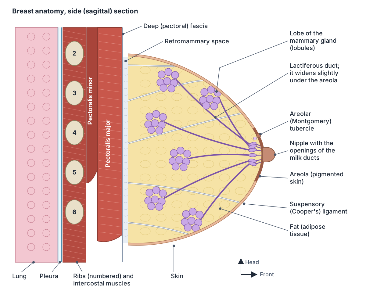
Parts of the breast and what they do
| Part | What it is | Function or key fact |
|---|---|---|
| Lobes | 15 to 20 sections of glandular tissue (classical count) arranged like spokes around the nipple | Each drains through its own lactiferous duct |
| Lobules and alveoli (acini) | Clusters of sacs lined by milk-secreting lactocytes and wrapped in myoepithelial cells | Make milk; myoepithelial cells squeeze it out under oxytocin |
| Lactiferous (milk) ducts | Tubes from the lobules to the nipple, about 2 mm wide near the nipple when lactating | Carry milk; about 9 open on each nipple (range 4 to 18) |
| Lactiferous sinuses | In the classical description, widened duct ends beneath the areola | Ultrasound shows no sac-like sinuses in lactating women |
| Nipple | Central projection made mostly of smooth muscle | Carries the duct openings; supplied mainly by the 4th intercostal nerve |
| Areola | Pigmented skin around the nipple | Darkens and enlarges in pregnancy |
| Areolar (Montgomery) glands | Glands opening as small bumps (tubercles) on the areola | Oily secretion protects the nipple during breastfeeding |
| Fat (adipose tissue) | Fills the spaces around the glandular tissue | Main component outside lactation; sets breast size |
| Suspensory (Cooper's) ligaments | Fibrous bands from the pectoral fascia to the skin | Support the breast; shortening causes skin dimpling |
| Retromammary space | Loose connective tissue behind the breast | Separates the breast from the pectoral fascia; used as a surgical plane |
Sources: OpenStax, TeachMeAnatomy, Kenhub, Radiopaedia, Cleveland Clinic, Ramsay et al. 2005 and Wysocki et al. 2020, as cited in the sections.
Lobes, lobules and milk ducts: how many are there?
Classical anatomy describes 15 to 20 secretory lobes separated by the suspensory ligaments. Each is made of many lobules of tubuloalveolar glands and drains through a lactiferous duct that, in the traditional description, widens into a lactiferous sinus before opening on the nipple. (Kenhub: Female breast anatomy.)
An ultrasound study of breastfeeding women found far fewer main ducts at the base of the nipple: on average 9.6 in the left breast and 9.2 in the right (range 4 to 18), and almost every woman had fewer than the 15 to 20 usually quoted. The ducts were about 2 mm across, lay only about 4.6 mm under the skin, first branched about 8 mm from the nipple, always beneath the areola, and no sac-like lactiferous sinuses were seen. (Ramsay DT et al. Anatomy of the lactating human breast redefined with ultrasound imaging. J Anat. 2005.)
Cleveland Clinic likewise gives about nine milk ducts in each nipple, along with hundreds of nerve endings that make it very sensitive to touch. (Cleveland Clinic: Breast Anatomy.)
OpenStax describes 12 to 20 lobules arranged radially around the nipple of a lactating breast, with milk leaving through 4 to 18 nipple pores. (OpenStax Anatomy and Physiology 2e, 28.6: Lactation.)
The milk ducts of the breast in the 3D Female body. Lactiferous ducts.
Nipple anatomy: nipple, areola and Montgomery glands
The nipple sits at the centre of the breast and is made mostly of smooth muscle fibres. The areola around it is pigmented skin containing many sebaceous glands, which enlarge in pregnancy and secrete an oily lubricant. (TeachMeAnatomy: The Breasts.)
The small bumps on the areola are Montgomery tubercles, the openings of the areolar (Montgomery) glands. Cleveland Clinic describes these as oil-producing glands, separate from the milk-producing mammary glands and found in both male and female breasts. Their secretion protects the nipple and areola during breastfeeding and keeps the skin slightly acidic, and its scent may help a baby find the nipple. Their number and size vary widely between people, and they start to enlarge in the first trimester of pregnancy. (Cleveland Clinic: Montgomery Glands.)
Kenhub instead describes the areolar glands as modified mammary glands with an antimicrobial secretion, and notes that the areola becomes more pigmented and prominent at puberty. (Kenhub: Female breast anatomy.)
Sensation in the nipple depends almost entirely on the fourth intercostal nerve. Surgical anatomists estimate that over 90% of nipple innervation comes from deep branches of its lateral cutaneous branch, which cross the breast tissue and reach the nipple from behind, and about 7% from its superficial branches. (Wysocki WM, Libondi G, Juszczak A. Surgical anatomy of the breast revisited. Biuletyn PTO Nowotwory. 2020.)
In a dissection study of 22 breasts, three to five branches of the fourth intercostal nerve were the main supply to the nipple in 18; the third or fifth intercostal nerve took over in the others. The nerve pierced the chest fascia above the fifth rib about 3 cm lateral to the border of pectoralis major. (Riccio CA et al. Plastic surgery of the breast: keeping the nipple sensitive. Eplasty. 2015.)
The nipple in the 3D Female body, with the areola and areolar tubercles. Nipple.
Cooper's ligaments, fat and breast shape
The fibrous stroma of the breast condenses into the suspensory ligaments (of Cooper), which attach the breast to the dermis of the skin and to the pectoral fascia and separate the lobules. A cancer that invades and shortens these ligaments pulls the skin in, causing dimpling; blocked lymph drainage makes the skin swell around its pores (peau d'orange, orange-peel skin) and can pull the nipple in or to one side. (TeachMeAnatomy: The Breasts.)
Breast density describes how the breast looks on a mammogram. Dense breasts have more glandular and fibrous tissue and less fat; density is not related to the size, appearance or feel of the breast. Up to half of women aged 40 to 74 have dense breasts, and because dense tissue and tumours both look white on a mammogram, cancers are harder to see. (Cleveland Clinic: Breast Anatomy.)
Cooper's ligaments in the 3D Female body. Suspensory ligaments of breast.
Blood supply of the breast
The medial part of the breast is supplied by the internal thoracic (internal mammary) artery, a branch of the subclavian artery. The lateral part receives the lateral thoracic and thoracoacromial branches of the axillary artery, lateral mammary branches of the posterior intercostal arteries in the 2nd to 4th intercostal spaces, and a mammary branch of an anterior intercostal artery. The veins follow the arteries and drain into the axillary and internal thoracic veins. (TeachMeAnatomy: The Breasts.)
Surgical anatomists estimate that the internal mammary artery delivers about 60% of the breast's blood and the lateral thoracic artery about 30%, with the rest from the 2nd to 6th intercostal perforators; the 5th and 6th supply the lower part of the breast. Internal mammary perforators emerge about 2 cm from the edge of the sternum in the 2nd to 4th spaces and run 0.5 to 1 cm under the skin, while lateral thoracic branches lie deeper, 1 to 2.5 cm below the surface. (Wysocki WM, Libondi G, Juszczak A. Surgical anatomy of the breast revisited. Biuletyn PTO Nowotwory. 2020.)
GPnotebook gives the same split, about 60% from the internal mammary artery and 30% from the lateral thoracic artery. (GPnotebook: Breast anatomy.)
Radiopaedia lists the internal thoracic perforators of the 2nd to 5th spaces, the thoracoacromial and lateral thoracic arteries, vessels to serratus anterior and branches of the 3rd to 8th intercostal perforators, with veins draining to the axillary, internal thoracic and posterior intercostal veins. (Radiopaedia: Breast.)
The main artery of the medial breast, in 3D. Internal thoracic artery.
Arteries of the breast
| Artery | Comes from | Area supplied |
|---|---|---|
| Internal thoracic (internal mammary) perforators, 2nd to 4th spaces | Subclavian artery | Medial breast and nipple; about 60% of total blood flow |
| Lateral thoracic artery | Axillary artery | Lateral and upper outer breast; about 30% of total blood flow |
| Pectoral branch of the thoracoacromial artery | Axillary artery | Small supply through pectoralis major |
| Intercostal perforators, 2nd to 6th spaces | Posterior intercostal arteries from the aorta | Remainder; the 5th and 6th supply the lower part of the breast |
Percentages from Wysocki et al. 2020 and GPnotebook; other studies give different splits, but the internal thoracic artery is always the largest supplier.
Lymph nodes and lymph drainage of the breast
Lymph from the breast drains to three groups of nodes: about 75% to the axillary nodes in the armpit, 20% to the parasternal nodes beside the sternum and 5% to the posterior intercostal nodes. The skin drains to the axillary, inferior deep cervical and infraclavicular nodes, and the nipple and areola drain into the subareolar lymphatic plexus. All five groups of axillary nodes drain on to the apical nodes. (TeachMeAnatomy: The Breasts.)
Kenhub gives 75 to 90% to the axillary nodes, usually through a sentinel node at the lateral border of pectoralis major, and 10 to 25% to the parasternal (internal mammary) nodes, mainly from the deep medial breast. The axillary nodes form pectoral (anterior), lateral, posterior (subscapular), central and apical groups, and one to four interpectoral (Rotter's) nodes lie between pectoralis major and minor. Surgeons describe level I nodes lateral to pectoralis minor, level II behind it and level III medial to it. (Kenhub: Lymphatic drainage of the breast.)
Because about 75% of the lymph from the breast drains into the axillary nodes, these nodes can be biopsied when breast cancer is suspected. Removing them (axillary clearance) can injure the long thoracic nerve and cause a winged scapula. (TeachMeAnatomy: The Axilla.)
In sentinel node biopsy, the first node downstream of a tumour is identified in more than 97% of cases and predicts the state of the remaining nodes more than 95% of the time. Lymphoedema of the arm affects an estimated 20% of patients after full axillary node dissection. (Kenhub: Lymphatic drainage of the breast.)
Lymph nodes and lymph vessels throughout the body. Lymphatic system.
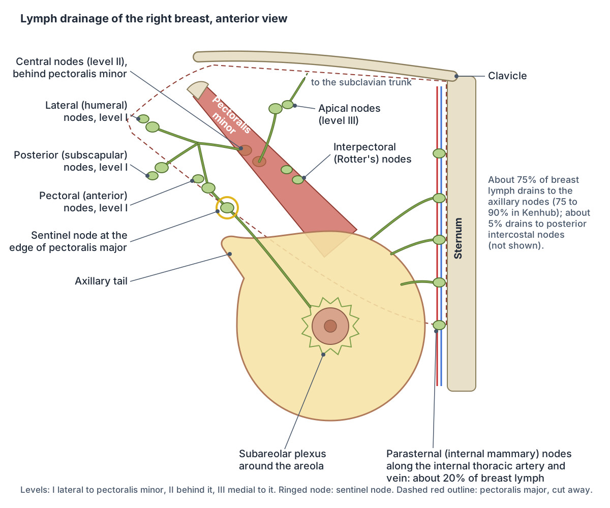
Where lymph from the breast drains
| Node group | Share of breast lymph | Notes |
|---|---|---|
| Axillary nodes | About 75% (TeachMeAnatomy); 75 to 90% (Kenhub) | Mainly from the lateral breast; usually through a sentinel node at the edge of pectoralis major |
| Parasternal (internal mammary) nodes | About 20% (TeachMeAnatomy); 10 to 25% (Kenhub) | Mainly from the deep medial breast; lie along the internal thoracic vessels |
| Posterior intercostal nodes | About 5% (TeachMeAnatomy) | Lie near the heads of the ribs |
| Subareolar plexus | Collects lymph from the nipple and areola | Drains on into the axillary pathway |
| Interpectoral (Rotter's) nodes | Small, variable | One to four nodes between pectoralis major and minor |
Sources: TeachMeAnatomy (The Breasts) and Kenhub (lymphatic drainage of the breast). Percentages are approximate.
Nerves of the breast
TeachMeAnatomy gives the nerve supply of the breast as the anterior and lateral cutaneous branches of the 4th to 6th intercostal nerves, which carry sensory and autonomic fibres. These nerves do not control milk production, which is regulated by prolactin and oxytocin from the pituitary gland. (TeachMeAnatomy: The Breasts.)
Kenhub widens the range to the 2nd to 6th intercostal nerves and names the 4th intercostal nerve as the supply of the nipple. (Kenhub: Female breast anatomy.)
Radiopaedia gives the intercostal nerves T3 to T5 together with the supraclavicular nerve from the cervical plexus. (Radiopaedia: Breast.)
A surgical review reconciles these: the 2nd and 3rd intercostal nerves give only small skin branches to the top of the breast, where branches of the supraclavicular nerves of the cervical plexus also reach, and the rest of the breast is supplied mainly by the 4th, 5th and 6th intercostal nerves. (Wysocki WM, Libondi G, Juszczak A. Surgical anatomy of the breast revisited. Biuletyn PTO Nowotwory. 2020.)
The skin at the level of the nipple lies in the T4 dermatome. (Kenhub: Dermatomes.)
Breast development at puberty and through the menstrual cycle
Breast tissue first appears as ridges of ectoderm in the 6th week of embryonic life, and ducts form around the 5th month. The glandular tissue depends on oestrogen: it becomes more glandular in adolescence and gains acini in pregnancy and lactation. (Radiopaedia: Breast.)
The embryonic milk line runs from the armpit to the inner thigh, and breast glandular tissue normally forms near the 4th and 5th ribs. Extra nipples along this line or beyond it (polythelia, also called supernumerary or accessory nipples) are a common congenital variation, and an extra nipple can develop into a complete extra breast (polymastia). (Radiopaedia: Polythelia.)
Thelarche, the start of breast development, is the first clinical sign of puberty in girls and is defined as Tanner stage B2: the first elevation of the breast and nipple with a wider areola. A meta-analysis of 30 studies found typical ages at B2 between about 8.8 and 13.2 years depending on the region, and that the age at thelarche fell by about 0.24 years (almost 3 months) per decade between 1977 and 2013. (Eckert-Lind C et al. Worldwide secular trends in age at pubertal onset assessed by breast development among girls. JAMA Pediatr. 2020.)
Hormones shape the breast throughout life. Oestrogen stretches the milk ducts and forms their side branches, progesterone increases the number and size of lobules after ovulation, prolactin prepares the glands to make milk and oxytocin releases it. (Cleveland Clinic: Breast Anatomy.)
Breast tissue responds to oestrogen and progesterone during the menstrual cycle, which often makes the breasts swell and feel tender in the second half of the cycle. (OpenStax Anatomy and Physiology 2e, 27.2: Anatomy and Physiology of the Ovarian Reproductive System.)
Breastfeeding anatomy: the breast in pregnancy and lactation
Before pregnancy the breast is mostly fat and collagen-rich tissue, and the mammary glands make up a very small part of its volume. In pregnancy the ducts lengthen and branch under oestrogen, growth hormone, cortisol and prolactin, and progesterone makes clusters of alveoli bud from them. Each alveolus is lined with milk-secreting cells (lactocytes) and wrapped in contractile myoepithelial cells. (OpenStax Anatomy and Physiology 2e, 28.6: Lactation.)
In pregnancy the milk ducts multiply and enlarge, the nipples darken, the areolas enlarge and the Montgomery glands secrete oil to lubricate the nipple. Milk production starts in stages: from around the 16th week of pregnancy the breast makes colostrum; two or three days after birth, once the placenta is delivered and pregnancy hormones fall, prolactin takes over and the milk comes in; after that, production continues as long as milk is removed. (Cleveland Clinic: Lactation.)
When a baby suckles, nerves in the areola trigger the release of oxytocin from the posterior pituitary, which makes the myoepithelial cells squeeze milk from the alveoli into the ducts and out through the nipple pores. This let-down reflex starts less than a minute after suckling begins, and prolactin rises in a spike during each feed to keep milk production going. (OpenStax Anatomy and Physiology 2e, 28.6: Lactation.)
Colostrum, a thick yellowish fluid rich in protein and antibodies, is secreted for the first 48 to 72 hours; transitional milk follows from about the third day and mature milk from about day 10. A single baby takes about 1.5 litres of milk a day. Milk at the start of a feed (foremilk) is watery and rich in lactose, and milk at the end (hindmilk) is creamier and richer in fat. (OpenStax Anatomy and Physiology 2e, 28.6: Lactation.)
Ultrasound of lactating breasts shows how much the gland grows. Glandular tissue made up 63 to 65% of the breast, a glandular-to-fat ratio of almost 2 to 1 compared with about 1 to 1 outside lactation, and about 70% of the tissue within 30 mm of the nipple. (Ramsay DT et al. Anatomy of the lactating human breast redefined with ultrasound imaging. J Anat. 2005.)
The gland that releases prolactin and oxytocin. Pituitary gland.
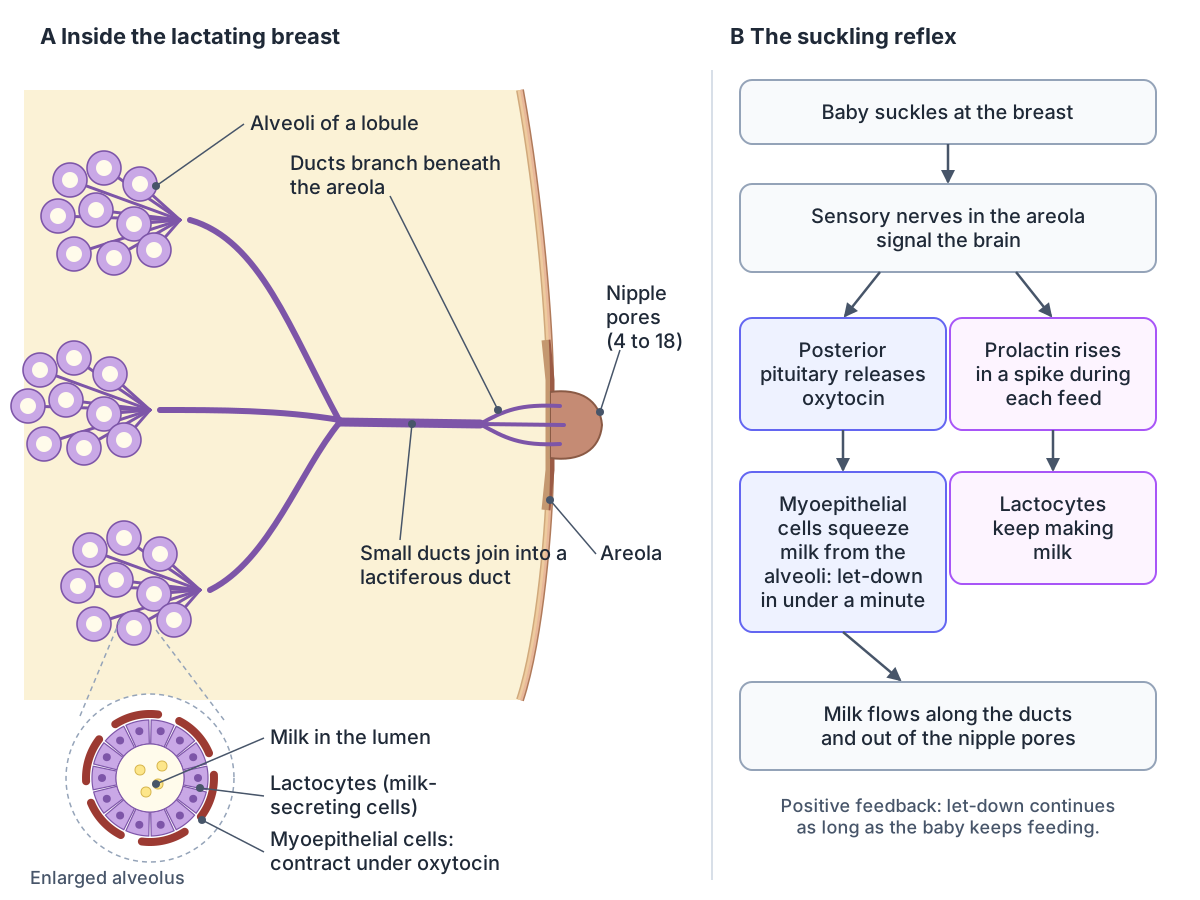
Breast changes after childbirth and with age
The glandular tissue begins to shrink in adulthood and shrinks faster at menopause, when the proportion of fat increases. The breast of a woman who has had children is less glandular than that of a woman of the same age who has not. (Radiopaedia: Breast.)
Outside the period after childbirth, most breast tissue is fat. In women who have had several children the breast often becomes larger and more pendulous, and in older women it is usually smaller because fat decreases and the glandular tissue shrinks. The suspensory ligaments help maintain the breast's non-ptotic (non-sagging) shape. (Kenhub: Female breast anatomy.)
Male breast anatomy
The breast is a gland in both sexes, but in males it is underdeveloped and has no function. (Radiopaedia: Breast.)
Testosterone usually stops breast development at puberty. Males have nipples and areolas but, according to Cleveland Clinic, no glandular tissue and undeveloped milk ducts. Gynaecomastia, a benign enlargement of breast tissue, is the most common male breast problem, and male breast cancer is very rare. (Cleveland Clinic: Breast Anatomy.)
Gynaecomastia (gynecomastia) is a benign, usually reversible growth of ducts and stroma centred under the nipple. Radiopaedia gives its prevalence as 60 to 90% of newborns (from maternal oestrogens), 50 to 60% of adolescents and up to 70% of men aged 50 to 69, though far fewer have symptoms. Pseudogynaecomastia is enlargement from fat alone. A male breast cancer is typically a hard, painless, irregular lump that may lie away from the areola, which gynaecomastia does not. (Radiopaedia: Gynaecomastia.)
Kenhub gives a higher figure, 70%, for slight temporary breast enlargement in boys at puberty, and lists anabolic steroids among the drugs that can cause gynaecomastia later in life. (Kenhub: Female breast anatomy.)
Pectoralis major, which shapes the male chest. Chest muscle anatomy.
Breast anatomy in the 3D model
- Abdominal part of pectoralis major
- The lowest fibres of pectoralis major, arising from the abdominal wall.
- Adipose tissue of breast
- Fat fills the spaces between the lobes of the mammary gland and gives the breast most of its size and shape.
- Areola
- The areola is the circle of pigmented skin around the nipple.
- Areolar tubercles
- Areolar tubercles are the small raised bumps on the areola.
- Clavicular part of pectoralis major
- The upper fibres of pectoralis major.
- Internal thoracic artery
- The internal thoracic artery supplies the anterior chest wall.
- Lactiferous ducts
- Lactiferous ducts carry milk from the lobes to the nipple.
- Lactiferous sinuses
- The lactiferous sinuses are the widened ends of the lactiferous ducts beneath the areola.
- Lateral thoracic artery
- A branch of the axillary artery that runs along the lateral chest wall.
- Lobes of mammary gland
- The mammary gland is divided into 15–20 lobes that produce milk.
- Nipple
- The nipple is the conical projection at the centre of the areola where the lactiferous ducts open.
- Sternocostal part of pectoralis major
- The large lower part of pectoralis major arising from the sternum and ribs.
- Suspensory ligaments of breast
- The suspensory ligaments are fibrous bands that support the breast.
Frequently asked questions
What is the breast made of?
The breast is made of glandular tissue (lobes divided into lobules of milk-producing alveoli, drained by milk ducts to the nipple), fat and fibrous tissue, including the suspensory (Cooper's) ligaments, covered by skin with the nipple and areola. It also contains blood vessels, nerves and lymph vessels. Fat is the main component outside breastfeeding and largely sets breast size.
How many milk ducts are in the nipple?
Textbooks traditionally say 15 to 20, one for each lobe. Ultrasound of breastfeeding women found about nine main ducts at the base of each nipple, with a range of 4 to 18, and Cleveland Clinic also gives about nine. OpenStax describes 4 to 18 nipple pores through which milk leaves the breast.
What are the bumps on the areola?
The small bumps on the areola are Montgomery tubercles, the openings of the areolar (Montgomery) glands. Their oily secretion lubricates and protects the nipple and areola, especially during breastfeeding, and its scent may help a baby find the nipple. They vary a lot in number between people and become larger and more visible in pregnancy.
Where does lymph from the breast drain?
Most lymph from the breast, about 75% (Kenhub gives 75 to 90%), drains to the axillary lymph nodes in the armpit, usually through a sentinel node at the edge of pectoralis major. Most of the rest, mainly from the deep medial breast, drains to the parasternal nodes beside the sternum, and a small share to the posterior intercostal nodes.
What muscle is under the breast?
The breast lies mainly on pectoralis major, with its lower outer part on serratus anterior. It rests on the deep pectoral fascia and is separated from it by the retromammary space, a layer of loose connective tissue. Pectoralis minor lies deeper, under pectoralis major, and the ribs and intercostal muscles lie beneath the muscles.
What are Cooper's ligaments?
Cooper's ligaments, or the suspensory ligaments of the breast, are bands of fibrous tissue that run from the pectoral fascia through the breast to the skin. They divide the glandular tissue into compartments and help hold the breast's shape. When a cancer invades and shortens them, they pull on the skin and cause dimpling.
What happens to the breast during breastfeeding?
In pregnancy the ducts branch and alveoli bud from them, so the glandular tissue grows to almost twice the amount of fat. After birth prolactin drives milk production, and suckling triggers oxytocin release, which makes the cells around the alveoli squeeze milk into the ducts and out of the nipple. Colostrum comes first, then mature milk from about day 10.
Do men have breast tissue?
Yes, men have a small, undeveloped breast with a nipple and areola, but testosterone usually stops its development at puberty, so there is little or no glandular tissue and the ducts stay rudimentary. Temporary enlargement (gynaecomastia) is common in newborns, adolescent boys and older men. Male breast cancer is rare.
References
- TeachMeAnatomy. The Breasts: structure, vasculature, lymphatics.
- TeachMeAnatomy. The Axilla.
- Kenhub. Female breast anatomy, blood supply and mammary glands.
- Kenhub. Breast lymph nodes and lymphatic drainage: clinical role.
- Kenhub. Dermatomes.
- Radiopaedia. Breast.
- Radiopaedia. Gynaecomastia.
- Radiopaedia. Polythelia (supernumerary nipples).
- OpenStax. Anatomy and Physiology 2e, §27.2 Anatomy and Physiology of the Ovarian Reproductive System. Rice University, 2022.
- OpenStax. Anatomy and Physiology 2e, §28.6 Lactation. Rice University, 2022.
- Cleveland Clinic. Breast Anatomy: Milk Ducts, Tissue, Conditions & Physiology.
- Cleveland Clinic. Montgomery Glands: What They Are & Function During Pregnancy.
- Cleveland Clinic. Lactation (Breast Milk Production): How It Works.
- GPnotebook. Breast anatomy.
- Ramsay DT, Kent JC, Hartmann RA, Hartman PE. Anatomy of the lactating human breast redefined with ultrasound imaging. J Anat. 2005;206(6):525-534.
- Riccio CA, Zeiderman MR, Chowdhry S, et al. Plastic surgery of the breast: keeping the nipple sensitive. Eplasty. 2015;15:e28.
- Wysocki WM, Libondi G, Juszczak A. Surgical anatomy of the breast revisited. Biuletyn PTO Nowotwory. 2020;5(1):28-30.
- Eckert-Lind C, Busch AS, Petersen JH, et al. Worldwide secular trends in age at pubertal onset assessed by breast development among girls: a systematic review and meta-analysis. JAMA Pediatr. 2020;174(4):e195881.
- Mitsuhashi N. et al. (2009). BodyParts3D: 3D structure database for anatomical concepts. Nucleic Acids Research 37, D782–D785. Data © The Database Center for Life Science, CC BY 4.0.
- Browne K., Schlehlein H. 3D Reference Organ Set for United, Female (Visible Human Female). Human Reference Atlas 3D Reference Object Library, HuBMAP / NIH. Data from the NLM Visible Human Project. CC BY 4.0.
Related anatomy
- Chest anatomyChest anatomy with labelled diagrams and a 3D model: chest wall layers, ribs and sternum, chest muscles, heart, lungs…
- Chest musclesChest muscle anatomy with a 3D model and diagrams: pectoralis major and its three parts, pectoralis minor, serratus…
- Female anatomyFemale anatomy explained with a labelled body diagram and 3D model: reproductive organs, vulva, breasts, pelvis…
- Female reproductive systemLabelled female reproductive system diagrams: uterus anatomy and layers, uterine tube parts, ovaries, ligaments…
- Lymphatic systemLymph node anatomy with labelled diagrams: node structure, main node groups, lymph vessels, thoracic duct, spleen…
- Pituitary glandPituitary gland (hypophyseal gland) anatomy: the hypophyseal fossa and sella turcica, relations, lobes, portal blood…
Explore the whole body in the interactive 3D human anatomy model, or browse the A–Z list of human structures.