

What are the pectoral (chest) muscles?
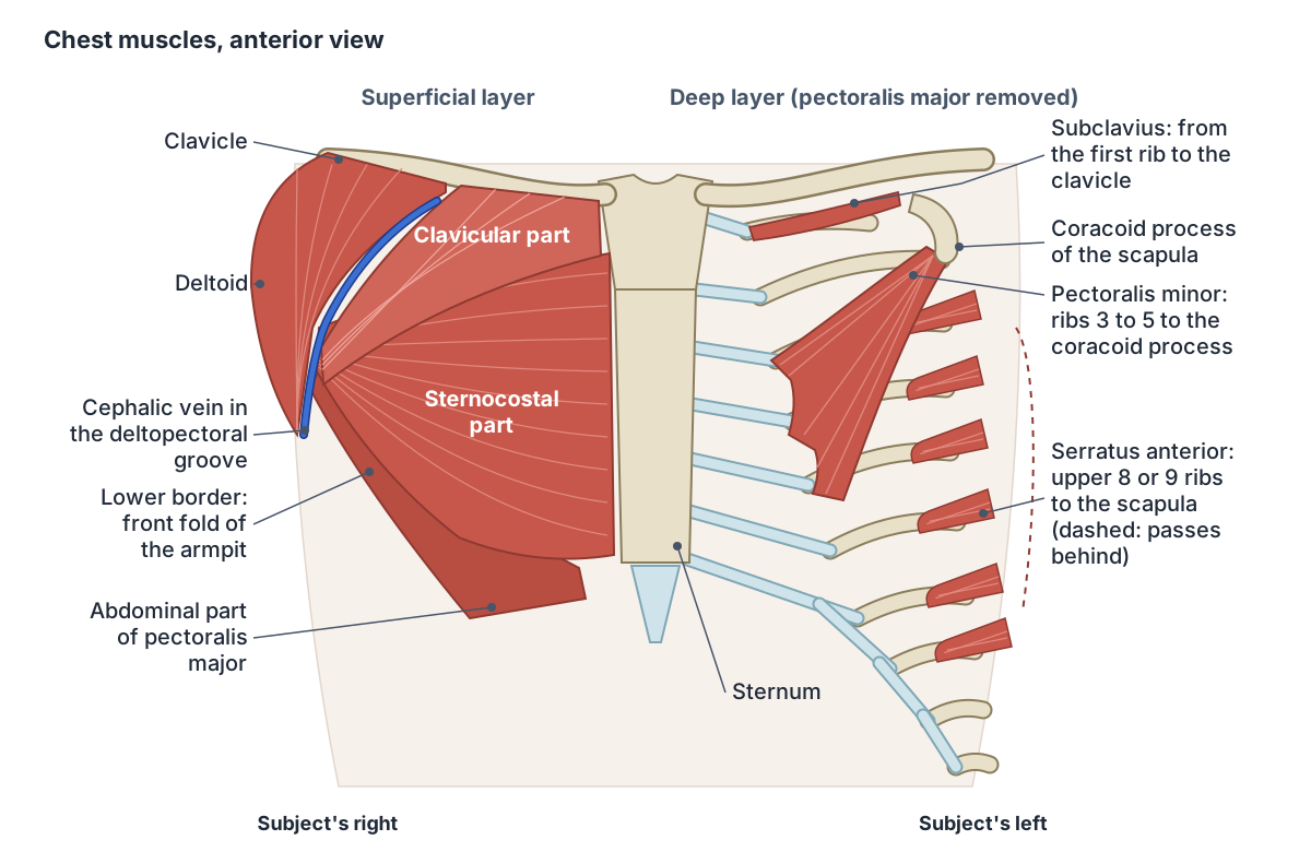
The pectoral region is the front of the upper chest. Its muscles connect the upper limb to the chest wall: pectoralis major, the most superficial, with pectoralis minor and subclavius beneath it, and serratus anterior on the side of the chest wall. (TeachMeAnatomy: Muscles of the Pectoral Region.)
Pectoralis major, pectoralis minor and subclavius form the front wall of the axilla (armpit), and serratus anterior, lying on the ribs and intercostal muscles, forms its medial wall. (TeachMeAnatomy: The Axilla.)
These muscles act on the shoulder, so they are grouped with the upper limb rather than with the muscles that form the chest wall itself (the intercostals, subcostals and transversus thoracis). They attach to the chest wall, though, and pectoralis major, pectoralis minor and serratus anterior can also act as accessory muscles of breathing. (TeachMeAnatomy: Muscles of the Thoracic Cage.)
The whole chest: wall layers, bones, organs, mediastinum and landmarks. Chest anatomy.
Muscles of the chest: origin, insertion, nerve and action
| Muscle | Origin | Insertion | Nerve (roots) | Main actions |
|---|---|---|---|---|
| Pectoralis major | Clavicular part: medial half of the clavicle. Sternocostal part: front of the sternum and costal cartilages 1 to 6. Abdominal part: anterior rectus sheath (external oblique aponeurosis) | Lateral lip of the intertubercular (bicipital) groove of the humerus | Lateral and medial pectoral nerves (C5 to T1) | Adducts and medially rotates the arm; clavicular part flexes it; sternocostal part extends the flexed arm |
| Pectoralis minor | Front of ribs 3 to 5 near their costal cartilages | Medial border and upper surface of the coracoid process of the scapula | Medial pectoral nerve (C8, T1), plus lateral pectoral fibres via the ansa pectoralis | Draws the scapula forwards and downwards against the chest; lifts ribs 3 to 5 in forced inspiration |
| Subclavius | First rib at its junction with the costal cartilage | Groove on the underside of the middle third of the clavicle | Nerve to subclavius (C5, C6), from the superior trunk | Steadies the clavicle at the sternoclavicular joint and depresses its sternal end |
| Serratus anterior | Outer surfaces of ribs 1 to 8 (or 9) | Costal surface of the medial border of the scapula, superior to inferior angle | Long thoracic nerve (C5 to C7) | Protracts the scapula, rotates it upwards so the arm can rise above 90 degrees, holds it against the chest |
Sources: Kenhub (pectoralis major, pectoralis minor, serratus anterior, subclavius), TeachMeAnatomy (pectoral region) and Radiopaedia (pectoralis major). Sources differ slightly on the lowest rib of serratus anterior and on the origin of the abdominal part.
Pectoralis major anatomy
Pectoralis major is a large, fan-shaped muscle. Its clavicular part arises from the front of the medial half of the clavicle, its sternocostal part from the front of the sternum and the costal cartilages of ribs 1 to 6, and its small abdominal part from the anterior layer of the rectus sheath. The fibres of all three parts run laterally and converge on the lateral lip of the intertubercular sulcus of the humerus. (Kenhub: Pectoralis major muscle.)
Radiopaedia notes that texts describe two or three parts. It gives the origin of the abdominal part as the aponeurosis of the external oblique and says some authors count it with the sternocostal head, which makes up about 80% of the muscle's volume. The costal origin covers most of the true ribs but often leaves out the first or seventh cartilage. (Radiopaedia: Pectoralis major muscle.)
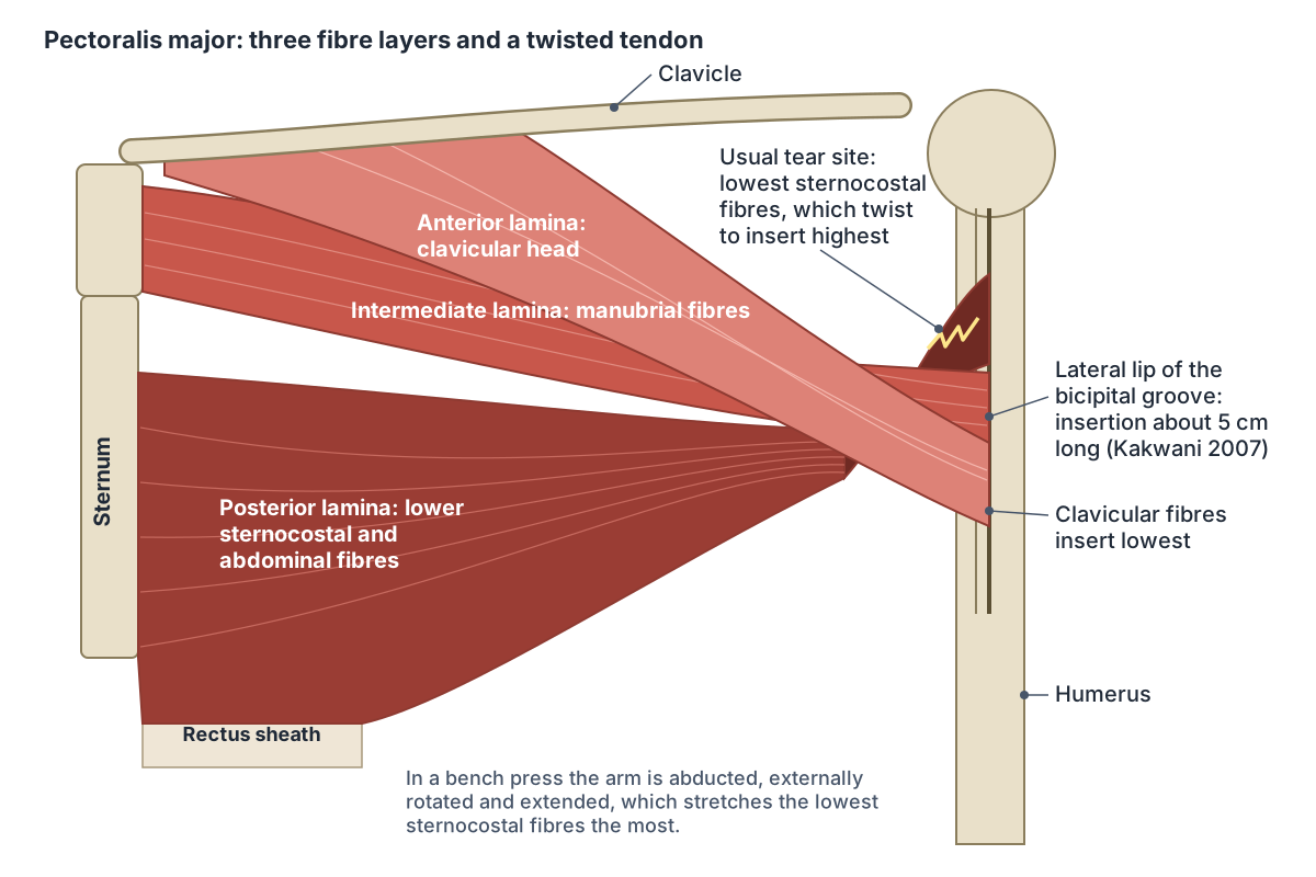
The fibres end in a U-shaped tendon about 40 to 60 mm wide and 3 to 5 mm thick on the lateral lip of the bicipital (intertubercular) groove. The main artery is the thoracoacromial artery, with segmental perforators from the internal thoracic artery that continue on to the breast. Radiopaedia gives the nerve roots as C5 and C6 for the clavicular part and C7 to T1 for the sternocostal part. (Radiopaedia: Pectoralis major muscle.)
The muscle is supplied by the lateral and medial pectoral nerves of the brachial plexus (C5 to T1). Its blood comes from the pectoral branches of the thoracoacromial artery and perforating branches of the internal thoracic artery. (Kenhub: Pectoralis major muscle.)
The largest part of the muscle, in 3D. Sternocostal part of pectoralis major.
Clavicular, sternocostal and abdominal parts: what each does
As a whole, pectoralis major is a strong adductor and medial (internal) rotator of the arm. Its clavicular part also flexes the arm, raising it forwards up to about 90 degrees, while its sternocostal part extends the flexed arm, pulling it back down towards the side. With the arm fixed, as in climbing, it works with latissimus dorsi to pull the trunk upwards. (Kenhub: Pectoralis major muscle.)
The tendon is built in three layers (laminae). The anterior lamina comes from the clavicular head and also attaches to the deltoid tuberosity and the deep fascia of the arm; the intermediate lamina comes from the manubrial fibres of the sternocostal head; and the posterior lamina comes from the sternocostal fibres below the sternal angle, including the abdominal part. (Radiopaedia: Pectoralis major muscle.)
The posterior lamina rotates through about 180 degrees before it inserts, so the lowest sternocostal fibres attach highest on the humerus, while the clavicular fibres insert further down. Those lowest sternocostal fibres are stretched most when the arm is abducted, externally rotated and extended, as at the bottom of a bench press, which is why the lower part of the tendon is usually the first to fail. (Kakwani RG et al. Rupture of the pectoralis major muscle: surgical treatment in athletes. Int Orthop. 2007.)
The three parts of pectoralis major
| Part | Origin | Where its fibres insert | Extra action | Nerve roots (Radiopaedia) |
|---|---|---|---|---|
| Clavicular (upper chest) | Front of the medial half of the clavicle | Anterior lamina, lowest on the lateral lip of the bicipital groove, also deltoid tuberosity and arm fascia | Flexes the arm up to about 90 degrees | C5, C6 |
| Sternocostal (middle and lower chest) | Front of the sternum and costal cartilages 1 to 6 | Intermediate and posterior laminae; the lowest fibres twist to insert highest | Extends the flexed arm back to the side | C7 to T1 |
| Abdominal | External oblique aponeurosis or anterior rectus sheath | Posterior lamina with the lower sternocostal fibres | Often counted as part of the sternocostal head | C7 to T1 |
Sources: Kenhub, Radiopaedia and Kakwani et al. 2007. All three parts adduct and medially rotate the arm.
Upper chest vs lower chest: which part is which?
The upper chest is the clavicular part of pectoralis major, which arises from the clavicle and flexes the arm. The middle and lower chest are the sternocostal fibres from the sternum and costal cartilages, with the small abdominal part at the bottom; they bring the raised arm down and, with the rest of the muscle, adduct it across the body. (Kenhub: Pectoralis major muscle.)
In an electromyography study, 30 trained young adults bench pressed 60% of their one-repetition maximum at five bench angles. The upper (clavicular) portion was most active at a 30 degree incline, while the middle and lower portions were most active on a flat bench and less active as the incline increased. At 45 and 60 degrees the anterior deltoid became the most active of the muscles measured. (Rodríguez-Ridao D et al. Effect of five bench inclinations on the electromyographic activity of the pectoralis major, anterior deltoid, and triceps brachii. Int J Environ Res Public Health. 2020.)
The upper chest fibres from the clavicle, in 3D. Clavicular part of pectoralis major.
Pectoralis minor
Pectoralis minor lies under pectoralis major. It arises as three slips from the front of the 3rd, 4th and 5th ribs near their costal cartilages, and from the fascia over the intercostal muscles, and inserts by a flat tendon on the medial border and upper surface of the coracoid process of the scapula. (Kenhub: Pectoralis minor muscle.)
It is supplied mainly by the medial pectoral nerve (C8, T1), with fibres from the lateral pectoral nerve through a loop called the ansa pectoralis, so its roots span C5 to T1. With serratus anterior it protracts the scapula, it pulls the coracoid process down to rotate the scapula downwards, and when the scapula is fixed it helps lift ribs 3 to 5 in deep, forced breathing. (Kenhub: Pectoralis minor muscle.)
Pectoralis minor crosses in front of the axillary artery and divides it into three parts: medial to, behind and lateral to the muscle. The space under its tendon near the coracoid process is one of the sites where the nerves and vessels to the arm can be compressed in thoracic outlet syndrome. (Kenhub: Pectoralis minor muscle.)
Surgeons also use it to define three levels of axillary lymph nodes in breast cancer surgery: level I lateral to pectoralis minor, level II behind it and level III medial to it. (Kenhub: Lymphatic drainage of the breast.)
The artery that pectoralis minor divides into three parts. Axillary artery.
Serratus anterior
Serratus anterior covers the side of the chest. It arises by separate slips from the outer surfaces of the upper ribs and runs back around the chest wall, under the scapula, to insert along the front (costal) surface of the medial border of the scapula from its superior to its inferior angle. (Kenhub: Serratus anterior muscle.)
TeachMeAnatomy gives its origin as the lateral aspects of ribs 1 to 8 and stresses its role in rotating the scapula so that the arm can be raised above 90 degrees. (TeachMeAnatomy: Muscles of the Pectoral Region.)
Kenhub gives ribs 1 to 8 or 9, with a slip from rib 10 in about 10% of people, and divides the muscle into superior, intermediate and inferior parts, the inferior part being the largest and strongest. Serratus anterior protracts the scapula (pulls it forwards around the chest), helps rotate it upwards and holds it against the chest wall. It is supplied by the long thoracic nerve (C5 to C7) and by the superior and lateral thoracic and thoracodorsal arteries. (Kenhub: Serratus anterior muscle.)
Damage to the long thoracic nerve, from axillary surgery, carrying heavy loads or other trauma, paralyses the muscle. Raising the arm becomes difficult and the medial border of the scapula lifts away from the chest wall, a winged scapula (scapula alata). (Kenhub: Serratus anterior muscle.)
The muscle that holds the scapula to the chest wall, in 3D. Serratus anterior.
Subclavius
Subclavius is a short, triangular muscle under the clavicle. It arises by a strong tendon from the first rib where it meets its costal cartilage and inserts into a groove on the underside of the middle third of the clavicle. It is supplied by the nerve to subclavius (C5, C6), a small branch of the superior trunk of the brachial plexus, and by the clavicular branch of the thoracoacromial artery. (Kenhub: Subclavius muscle.)
It steadies the clavicle at the sternoclavicular joint during shoulder movements by depressing the sternal end of the clavicle. The brachial plexus and the subclavian artery and vein pass deep to it, so it helps protect them when the clavicle breaks; they can also be squeezed between the clavicle and the first rib in costoclavicular syndrome, one type of thoracic outlet syndrome. (Kenhub: Subclavius muscle.)
The collarbone that subclavius anchors. Clavicle.
Nerves and blood supply of the chest muscles
The lateral pectoral nerve is usually the first branch of the lateral cord of the brachial plexus and carries fibres from C5, C6 and C7. It crosses in front of the axillary artery and vein, pierces the clavipectoral fascia with the thoracoacromial artery and ends on the deep surface of pectoralis major. A communicating branch to the medial pectoral nerve forms the ansa pectoralis, which carries fibres on to pectoralis minor. (Kenhub: Lateral pectoral nerve.)
The medial pectoral nerve (C8, T1) arises from the medial cord behind the axillary artery. It runs forwards, receives the communicating branch from the lateral pectoral nerve, ends partly by piercing pectoralis minor and sends fibres on to part of pectoralis major. (Kenhub: Medial pectoral nerve.)
Pectoralis minor receives blood from the thoracoacromial, superior thoracic and lateral thoracic arteries. The lateral pectoral nerve and branches of the thoracoacromial artery run in the plane between pectoralis minor and pectoralis major. (Kenhub: Pectoralis minor muscle.)
Perforating branches of the internal thoracic artery pass forwards through the intercostal spaces to the pectoral muscles and the skin over them. (TeachMeAnatomy: Thoracic Wall Vasculature.)
The brachial plexus and its branches to the shoulder and arm. Nerves of the arm.
Nerves and arteries of the chest muscles
| Muscle | Nerve and origin | Main arteries |
|---|---|---|
| Pectoralis major | Lateral pectoral nerve (lateral cord, C5 to C7) and medial pectoral nerve (medial cord, C8, T1) | Pectoral branches of the thoracoacromial artery; perforators of the internal thoracic artery |
| Pectoralis minor | Medial pectoral nerve, plus lateral pectoral fibres through the ansa pectoralis | Thoracoacromial, superior thoracic and lateral thoracic arteries |
| Subclavius | Nerve to subclavius from the superior trunk (C5, C6) | Clavicular branch of the thoracoacromial artery, suprascapular artery |
| Serratus anterior | Long thoracic nerve (C5 to C7) | Superior and lateral thoracic arteries, thoracodorsal artery |
Sources: Kenhub pages on each muscle and on the lateral and medial pectoral nerves.
Chest muscle anatomy in men and women
Pectoralis major has the same attachments in both sexes. In males it lies just under deep fascia, subcutaneous tissue and skin, so its outline shapes the chest; in females the breast lies on top of it. (Kenhub: Pectoralis major muscle.)
The breast rests on the pectoral fascia over pectoralis major and serratus anterior, and its axillary tail runs along the lower outer edge of pectoralis major towards the armpit. A potential space of loose connective tissue, the retromammary space, separates the breast from the pectoral fascia and is used in reconstructive surgery. (TeachMeAnatomy: The Breasts.)
On mammograms pectoralis major lies behind the breast and is checked on the mediolateral oblique (MLO) view. (Radiopaedia: Pectoralis major muscle.)
How the breast sits on the chest muscles, with lymph drainage. Breast anatomy.
Deltopectoral groove, clavipectoral fascia and the armpit
The upper border of pectoralis major meets deltoid at the deltopectoral groove, where the cephalic vein runs up the arm. Near the clavicle the vein pierces the clavipectoral fascia to drain into the first part of the axillary vein. (Radiopaedia: Cephalic vein.)
Pectoralis major is enclosed between two layers of deep fascia, the pectoral fascia. Under it lies the clavipectoral fascia, which covers subclavius and pectoralis minor. (Radiopaedia: Pectoralis major muscle.)
Between pectoralis major, deltoid and the clavicle lies a small hollow, the infraclavicular (Mohrenheim's) fossa, used as a landmark in surgery on the subclavian artery. (Kenhub: Pectoralis major muscle.)
The shoulder joint and the muscles around it. Shoulder anatomy.
Pectoralis major tear (torn pec)
Pectoralis major tears mostly affect young, physically active men aged 20 to 40, and most happen in weight training, especially the bench press. The usual mechanism is eccentric loading: the muscle is working while it lengthens as the bar is lowered with the shoulder abducted and externally rotated, and the lowest fibres of the sternal head are injured most often. Anabolic steroid use has been linked to these tears, though not consistently. (Radiopaedia: Pectoralis major injury.)
Radiopaedia places about 60% of tears in the tendon itself or at its insertion on the humerus, sometimes pulling off a flake of bone, and about 25% at the musculotendinous junction; the rest are in the muscle belly or at the origin. (Radiopaedia: Pectoralis major injury.)
Not every source agrees: a 2016 case report states that most lesions lie at the musculotendinous junction. It also notes that almost half of ruptures happen during weightlifting, particularly the bench press. (Kadu VV et al. Pectoralis major tear: an unusual and rare presentation. J Orthop Case Rep. 2016.)
In a surgical series of 13 male athletes (average age 28.6 years), 10 had complete avulsion of the tendon from the humerus. Patients described a tearing feeling in the armpit, with bruising, loss of the muscle's contour and of the normal anterior axillary fold, and weak adduction and internal rotation; MRI confirmed the site of the tear in 11 of the 13. (Kakwani RG et al. Rupture of the pectoralis major muscle: surgical treatment in athletes. Int Orthop. 2007.)
The arm bone where the pectoralis major tendon inserts. Humerus.
Variations: Poland syndrome and the sternalis muscle
In Poland syndrome, pectoralis major and often pectoralis minor are missing or underdeveloped on one side from birth. Radiopaedia gives an estimated incidence of 1 in 36,000 to 50,000, with no side or sex preference. The ribs 2 to 5 on that side, the breast and nipple can also be affected, and webbed fingers (syndactyly) on the same side are the most frequent associated finding. (Radiopaedia: Poland syndrome.)
The sternalis is an extra strap of muscle that runs vertically beside the sternum, next to the medial edge of pectoralis major. Dissection studies find it in about 5% of people (range 1 to 8%), twice as often on one side as on both. On a mammogram it can look like a small lump against the chest wall and should not be mistaken for disease. (Radiopaedia: Sternalis muscle.)
Do the chest muscles help you breathe?
The main muscles of quiet breathing are the diaphragm and the external intercostal muscles between the ribs, and quiet breathing out is passive. Extra muscles join in only when breathing is forced. (OpenStax Anatomy and Physiology 2e, 22.3: The Process of Breathing.)
Pectoralis major, pectoralis minor and serratus anterior, with the scalene muscles of the neck, are listed as accessory muscles of respiration: they do not form the chest wall but attach to it. (TeachMeAnatomy: Muscles of the Thoracic Cage.)
Acting from its attachment on the humerus, pectoralis major helps inspiration, especially in forced breathing. (Kenhub: Pectoralis major muscle.)
The ribs, sternum and the muscles between them that drive breathing. Rib cage and intercostal muscles.
Chest muscles in the 3D model
- Abdominal part of pectoralis major
- The lowest fibres of pectoralis major, arising from the abdominal wall.
- Clavicular part of pectoralis major
- The upper fibres of pectoralis major.
- Pectoralis minor
- A thin triangular muscle lying deep to pectoralis major on the upper chest wall.
- Serratus anterior
- A broad muscle on the side of the thorax that holds the scapula against the chest wall.
- Sternocostal part of pectoralis major
- The large lower part of pectoralis major arising from the sternum and ribs.
- Subclavius
- A small muscle between the clavicle and the first rib.
Frequently asked questions
What are the four chest muscles?
The four pectoral muscles are pectoralis major, the large fan-shaped muscle under the skin; pectoralis minor and subclavius beneath it; and serratus anterior on the side of the chest. They attach the arm and shoulder blade to the chest wall. The intercostal muscles between the ribs and the diaphragm are breathing muscles of the chest wall itself.
What is the difference between pectoralis major and pectoralis minor?
Pectoralis major is the large superficial muscle from the clavicle, sternum and costal cartilages to the humerus; it moves the arm. Pectoralis minor is a smaller muscle beneath it, from ribs 3 to 5 to the coracoid process of the scapula; it moves and steadies the shoulder blade. Both form the front wall of the armpit.
Which nerves supply the pectoralis major?
Pectoralis major is supplied by the lateral pectoral nerve from the lateral cord of the brachial plexus (C5 to C7) and the medial pectoral nerve from the medial cord (C8, T1). Radiopaedia gives the roots as C5 and C6 for the clavicular part and C7 to T1 for the sternocostal part. The two nerves are joined by a loop, the ansa pectoralis.
What does the pectoralis major do?
Pectoralis major adducts the arm (brings it towards the body) and rotates it medially. Its clavicular part also flexes the arm forwards up to about 90 degrees, and its sternocostal part pulls the raised arm back down. With the arm fixed, as in climbing, it helps pull the trunk up, and it can assist forced breathing.
Which part of the chest muscle is the upper chest?
The upper chest is the clavicular part of pectoralis major, the fibres that arise from the medial half of the clavicle. In one electromyography study this part was most active in a bench press inclined at 30 degrees, whereas the sternocostal fibres of the middle and lower chest were most active on a flat bench.
What is a torn pec?
A torn pec is a tear of pectoralis major, usually of its tendon near the humerus or at the junction of muscle and tendon. It mostly affects active men aged 20 to 40, typically while lowering a heavy bench press. Signs include a tearing feeling in the armpit, bruising, loss of the front fold of the armpit and weak adduction.
What muscle is under the pectoralis major?
Under pectoralis major lie pectoralis minor, subclavius below the clavicle and the front of serratus anterior, together with the front of the upper six ribs and the intercostal muscles. The clavipectoral fascia covers subclavius and pectoralis minor, and the thoracoacromial vessels and lateral pectoral nerve run between the two pectoral muscles.
References
- TeachMeAnatomy. Muscles of the Pectoral Region.
- TeachMeAnatomy. The Axilla.
- TeachMeAnatomy. Muscles of the Thoracic Cage.
- TeachMeAnatomy. Thoracic Wall Vasculature.
- TeachMeAnatomy. The Breasts.
- Kenhub. Pectoralis major: origin, insertion, innervation, function.
- Kenhub. Pectoralis minor muscle.
- Kenhub. Serratus anterior muscle: origin, insertion and action.
- Kenhub. Subclavius muscle.
- Kenhub. Lateral pectoral nerve: origin, course and function.
- Kenhub. Medial pectoral nerve.
- Kenhub. Breast lymph nodes and lymphatic drainage.
- Radiopaedia. Pectoralis major muscle.
- Radiopaedia. Pectoralis major injury.
- Radiopaedia. Poland syndrome.
- Radiopaedia. Sternalis muscle.
- Radiopaedia. Cephalic vein.
- OpenStax. Anatomy and Physiology 2e, §22.3 The Process of Breathing. Rice University, 2022.
- Kakwani RG, Matthews JJ, Kumar KM, Pimpalnerkar A, Mohtadi N. Rupture of the pectoralis major muscle: surgical treatment in athletes. Int Orthop. 2007;31(2):159-163.
- Kadu VV, Saindane KA, Godghate N, Godghate N. Pectoralis major tear: an unusual and rare presentation. J Orthop Case Rep. 2016;6(4):17-19.
- Rodríguez-Ridao D, Antequera-Vique JA, Martín-Fuentes I, Muyor JM. Effect of five bench inclinations on the electromyographic activity of the pectoralis major, anterior deltoid, and triceps brachii during the bench press exercise. Int J Environ Res Public Health. 2020;17(19):7339.
- Mitsuhashi N. et al. (2009). BodyParts3D: 3D structure database for anatomical concepts. Nucleic Acids Research 37, D782–D785. Data © The Database Center for Life Science, CC BY 4.0.
Related anatomy
- Chest anatomyChest anatomy with labelled diagrams and a 3D model: chest wall layers, ribs and sternum, chest muscles, heart, lungs…
- Shoulder jointShoulder anatomy with labelled diagrams and a 3D model: clavicle, scapula and humerus, the glenohumeral, AC and SC…
- Breast anatomyBreast anatomy with labelled diagrams and a 3D model: lobes, milk ducts, Cooper's ligaments, nipple, areola, blood…
- Rib cageRib cage anatomy: 12 pairs of ribs, true, false and floating ribs, parts of a rib, sternum and jugular notch, costal…
- Arm musclesArm muscles labeled in 3D: biceps brachii, brachialis, coracobrachialis, triceps brachii, deltoid and anconeus, with…
- Upper limb nervesBrachial plexus diagram with roots, trunks, cords and branches; axillary, musculocutaneous, median, ulnar and radial…
Explore the whole body in the interactive 3D human anatomy model, or browse the A–Z list of human structures.