

What is the chest (thorax)?
The chest, properly called the thorax, is the region between the neck and the abdomen. It runs from the thoracic inlet at the root of the neck down to the diaphragm, the dome-shaped muscle that separates it from the abdomen. (Cleveland Clinic: Chest (Thorax).)
Its upper opening, the superior thoracic aperture, opens into the neck and is bounded by the manubrium of the sternum, the first pair of ribs and the body of the first thoracic vertebra (T1). The inferior thoracic aperture is almost completely closed by the diaphragm. (Kenhub: Thorax.)
The chest protects the organs inside it, changes its volume so that you can breathe, gives the upper body a stable but flexible frame, supports the head and neck and anchors the arms. The kidneys, liver, spleen and stomach lie below the diaphragm, in the abdomen, but the lower chest wall partly protects them. (Cleveland Clinic: Chest (Thorax).)
Chest anatomy at a glance
| Part | What it includes | Key fact |
|---|---|---|
| Chest wall | Skin, subcutaneous fat, deep fascia, chest muscles, ribs and intercostal muscles, endothoracic fascia, parietal pleura | Each of the 11 intercostal spaces carries a vein, artery and nerve under the rib above |
| Bones | Sternum (manubrium, body, xiphoid process), 12 pairs of ribs and costal cartilages, 12 thoracic vertebrae | The sternal angle marks the second costal cartilage |
| Muscles | Pectoralis major and minor, subclavius, serratus anterior, intercostals, diaphragm | The diaphragm and external intercostals drive quiet breathing |
| Pleural cavities | The right lung (3 lobes) and left lung (2 lobes), each in its pleura | The lung apex rises above the first rib into the neck |
| Mediastinum | Heart and pericardium, great vessels, trachea and bronchi, oesophagus, thymus, thoracic duct, vagus and phrenic nerves | Divided into superior and inferior parts at the sternal angle |
| Diaphragm | Dome-shaped muscle with a central tendon | Openings at about T8, T10 and T12; phrenic nerve C3 to C5 |
| Breasts | Glandular and fatty tissue on pectoralis major and serratus anterior | Span the 2nd to 6th costal cartilages; more prominent in females after puberty |
Summary of the sections below. Sources: Kenhub, TeachMeAnatomy, OpenStax and Cleveland Clinic as cited in each section.
Layers of the chest wall
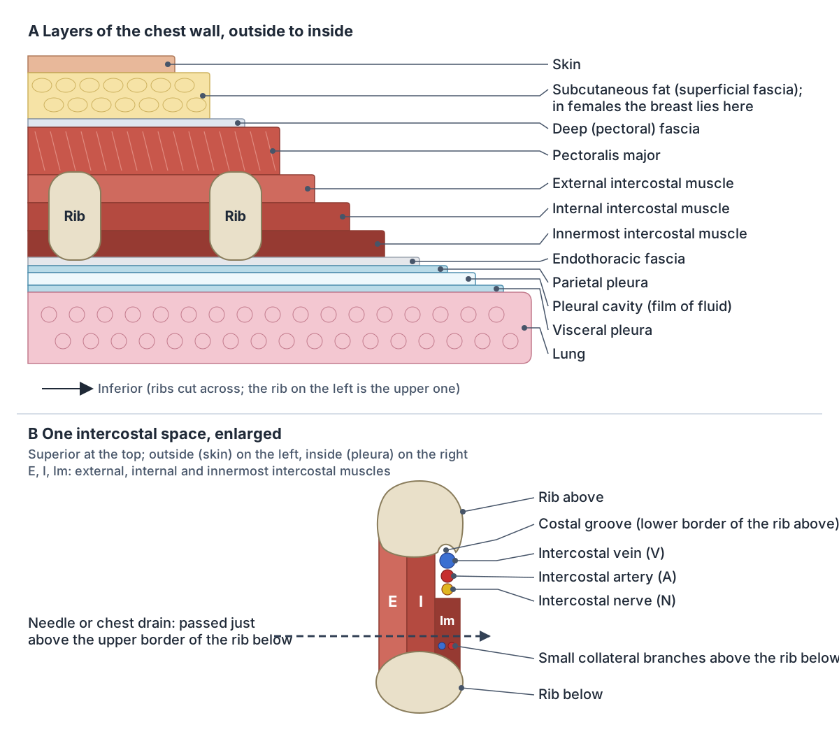
The chest wall is built in layers. Between each pair of ribs lies an intercostal space holding three layers of intercostal muscle (external, internal and innermost), with the deep fascia of the thorax on its outer side and the endothoracic fascia and pleura on its inner side. (Kenhub: Intercostal spaces.)
Outside the ribs lie the chest muscles. Pectoralis major is the most superficial: in males it is covered only by deep fascia, subcutaneous tissue and skin, while in females the breast lies over it. Its deep surface covers pectoralis minor, serratus anterior and the front of the upper six ribs. (Kenhub: Pectoralis major muscle.)
The external intercostals run downwards and forwards from each rib to the rib below; the internal intercostals lie deep to them and run the opposite way, downwards and backwards. The innermost intercostals are the deepest layer and are separated from the internal intercostals by the intercostal neurovascular bundle. (TeachMeAnatomy: Muscles of the Thoracic Cage.)
The bundle of intercostal vein, artery and nerve runs in the costal groove along the lower border of each rib, in the order vein, artery, nerve from top to bottom (VAN). For this reason a needle or chest drain is passed just above the upper border of a rib; going in along the lower border risks bleeding and nerve damage. Drains are usually placed in the safe triangle bounded by the lateral border of pectoralis major, the anterior border of latissimus dorsi and a line above the fifth intercostal space. (TeachMeAnatomy: Thoracic Wall Vasculature.)
Bones of the chest: rib cage and sternum
The skeleton of the chest, the thoracic cage, is formed by the sternum in front, 12 pairs of ribs at the sides and the 12 thoracic vertebrae (T1 to T12) behind. Ribs 1 to 7 are true ribs that reach the sternum through their own costal cartilages; ribs 8 to 10 are false ribs whose cartilages join the rib above; and ribs 11 and 12 are floating ribs that end in the abdominal muscles. (Cleveland Clinic: Rib Cage.)
The sternum (breastbone) has three parts: the manubrium at the top, the body and the small xiphoid process at the bottom. The manubrium joins the clavicles at the sternoclavicular joints and takes the first costal cartilage; the body takes the cartilages of ribs 3 to 6 and parts of ribs 2 and 7. (TeachMeAnatomy: The Sternum.)
Between the 12 ribs on each side lie 11 intercostal spaces, each bordered by the rib above and the rib below and filled by the intercostal muscles, nerves, arteries and veins. (Kenhub: Intercostal spaces.)
Every rib, the sternum, the costal cartilages and their joints, in 3D. Rib cage anatomy.
Chest muscles
The muscles on the front of the chest are pectoralis major, pectoralis minor, subclavius and serratus anterior. Pectoralis major is the large, fan-shaped and most superficial one; it adducts and medially rotates the arm, and its clavicular head also flexes it. Pectoralis minor lies beneath it and steadies the scapula against the chest wall, subclavius anchors the clavicle, and serratus anterior on the side of the chest protracts the scapula and rotates it so that the arm can be raised above 90 degrees. (TeachMeAnatomy: Muscles of the Pectoral Region.)
These muscles do not form the chest wall itself but attach to it. Pectoralis major, pectoralis minor and serratus anterior, together with the scalene muscles of the neck, act as accessory muscles of breathing. Inside the wall, transversus thoracis and the subcostal muscles weakly pull the ribs down in expiration. (TeachMeAnatomy: Muscles of the Thoracic Cage.)
Quiet breathing uses two muscle groups: the diaphragm, which contracts and moves down, and the external intercostals, which move the ribs upwards and outwards. Breathing out at rest is passive. In forced expiration the internal intercostals compress the rib cage and the abdominal muscles push the abdominal organs up against the diaphragm. (OpenStax Anatomy and Physiology 2e, 22.3: The Process of Breathing.)
Pectoralis major and minor, serratus anterior and subclavius with origins, insertions and nerves. Chest muscle anatomy.
What is inside the chest? The thoracic cavity
The space inside the chest wall is the thoracic cavity, the second-largest hollow space in the body after the abdominal cavity. It has three parts: the right and left pleural cavities and the mediastinum between them. Cleveland Clinic lists five organs in it: the heart, lungs, oesophagus (esophagus), trachea and thymus. (Cleveland Clinic: Thoracic Cavity.)
Each lung is wrapped in pleura, a serous membrane with two layers. The visceral pleura covers the lung and dips into its fissures; the parietal pleura lines the inside of the ribs and intercostal muscles, the mediastinum and the top of the diaphragm, and reaches up into the root of the neck. A thin film of fluid between the layers lets them slide over each other, and its surface tension makes the lung follow the chest wall as it expands. (TeachMeAnatomy: The Pleurae.)
Where two parts of the parietal pleura meet they form recesses, such as the costodiaphragmatic recess at the bottom of the chest, where fluid can collect. Air in the pleural space, a pneumothorax, breaks the surface tension, so the lung no longer expands with the chest wall. (TeachMeAnatomy: The Pleurae.)
Where every major organ sits in the chest, abdomen and pelvis. Organs of the body.
The mediastinum: the middle of the chest
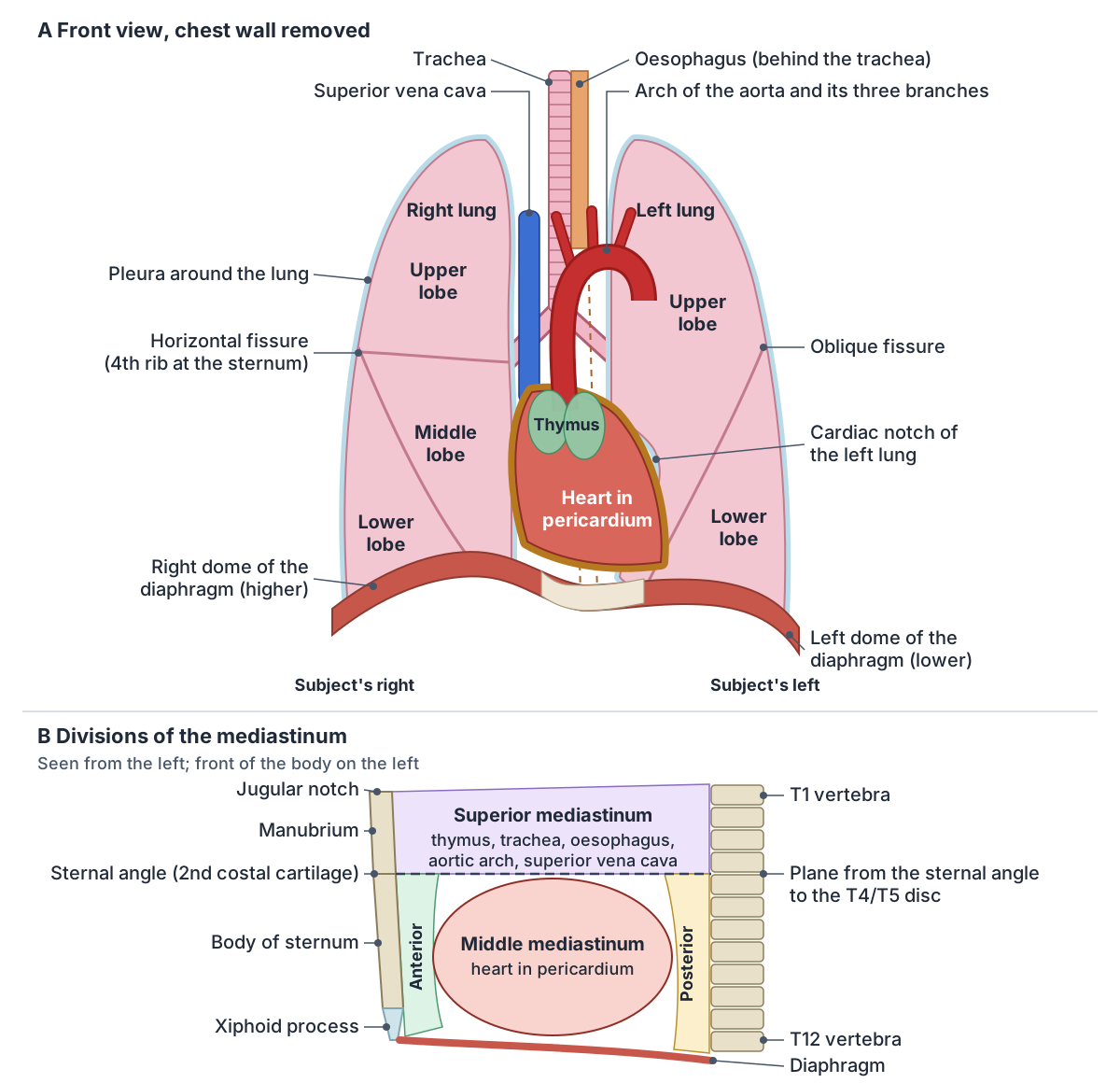
The mediastinum is the central compartment of the thoracic cavity, between the two pleural sacs. It reaches from the superior thoracic aperture down to the diaphragm and from the sternum back to the bodies of the thoracic vertebrae. (Kenhub: Mediastinum.)
A horizontal plane from the sternal angle back to the disc between T4 and T5 divides it into a superior and an inferior mediastinum. The larger inferior part is subdivided into an anterior mediastinum between the sternum and the pericardium, a middle mediastinum made of the pericardium and heart, and a posterior mediastinum between the pericardium and the spine. (Kenhub: Mediastinum.)
TeachMeAnatomy draws the same dividing line from the sternal angle to the T4 vertebra. The superior mediastinum lies behind the manubrium and in front of the bodies of T1 to T4; its contents, and those of the other divisions, are listed in the table below. (TeachMeAnatomy: The Superior Mediastinum.)
The middle mediastinum contains the heart inside the pericardium, the roots of the great vessels (ascending aorta, pulmonary trunk and superior vena cava), the tracheal bifurcation with the left and right main bronchi, and the phrenic nerves on their way to the diaphragm. (TeachMeAnatomy: The Middle Mediastinum.)
The posterior mediastinum holds the oesophagus, the descending thoracic aorta, the azygos and hemiazygos veins, the thoracic duct, the vagus nerves and the splanchnic nerves. Radiologists now often use a three-part model based on CT instead: a prevascular compartment in front (mainly the thymus and left brachiocephalic vein), a visceral compartment (heart, great vessels, trachea and oesophagus) and a paravertebral compartment beside the spine. (Kenhub: Mediastinum.)
On a chest X-ray a mediastinum wider than about 8 cm can be a sign of a tumour or of mediastinitis, an infection or inflammation of this space. (Cleveland Clinic: Mediastinum.)
Divisions of the mediastinum
| Division | Boundaries | Main contents |
|---|---|---|
| Superior | Superior thoracic aperture to the plane of the sternal angle; manubrium in front, T1 to T4 behind | Thymus, trachea, oesophagus, aortic arch and its branches, brachiocephalic veins, superior vena cava, vagus and phrenic nerves, thoracic duct |
| Anterior (inferior) | Between the body of the sternum and the pericardium | Lower part of the thymus, fat, connective tissue, lymph nodes, branches of the internal thoracic vessels |
| Middle (inferior) | The pericardium, from the sternal angle plane to the diaphragm | Heart and pericardium, roots of the great vessels, tracheal bifurcation, main bronchi, phrenic nerves |
| Posterior (inferior) | Behind the pericardium, in front of the T4 to T12 vertebrae, down to the diaphragm | Oesophagus, descending thoracic aorta, azygos and hemiazygos veins, thoracic duct, vagus and splanchnic nerves |
Classical four-part division. Sources: Kenhub (Mediastinum) and TeachMeAnatomy (superior and middle mediastinum).
Where is the heart in the chest?
The heart lies in the mediastinum between the lungs, behind the sternum and costal cartilages and in front of the vertebral bodies. It is about the size of a fist: roughly 12 cm long, 8 cm wide and 6 cm thick. Its base, where the great vessels attach, is at the level of the third costal cartilage, and its apex points down and to the left, lying just left of the sternum near the junction of the fourth and fifth ribs. (OpenStax Anatomy and Physiology 2e, 19.1: Heart Anatomy.)
An adult female heart weighs about 250 to 300 g and a male heart about 300 to 350 g. It sits in the pericardium: a tough outer fibrous pericardium and an inner serous pericardium, whose two layers enclose the pericardial cavity, a thin space filled with lubricating fluid. (OpenStax Anatomy and Physiology 2e, 19.1: Heart Anatomy.)
Chambers, valves, wall layers and coronary vessels of the heart, in 3D. Heart anatomy.
Lungs and pleura
The two lungs fill most of the chest on either side of the mediastinum. The right lung has three lobes (superior, middle and inferior) and is shorter and wider than the left. The left lung has two lobes (superior and inferior) and a cardiac notch that makes room for the heart. (OpenStax Anatomy and Physiology 2e, 22.2: The Lungs.)
The apex of each lung rises above the level of the first rib into the floor of the neck, and the base rests on the diaphragm; the right base is more hollowed because the right dome of the diaphragm sits higher over the liver. On the right lung, the horizontal fissure runs from the sternum at the level of the fourth rib to meet the oblique fissure. (TeachMeAnatomy: The Lungs.)
Structures enter and leave each lung at its hilum on the mediastinal surface: the main bronchus, the pulmonary artery, two pulmonary veins, the bronchial vessels, the pulmonary plexus of nerves and lymphatic vessels. (TeachMeAnatomy: The Lungs.)
In the 3D model the five lung lobes are drawn see-through so that the heart and bronchi stay visible. They are built from the BodyParts3D chest cavity, with the fissures placed from radiological surface anatomy.
Lobes, fissures, hilum, bronchopulmonary segments and pleura. Lung anatomy.
Trachea, oesophagus and thymus
The trachea runs down the superior mediastinum in front of the oesophagus, held open by cartilage rings, and divides into the left and right main bronchi. Kenhub places this division at the level of the sternal angle. (Kenhub: Thorax.)
CT scans of living adults show that this textbook marking is often too high. In a study of 153 adults scanned lying down, the tracheal bifurcation, the arch of the aorta and the end of the azygos vein lay below the plane of the sternal angle, at T5 to T6 in most people, while the levels of the sternal angle, the xiphisternal joint and the aortic hiatus matched the standard descriptions. (Mirjalili SA et al. A reappraisal of adult thoracic surface anatomy. Clin Anat. 2012.)
The oesophagus is a fibromuscular tube from the pharynx to the stomach with cervical, thoracic and abdominal parts, and sphincters at each end help to prevent reflux. The thymus, a lymphoid organ in which T lymphocytes mature, lies in the superior and anterior mediastinum and starts to shrink after puberty. (Kenhub: Thorax.)
The oesophagus from the neck through the chest to the stomach. Oesophagus anatomy.
The diaphragm: the floor of the chest
The diaphragm is the main muscle of breathing and the floor of the thoracic cavity. Its fibres arise from the xiphoid process, the costal cartilages of ribs 7 to 10 and the 11th and 12th ribs, and the lumbar vertebrae and arcuate ligaments, and converge on a central tendon that is attached to the underside of the fibrous pericardium. (TeachMeAnatomy: The Diaphragm.)
When it contracts it flattens, making the chest taller, and it relaxes passively back into a dome in expiration. At rest the right dome sits slightly higher than the left. Its three main openings are the caval hiatus at T8 for the inferior vena cava, the oesophageal hiatus at T10 for the oesophagus and vagus nerves, and the aortic hiatus at T12 for the aorta, thoracic duct and azygos vein. Each half is supplied by the phrenic nerve (C3 to C5) of its own side. (TeachMeAnatomy: The Diaphragm.)
The CT study of 153 adults found that the inferior vena cava and the oesophagus usually cross the diaphragm at T11, lower than the classic levels, although the aortic hiatus was where textbooks place it. (Mirjalili SA et al. A reappraisal of adult thoracic surface anatomy. Clin Anat. 2012.)
The breathing muscle between the chest and abdomen, in 3D. Diaphragm.
Male and female chest anatomy: breasts and pectorals
Breasts are present in both sexes but are more prominent in females after puberty, when they contain the mammary glands. Each breast lies on pectoralis major and serratus anterior, spans the 2nd to 6th costal cartilages vertically and reaches from the side of the sternum to the midaxillary line, with an axillary tail that runs towards the armpit. (TeachMeAnatomy: The Breasts.)
In males, pectoralis major is covered only by deep fascia, subcutaneous tissue and skin, so the muscle itself shapes the front of the chest; in females the breast lies on top of it. (Kenhub: Pectoralis major muscle.)
Testosterone usually stops breast development at puberty. Males have nipples and areolas but, according to Cleveland Clinic, no glandular tissue and undeveloped milk ducts. (Cleveland Clinic: Breast Anatomy.)
The nipple typically lies at the level of the fourth intercostal space, just lateral to the midclavicular line, although its position changes with age. (GPnotebook: Breast anatomy.)
Lobes, ducts, nipple and areola, blood supply and lymph drainage, in 3D. Breast anatomy.
Chest surface anatomy: landmarks you can feel
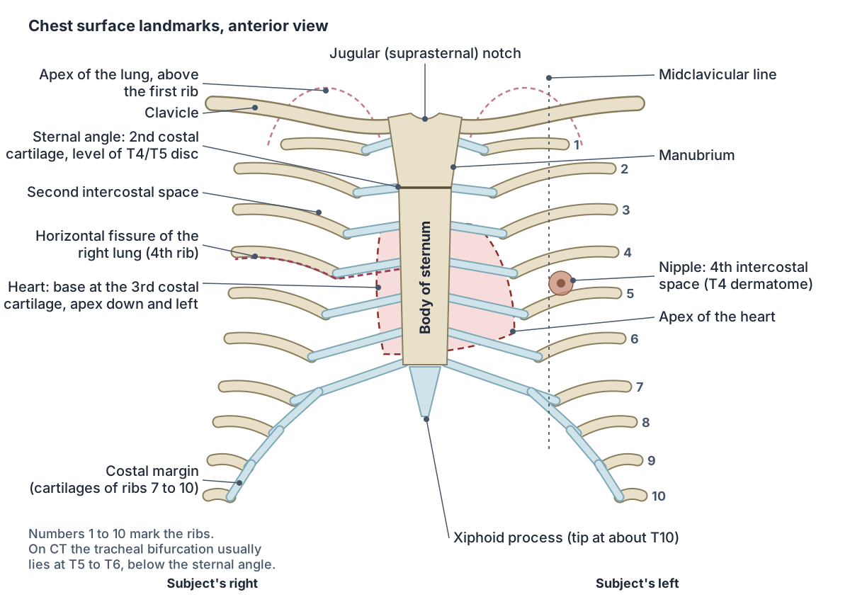
The sternal angle (angle of Louis) is the ridge you can feel where the manubrium meets the body of the sternum. It lies at the level of the second costal cartilage, so clinicians use it to find the second rib and count the ribs below, and it sits on a horizontal plane that passes back to the disc between T4 and T5. (Kenhub: Sternal angle.)
In classical descriptions this plane marks the upper limit of the pericardium, the start and end of the aortic arch, the division of the trachea and of the pulmonary trunk, and the boundary between the superior and inferior mediastinum. (Kenhub: Sternal angle.)
Above the manubrium lies the jugular (suprasternal) notch, between the inner ends of the clavicles. At the lower end of the sternum, the xiphoid process is largely cartilage, ossifies late (around 40 years of age) and has its tip at about the level of T10. (TeachMeAnatomy: The Sternum.)
Chest surface markings were first drawn from cadavers. CT in living adults confirms the levels of the sternal angle and xiphisternal joint, with wide variation between people, but places the tracheal bifurcation, aortic arch and azygos vein lower, as described above. (Mirjalili SA et al. A reappraisal of adult thoracic surface anatomy. Clin Anat. 2012.)
The skin of the chest is supplied in horizontal bands (dermatomes) from T2 downwards. The T4 dermatome lies at the level of the nipple and T10 at the umbilicus. (Kenhub: Dermatomes.)
Surface landmarks of the chest
| Landmark | Where to find it | What it marks |
|---|---|---|
| Jugular (suprasternal) notch | Top edge of the manubrium, between the clavicles | Upper end of the sternum |
| Sternal angle | Ridge between the manubrium and body of the sternum | Second costal cartilage; plane of the T4/T5 disc; superior vs inferior mediastinum |
| Fourth intercostal space | Count down from the second rib beside the sternal angle | Usual level of the male nipple, just lateral to the midclavicular line |
| Horizontal fissure of the right lung | Fourth rib at the sternum | Boundary between the right upper and middle lobes |
| Apex of the heart | Just left of the sternum near the junction of the fourth and fifth ribs (OpenStax) | Lowest, left-most point of the heart |
| Xiphoid process | Lower tip of the sternum | Tip at about T10 |
| Tracheal bifurcation | Behind the sternal angle in textbooks | On CT usually T5 to T6, below the sternal angle (Mirjalili 2012) |
Levels vary between people and with breathing and posture. Sources: Kenhub, TeachMeAnatomy, OpenStax, GPnotebook and Mirjalili et al. 2012.
Blood vessels and nerves of the chest wall
The internal thoracic artery, a branch of the subclavian artery, supplies the front of the chest wall. Its anterior intercostal branches supply intercostal spaces 1 to 6, its perforating branches pass forwards through the spaces to the pectoral muscles and the skin, and it ends as the musculophrenic artery, which supplies spaces 7 to 9, and the superior epigastric artery. (TeachMeAnatomy: Thoracic Wall Vasculature.)
Posterior intercostal arteries from the thoracic aorta supply spaces 3 to 11, while spaces 1 and 2 are supplied by the superior intercostal artery, and they join the anterior intercostal arteries near the costochondral junctions. The axillary artery adds the superior and lateral thoracic arteries to the upper and lateral wall. The veins follow the arteries: anterior intercostal veins drain to the internal thoracic veins and posterior intercostal veins to the azygos system. (TeachMeAnatomy: Thoracic Wall Vasculature.)
The intercostal nerves are the anterior rami of spinal nerves T1 to T11. They run in the intercostal spaces with the vessels; the first is short and usually has no branch to the skin. (Kenhub: Intercostal spaces.)
The artery behind the costal cartilages that supplies the front of the chest, in 3D. Internal thoracic artery.
Chest pain anatomy: which structure is where?
This section is educational and not a guide to diagnosis. Chest pain can come from the heart and large vessels (heart attack, pericarditis, aortic dissection), the lungs and pleura (pulmonary embolism, pneumonia, pleurisy, pneumothorax), the oesophagus and upper digestive organs (reflux, oesophageal spasm, gallstones, pancreatitis), the chest wall (rib fracture, a strained chest muscle, costochondritis) and nerves (shingles). Cleveland Clinic advises calling emergency services for chest pain that is new, sudden or lasts longer than five minutes after rest or medication. (Cleveland Clinic: Chest Pain.)
Pain from the heart is often felt somewhere else. Sensory fibres from the heart enter the spinal cord at the same levels as the nerves from the shoulder and arm, so strong sensations from the heart can be felt as pain in the left shoulder and left arm. In the same way, the phrenic nerve carries sensation from the diaphragm into the cord at C3 to C5, the levels that also serve the neck and shoulder, so irritation under the diaphragm can be felt in the shoulder. (OpenStax Anatomy and Physiology 2e, 15.2: Autonomic Reflexes and Homeostasis.)
The two layers of pleura behave differently. The parietal pleura lining the chest wall is supplied by the intercostal and phrenic nerves and gives sharp, well-localised pain, whereas the visceral pleura on the lung is not sensitive to pain, temperature or touch and responds only to stretch. (TeachMeAnatomy: The Pleurae.)
Costochondritis is inflammation of the cartilage joining the ribs to the sternum, usually at ribs two to five. Its pain worsens with deep breaths, coughing, twisting and reaching overhead, and Cleveland Clinic cites expert estimates that about a third of people who see a doctor with chest or rib pain have it. Tietze syndrome is similar but causes visible swelling, usually at the second or third rib. (Cleveland Clinic: Costochondritis.)
How chest structures signal pain
| Structure | Nerve supply | How pain is felt |
|---|---|---|
| Skin, muscles, ribs and costal cartilages | Intercostal nerves (T1 to T11) | Over the spot; costochondritis pain worsens with deep breaths, coughing and twisting |
| Parietal pleura | Intercostal and phrenic nerves | Sharp and well localised |
| Visceral pleura on the lung | Autonomic fibres from the pulmonary plexus | Not sensitive to pain; responds only to stretch |
| Heart | Visceral sensory fibres entering the cord at the levels of the shoulder and arm nerves | Can be referred to the left shoulder and left arm |
| Diaphragm | Phrenic nerve (C3 to C5) | Can be referred to the neck and shoulder |
Educational overview from OpenStax (15.2), TeachMeAnatomy (pleurae), Kenhub (intercostal spaces) and Cleveland Clinic (costochondritis); not for self-diagnosis.
Chest anatomy in the 3D model
- Abdominal part of pectoralis major
- The lowest fibres of pectoralis major, arising from the abdominal wall.
- Arch of aorta
- The aortic arch is the curved part of the aorta between the ascending and descending aorta, giving the great vessels to the head and arms.
- Body of sternum
- The long middle part of the sternum.
- Clavicular part of pectoralis major
- The upper fibres of pectoralis major.
- Diaphragm
- The dome-shaped muscle separating the thoracic and abdominal cavities and the main muscle of inspiration.
- Esophagus
- The esophagus is a muscular tube about 22 to 25 cm long that carries swallowed food from the pharynx to the stomach.
- External intercostal muscle
- The outermost layer of muscles between the ribs.
- Internal intercostal muscle
- The middle layer of intercostal muscles.
- Internal thoracic artery
- The internal thoracic artery supplies the anterior chest wall.
- Lobe of thymus
- The thymus lobes are the two lobes of the thymus gland, where T lymphocytes mature.
- Lower lobe of left lung
- The lower lobe of the left lung lies below and behind the oblique fissure. It forms the back and the base of the left lung, resting on the left dome of the diaphragm.
- Lower lobe of right lung
- The lower lobe of the right lung lies below and behind the oblique fissure. It forms the back and the base of the right lung, resting on the right dome of the diaphragm above the liver.
- Main bronchus
- The left main (primary) bronchus, one of the two airways formed by the division of the trachea.
- Manubrium
- The upper part of the sternum.
- Middle lobe of right lung
- The middle lobe is the wedge of the right lung that lies between the horizontal and oblique fissures, at the front and side of the lower chest. Only the right lung has one.
- Pectoralis minor
- A thin triangular muscle lying deep to pectoralis major on the upper chest wall.
- Pulmonary trunk
- The large artery arising from the right ventricle that carries blood to the lungs.
- Serratus anterior
- A broad muscle on the side of the thorax that holds the scapula against the chest wall.
- Sternocostal part of pectoralis major
- The large lower part of pectoralis major arising from the sternum and ribs.
- Subclavius
- A small muscle between the clavicle and the first rib.
- Superior vena cava
- The large vein draining the upper body into the right atrium.
- Trachea
- The trachea (windpipe) is a flexible air tube extending from the larynx to the bronchi.
- Upper lobe of left lung
- The upper lobe of the left lung is one of the left lung's two lobes. It lies above and in front of the oblique fissure, forms the apex of the lung and includes the lingula.
- Upper lobe of right lung
- The upper lobe of the right lung is the top of the right lung's three lobes. It lies above the horizontal fissure and above the upper part of the oblique fissure, and forms the apex of the lung.
- Wall of atrium
- The muscular walls of the right and left atria, the thin-walled receiving chambers of the heart.
- Wall of ventricle
- The muscular wall of a ventricle of the heart.
- Xiphoid process
- The small lowest part of the sternum.
Frequently asked questions
What organs are in the chest?
The chest (thoracic cavity) contains the heart, the two lungs, the trachea and main bronchi, the oesophagus and the thymus, together with the aorta, the venae cavae and pulmonary vessels, nerves and the thoracic duct. The lungs sit in the right and left pleural cavities, and everything else lies in the mediastinum between them.
What bones make up the chest?
The bony chest, or thoracic cage, is the sternum in front (manubrium, body and xiphoid process), 12 pairs of ribs with their costal cartilages at the sides, and the 12 thoracic vertebrae behind. Ribs 1 to 7 reach the sternum through their own cartilages, ribs 8 to 10 join the cartilage above, and ribs 11 and 12 are floating ribs.
What are the main muscles of the chest?
On the front of the chest are pectoralis major, the large fan-shaped muscle under the skin, and beneath it pectoralis minor and subclavius, with serratus anterior on the side of the chest. Between the ribs lie three layers of intercostal muscles, and the diaphragm forms the floor. The diaphragm and external intercostals do the work of quiet breathing.
Where is the heart located in the chest?
The heart lies in the middle of the chest, in the mediastinum between the lungs, behind the sternum and costal cartilages and in front of the spine. Its base, where the great vessels attach, is at the level of the third costal cartilage, and its apex points down and to the left, just left of the sternum near the fourth and fifth ribs.
What is the sternal angle and why is it important?
The sternal angle, or angle of Louis, is the ridge where the manubrium meets the body of the sternum. It marks the second costal cartilage, so it is the starting point for counting ribs and intercostal spaces, and it lies on the plane of the T4/T5 disc that separates the superior and inferior mediastinum.
What separates the chest from the abdomen?
The diaphragm, a dome-shaped sheet of muscle with a central tendon, separates the thoracic cavity from the abdominal cavity. The inferior vena cava, oesophagus and aorta pass through it at openings classically placed at T8, T10 and T12. It is supplied by the phrenic nerves (C3 to C5) and is the main muscle of breathing.
How is male chest anatomy different from female chest anatomy?
The skeleton, muscles and organs of the chest are the same in both sexes. The difference is the breast: after puberty the female breast develops glandular and fatty tissue that lies on pectoralis major and serratus anterior, while in males pectoralis major is covered only by fascia, fat and skin, and the nipple usually lies at the fourth intercostal space.
References
- Kenhub. Thorax: anatomy, wall, cavity, organs and neurovasculature.
- Kenhub. Mediastinum: definition, anatomy, borders and contents.
- Kenhub. Sternal angle: anatomy, location and function.
- Kenhub. Intercostal spaces: anatomy, contents and boundaries.
- Kenhub. Pectoralis major: origin, insertion, innervation, function.
- Kenhub. Dermatomes.
- TeachMeAnatomy. The Superior Mediastinum.
- TeachMeAnatomy. The Middle Mediastinum.
- TeachMeAnatomy. Muscles of the Thoracic Cage.
- TeachMeAnatomy. Thoracic Wall Vasculature.
- TeachMeAnatomy. The Pleurae.
- TeachMeAnatomy. The Lungs.
- TeachMeAnatomy. The Sternum.
- TeachMeAnatomy. The Diaphragm.
- TeachMeAnatomy. Muscles of the Pectoral Region.
- TeachMeAnatomy. The Breasts.
- OpenStax. Anatomy and Physiology 2e, §19.1 Heart Anatomy. Rice University, 2022.
- OpenStax. Anatomy and Physiology 2e, §22.2 The Lungs. Rice University, 2022.
- OpenStax. Anatomy and Physiology 2e, §22.3 The Process of Breathing. Rice University, 2022.
- OpenStax. Anatomy and Physiology 2e, §15.2 Autonomic Reflexes and Homeostasis. Rice University, 2022.
- Cleveland Clinic. Chest (Thorax): Anatomy & Function.
- Cleveland Clinic. Thoracic Cavity: Location and Function.
- Cleveland Clinic. What is the Mediastinum?
- Cleveland Clinic. Rib Cage (Thoracic Cage): What It Is, Anatomy & Function.
- Cleveland Clinic. Chest Pain: What It Feels Like, Causes & Treatment.
- Cleveland Clinic. Costochondritis: What It Is, Causes & Treatment.
- Cleveland Clinic. Breast Anatomy: Milk Ducts, Tissue, Conditions & Physiology.
- GPnotebook. Breast anatomy.
- Mirjalili SA, Hale SJM, Buckenham T, Wilson B, Stringer MD. A reappraisal of adult thoracic surface anatomy. Clin Anat. 2012;25(7):827-834.
- Mitsuhashi N. et al. (2009). BodyParts3D: 3D structure database for anatomical concepts. Nucleic Acids Research 37, D782–D785. Data © The Database Center for Life Science, CC BY 4.0.
- Radiopaedia. Oblique fissure (surface anatomy used to place the oblique fissures in the 3D lungs).
- Radiopaedia. Horizontal fissure (surface anatomy used to place the horizontal fissure in the 3D right lung).
Related anatomy
- Rib cageRib cage anatomy: 12 pairs of ribs, true, false and floating ribs, parts of a rib, sternum and jugular notch, costal…
- Chest musclesChest muscle anatomy with a 3D model and diagrams: pectoralis major and its three parts, pectoralis minor, serratus…
- Breast anatomyBreast anatomy with labelled diagrams and a 3D model: lobes, milk ducts, Cooper's ligaments, nipple, areola, blood…
- HeartHeart anatomy with labelled diagrams and a 3D model: location, pericardium, wall layers, four chambers, valves, great…
- LungsLung anatomy with labelled diagrams and a 3D model of all five lobes: fissures, hilum, segments, pleura, blood supply…
- DiaphragmDiaphragm muscle anatomy with a labelled diagram and 3D model: attachments, crura, central tendon, openings at T8, T10…
Explore the whole body in the interactive 3D human anatomy model, or browse the A–Z list of human structures.