

What is the lymphatic system?
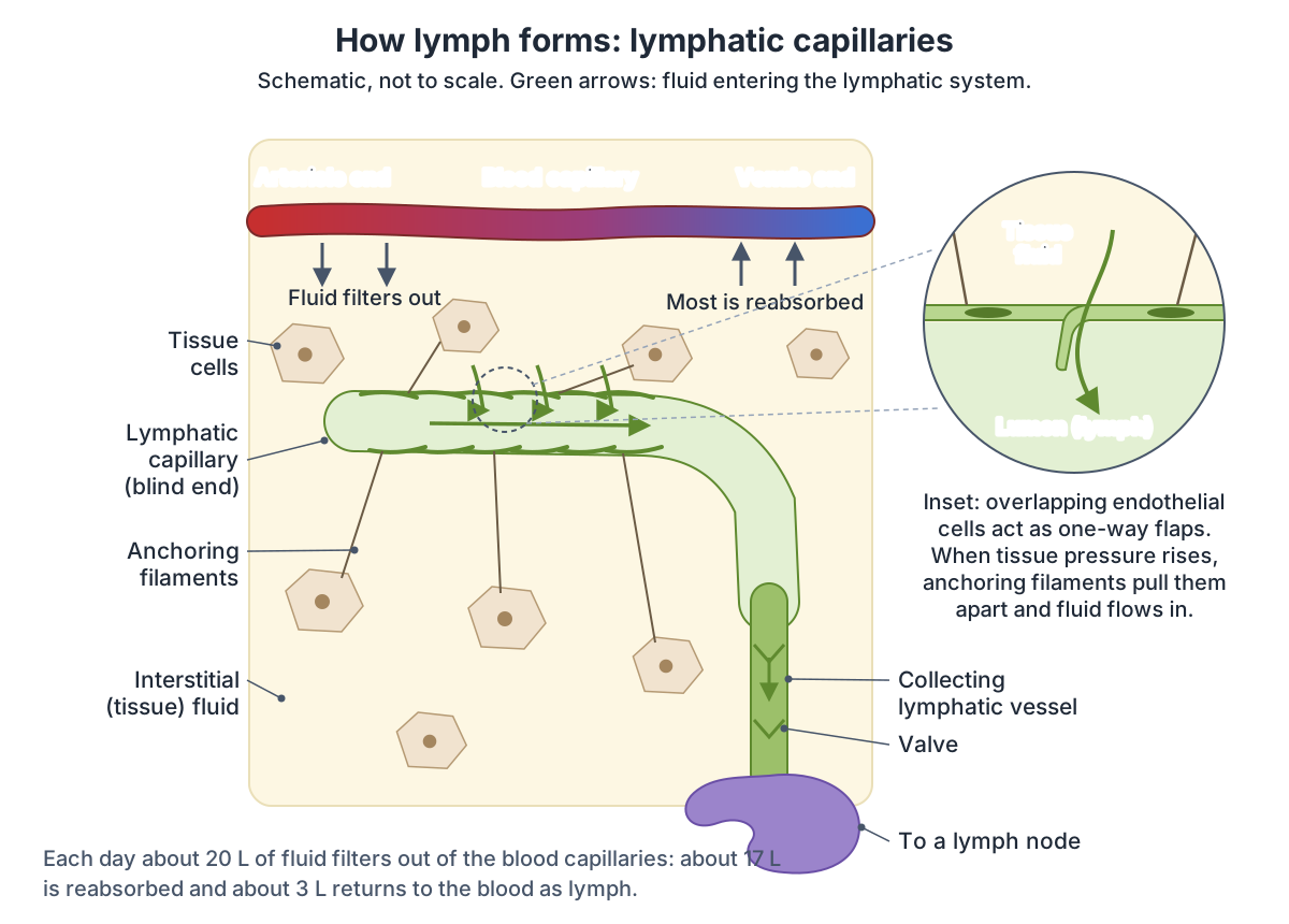
The lymphatic system drains excess fluid from the tissues and returns it to the blood. Each day about 20 litres of plasma filter out of the blood capillaries into the tissues; about 17 litres are reabsorbed by the blood vessels and the lymphatic system carries back the remaining 3 litres. It also carries immune cells, transports dietary fats and fat-soluble vitamins from the gut, and its lymph nodes are the main staging areas for immune responses. (OpenStax Anatomy and Physiology 2e, 21.1: Anatomy of the Lymphatic and Immune Systems.)
Cleveland Clinic lists its parts as lymph (the fluid), lymphatic vessels, two collecting ducts and the lymphoid organs and tissues: bone marrow, thymus, lymph nodes, spleen, the tonsils and adenoids, and mucosa-associated lymphoid tissue (MALT). (Cleveland Clinic: Lymphatic System.)
Unlike blood vessels, which form a circuit, lymphatic vessels only carry fluid away from the tissues. Lymph capillaries begin as blind-ended sacs whose walls work as a simple one-way valve, letting fluid in but not out. (NCI SEER Training: Components of the Lymphatic System.)
The heart, arteries and veins that lymph returns to. Circulatory system.
What is lymph and how does it move?
Lymph is a clear, yellowish fluid that forms when fluid leaves the capillary beds under pressure. It is about 95% water, with proteins, lipids, glucose, ions and cells, mostly lymphocytes. Lymph from the intestine, called chyle, is rich in fat. TeachMeAnatomy estimates that an adult makes roughly 3 to 4 litres a day. (TeachMeAnatomy: The Lymphatic System.)
Kenhub gives a lower figure of roughly 2 litres a day in a healthy adult, while OpenStax and Cleveland Clinic give about 3 litres. Kenhub also notes that lymph from different parts of the body differs, gut lymph being the fattiest. (Kenhub: Lymphatic system.)
The heart does not pump lymph. It is pushed along by body movement, the contraction of skeletal muscles and breathing, and closely spaced one-way valves keep it moving towards the heart. (OpenStax Anatomy and Physiology 2e, 21.1: Anatomy of the Lymphatic and Immune Systems.)
Lymphatic capillaries and vessels
Lymphatic capillaries begin as blind-ended vessels in almost every tissue; the exceptions are the central nervous system, bone marrow, bones, teeth and the cornea. Their wall is one layer of endothelial cells whose edges overlap like flaps. When pressure in the tissue rises, collagen anchoring filaments pull the flaps open and fluid flows in; when it falls, the flaps close. In the small intestine, special capillaries called lacteals absorb fats as milky chyle. (OpenStax Anatomy and Physiology 2e, 21.1: Anatomy of the Lymphatic and Immune Systems.)
Capillaries join into larger lymphatic vessels that resemble veins, with three layers in their walls and many valves, which give them a beaded look. Superficial vessels generally follow veins and deep vessels generally follow arteries. They pass through lymph nodes and then merge into lymphatic trunks. (OpenStax Anatomy and Physiology 2e, 21.1: Anatomy of the Lymphatic and Immune Systems.)
The upper limb shows the pattern. Superficial vessels from the skin of the hand climb with the basilic and cephalic veins, those along the basilic vein passing through the cubital nodes at the elbow, while deep vessels follow the radial, ulnar and brachial veins to the axillary nodes. (TeachMeAnatomy: Lymphatic Drainage of the Upper Limb.)
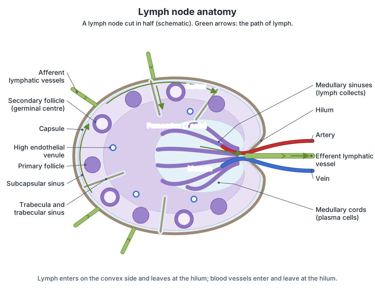
Lymph node anatomy: the structure of a lymph node
A lymph node is a small ellipsoid, often kidney-shaped organ from about 0.1 to 2.5 cm long. Blood vessels enter and leave at an indentation called the hilum. Afferent lymph vessels enter through the capsule, not at the hilum, and the efferent vessel leaves at the hilum. Each node has a peripheral cortex with spherical follicles, each with a paler germinal centre, and a central medulla. (Radiopaedia: Lymph node.)
A tough connective tissue capsule surrounds the node and sends trabeculae inwards, dividing it into compartments supported by reticular fibres. Lymph arriving in the afferent vessels enters the subcapsular sinus. The cortex holds the follicles, whose germinal centres contain rapidly dividing B cells surrounded by T cells, and the medulla holds medullary cords of B cells and plasma cells and medullary sinuses where lymph collects before leaving. (OpenStax Anatomy and Physiology 2e, 21.1: Anatomy of the Lymphatic and Immune Systems.)
Between the cortex and the medulla lies the paracortex, which has no follicles but many T cells. It contains high endothelial venules, small veins with a cuboidal lining through which most lymphocytes enter the node from the blood. A follicle without a germinal centre is a primary follicle; one with a germinal centre, full of B cells activated by an antigen, is a secondary follicle. (Kenhub: Histology of lymph nodes.)
The path of lymph through a node is: afferent vessels, subcapsular sinus, cortical (trabecular) sinuses, medullary sinuses and out through the efferent vessel at the hilum. Arteries enter at the hilum and branch to capillaries, which are densest around the germinal centres, and veins leave through the hilum. (Kenhub: Histology of lymph nodes.)
SEER Training notes that lymph enters a node at several points on its convex side but leaves through a single efferent vessel at the hilum, and that nodes are usually less than 2.5 cm long. (NCI SEER Training: Lymph Nodes.)
Parts of a lymph node
| Part | Where it is | What it contains or does |
|---|---|---|
| Capsule and trabeculae | Outer covering, with partitions running inwards | Dense connective tissue; reticular fibres form the inner framework |
| Afferent lymphatic vessels | Convex surface, through the capsule | Several valved vessels bring lymph in |
| Subcapsular sinus | Just under the capsule | First space that incoming lymph enters |
| Cortex | Outer layer | Lymphoid follicles of B cells; secondary follicles have germinal centres where activated B cells divide |
| Paracortex | Deep cortex, between cortex and medulla | T cells (CD4 and CD8), no follicles; high endothelial venules where most lymphocytes enter from the blood |
| Medulla | Centre, towards the hilum | Medullary cords with plasma cells, B and T cells; medullary sinuses, the final filter |
| Hilum | Indentation on the concave side | Efferent lymphatic vessel leaves; artery enters and vein leaves |
From Kenhub, OpenStax and Radiopaedia.
What do lymph nodes do?
Lymph nodes act as filters of the lymph. Dendritic cells and macrophages inside them take up and kill many pathogens and remove debris, and lymphocytes in the follicles mount immune responses. This is why nodes swell during an infection. (OpenStax Anatomy and Physiology 2e, 21.1: Anatomy of the Lymphatic and Immune Systems.)
Cleveland Clinic compares a node to a security checkpoint: as lymph passes through, its B cells, T cells, macrophages and dendritic cells look for foreign invaders such as viruses and bacteria and remove damaged, abnormal or cancerous cells. (Cleveland Clinic: Lymph Nodes.)
Because cancer cells can travel in lymph, nodes are also where many cancers spread first. The first node that drains a tumour is called the sentinel lymph node, and finding cancer in it helps doctors stage the cancer and judge its outlook. Lymphatic tissue can also be the starting site of a cancer, lymphoma. (Kenhub: Lymphatic system.)
How many lymph nodes are in the body?
Sources disagree on the number. Cleveland Clinic says you have anywhere from 400 to 800 lymph nodes and stresses that there is no precise number that everyone has. (Cleveland Clinic: Lymph Nodes.)
OpenStax gives about 500 to 600 nodes in the body, concentrated near the groin, armpits, neck, chest and abdomen. (OpenStax Anatomy and Physiology 2e, 21.1: Anatomy of the Lymphatic and Immune Systems.)
TeachMeAnatomy gives a lower count of roughly 400 to 450 nodes in adults, most of them in the abdomen. (TeachMeAnatomy: The Lymphatic System.)
Where are the lymph nodes? Main lymph node groups
Lymph nodes lie in clusters throughout the body: in the head and neck (behind the ear, around the jaw, at the base of the skull, along the sides of the neck and above the collarbone), in the armpits (axillary), in the chest (mediastinal, aortic and hilar), in the abdomen and pelvis (para-aortic, mesenteric and iliac) and in the groin (inguinal). There are no lymph nodes in the brain or spinal cord. (Cleveland Clinic: Lymph Nodes.)
The cervical, axillary and inguinal nodes are the main superficial clusters on each side of the body. (NCI SEER Training: Lymph Nodes.)
In the neck, superficial nodes form a ring where the head meets the neck, and all lymph from the head and neck passes through the deep cervical chain along the internal jugular vein. Virchow's node, a supraclavicular node in the left supraclavicular fossa, receives lymph from the abdominal cavity, and its enlargement (Troisier's sign) can point to an abdominal cancer. (TeachMeAnatomy: Lymphatic Drainage of the Head and Neck.)
Every head and neck node group and the clinical neck levels I to VI. Lymph nodes of the head and neck.
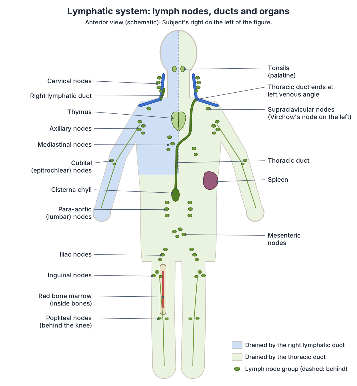
Main lymph node groups of the body
| Node group | Where | Main areas drained |
|---|---|---|
| Cervical (superficial ring and deep cervical chain) | Ring where the head meets the neck; deep chain along the internal jugular vein | Head and neck |
| Supraclavicular | Above the clavicle; Virchow's node on the left | Left node receives lymph from the abdominal cavity |
| Axillary (20 to 30, up to 40) | Armpit: pectoral, subscapular, humeral, central and apical groups | Upper limb, breast and trunk above the umbilicus |
| Cubital (epitrochlear) | Medial to the basilic vein above the medial epicondyle | Superficial vessels of the hand and forearm that follow the basilic vein |
| Thoracic: parasternal, intercostal, diaphragmatic, brachiocephalic, tracheobronchial, posterior mediastinal | Chest wall and mediastinum | Breast, chest wall, diaphragm, heart and lungs |
| Preaortic: coeliac, superior and inferior mesenteric | In front of the abdominal aorta | Digestive tract, liver, pancreas and spleen; form the intestinal trunks |
| Lateral aortic (para-aortic, lumbar) | Beside the abdominal aorta | Kidneys, adrenals, ureters, gonads, upper uterus and posterior abdominal wall; form the lumbar trunks |
| Iliac (pelvic) | Along the iliac vessels | Pelvis; the external iliac nodes receive the deep inguinal nodes |
| Superficial inguinal | Below the inguinal ligament and along the great saphenous vein | Lower limb, buttock, perineum, external genitalia, anal canal and abdominal wall below the umbilicus |
| Deep inguinal | Medial to the femoral vein | Deep vessels of the thigh, glans and superficial inguinal nodes; drain to the external iliac nodes |
| Popliteal (5 to 7) | Fat of the popliteal fossa behind the knee | Lateral foot and leg along the small saphenous vein, deep leg vessels and the knee |
Compiled from TeachMeAnatomy (neck, upper and lower limb), Kenhub (axillary, thorax and abdomen, pelvis and lower limb) and Cleveland Clinic. TeachMeAnatomy counts 1 to 20 inguinal nodes in all.
Axillary, inguinal and popliteal lymph nodes
The axillary nodes form five groups: pectoral (3 to 5 nodes, mainly draining the front of the chest wall and most of the breast), subscapular (6 to 7, back of the chest wall), humeral (4 to 6, most of the upper limb), central (3 to 4, receiving the first three groups) and apical, in the apex of the armpit. Efferents of the apical nodes form the subclavian lymph trunk. (TeachMeAnatomy: Lymphatic Drainage of the Upper Limb.)
Kenhub counts about 20 to 30 axillary nodes (up to 40 have been noted). In breast cancer the status of these nodes guides treatment: the sentinel node is identified and further nodes are removed if it contains cancer, which carries a risk of lymphoedema of the arm. (Kenhub: Axillary lymph nodes.)
The inguinal nodes lie in the upper femoral triangle and number 1 to 20. The superficial group drains the penis, scrotum, perineum, buttock and abdominal wall as far as the umbilicus, and the subinguinal nodes along the great saphenous vein take the superficial vessels of the leg. Five to seven popliteal nodes sit in the fat behind the knee. The testes drain directly to the para-aortic nodes, so testicular disease rarely enlarges the groin nodes. (TeachMeAnatomy: Lymphatic Drainage of the Lower Limb.)
Kenhub describes the deep inguinal nodes as lying medial to the femoral vein and draining into the external iliac nodes, the first group of the pelvic chain. (Kenhub: Lymph nodes of the pelvis and lower limb.)
Lymph nodes of the chest and abdomen
In the chest, parietal nodes (parasternal, intercostal and diaphragmatic) drain the chest wall, and visceral nodes (brachiocephalic, tracheobronchial and posterior mediastinal) drain the heart and lungs. The parasternal nodes along the internal thoracic vessels also receive lymph from the breast. (Kenhub: Lymph nodes of the thorax and abdomen.)
In the abdomen, the preaortic nodes in front of the aorta (coeliac, superior mesenteric and inferior mesenteric) collect lymph from the digestive organs and give rise to the intestinal trunks. The lateral aortic nodes beside the aorta drain the adrenal glands, kidneys, ureters, gonads, uterine tubes and posterior abdominal wall into the two lumbar trunks. Almost all abdominal lymph returns through the thoracic duct. (Kenhub: Lymph nodes of the thorax and abdomen.)
The abdominal organs and regions that these nodes drain. Abdomen anatomy.
Lymphatic trunks, the thoracic duct and the right lymphatic duct
Lymph from the nodes collects in five main lymphatic trunks: four paired trunks, the lumbar, bronchomediastinal, subclavian and jugular trunks, and one unpaired intestinal trunk, which drains most of the digestive organs. (Kenhub: Lymphatic system.)
Drainage is asymmetrical. The right lymphatic duct drains only the upper right part of the body (the right side of the head, thorax and right upper limb) into the right subclavian vein. The thoracic duct drains the rest into the left subclavian vein. It begins below the diaphragm in a sac called the cisterna chyli, which receives lymph from the lower abdomen, pelvis and lower limbs through the left and right lumbar trunks and the intestinal trunk. (OpenStax Anatomy and Physiology 2e, 21.1: Anatomy of the Lymphatic and Immune Systems.)
The thoracic duct is about 40 cm long and about 5 mm wide at its start, at T12 to L2. It passes through the aortic hiatus of the diaphragm at T12, climbs between the aorta and the azygos vein, crosses to the left between T4 and T6 and ends at the venous angle between the left subclavian and left internal jugular veins, where a valve stops blood flowing back. It carries about 75% of the body's lymph. If it is torn, chyle leaks into the chest (chylothorax). (Kenhub: Thoracic duct.)
Radiopaedia places the cisterna chyli at L1 (range T10 to L3), with the duct climbing behind the oesophagus. It notes that variants are common, in about 40% of people, including a double thoracic duct in about 20% and endings in other neck veins. (Radiopaedia: Thoracic duct.)
The cisterna chyli itself lies in front of the L1 and L2 vertebral bodies, just to the right of the aorta behind the right crus of the diaphragm, and is 5 to 7 cm long, although its size varies greatly with position. On CT it can mimic enlarged lymph nodes. (Radiopaedia: Cisterna chyli.)
The right lymphatic duct is only about 1 to 2 cm long. It usually forms from the right bronchomediastinal, jugular and subclavian trunks in front of scalenus anterior and empties at the junction of the right internal jugular and subclavian veins or into the right brachiocephalic vein, but its formation is highly variable. (Kenhub: Right lymphatic duct.)
Thoracic duct vs right lymphatic duct
| Feature | Thoracic duct | Right lymphatic duct |
|---|---|---|
| Length | About 40 cm | About 1 to 2 cm |
| Begins | Cisterna chyli, the junction of the lumbar and intestinal trunks, at about T12 to L2 | Union of the right jugular, subclavian and bronchomediastinal trunks |
| Course | Through the aortic hiatus, up between the aorta and azygos vein, crossing to the left at T4 to T6 | Short vessel in the root of the neck in front of scalenus anterior |
| Drains | Both lower limbs, abdomen, left side of the chest, left upper limb, left head and neck: about 75% of lymph | Right head and neck, right side of the chest, right upper limb |
| Ends | Left venous angle (left internal jugular and subclavian veins) | Right venous angle (right internal jugular and subclavian veins) or right brachiocephalic vein |
| Variants | In about 40% (double duct in about 20%) | Formation highly variable |
From Kenhub, Radiopaedia and OpenStax.
Primary lymphoid organs: bone marrow and thymus
Lymphoid organs are primary, where lymphocytes mature, or secondary, where immune responses are mounted. The primary organs are the red bone marrow, where blood cells form and most B cells develop, and the thymus, where T cells mature. The thymus lies above the heart between the sternum and the aorta. After puberty it shrinks, losing about 3% of its tissue a year until age 35 to 45 and then about 1% a year. (OpenStax Anatomy and Physiology 2e, 21.1: Anatomy of the Lymphatic and Immune Systems.)
Kenhub places the thymus in the superior and anterior mediastinum, from the lower poles of the thyroid gland down to the fourth costal cartilage, behind the sternum and in front of the great vessels. Its two lobes are joined by an isthmus and divided into lobules with an outer cortex and an inner medulla containing Hassall's corpuscles. Developing T cells that cannot recognise the body's own MHC proteins, or that react strongly against self, are eliminated there. (Kenhub: Thymus.)
The thymus lobes in the 3D model. Thymus.
The spleen
The spleen is about 12 cm long and dark red because of its rich blood supply. Its red pulp, mainly red blood cells with macrophages, filters the blood and removes microbes and dying red cells. Its white pulp, arranged around central arterioles, contains B-cell germinal centres surrounded by T cells and mounts immune responses to pathogens in the blood. A marginal zone between the two captures particles for the lymphocytes. (OpenStax Anatomy and Physiology 2e, 21.1: Anatomy of the Lymphatic and Immune Systems.)
Cleveland Clinic calls it the largest lymphatic organ. It sits under the left ribs, filters the blood and removes old or faulty blood cells. (Cleveland Clinic: Lymphatic System.)
Spleen location, size, relations, blood supply and function in 3D. Spleen.
Tonsils, MALT and Peyer's patches
Waldeyer's ring is a ring of lymphoid tissue around the upper throat. The pharyngeal tonsil sits in the roof of the nasopharynx (when enlarged it is called the adenoids), the tubal tonsils surround the openings of the Eustachian tubes, the palatine tonsils lie in the side walls of the oropharynx between the palatoglossal and palatopharyngeal arches, and the lingual tonsil lies in the back third of the tongue. The palatine tonsils drain to the jugulodigastric and upper deep cervical nodes. (TeachMeAnatomy: The Tonsils and Adenoids (Waldeyer's Ring).)
Tonsils are lymphoid nodules without a complete capsule, with deep folds of lining called crypts. Similar unencapsulated lymphoid tissue in mucous membranes is called MALT: it is found in the digestive tract, breast tissue, lungs and eyes. Peyer's patches are MALT in the small intestine, where M cells sample the gut contents, and BALT lies along the branching bronchi. (OpenStax Anatomy and Physiology 2e, 21.1: Anatomy of the Lymphatic and Immune Systems.)
Kenhub counts the spleen, tonsils, vermiform appendix, lymph nodes and MALT as the secondary lymphoid organs, where lymphocytes are finally activated after meeting antigens. (Kenhub: Lymphatic system.)
The palatine, pharyngeal, tubal and lingual tonsils of Waldeyer's ring. Tonsils.
Swollen lymph nodes: what size is normal?
Lymphadenopathy means lymph nodes that are abnormal in size, for example larger than 1 cm, or in consistency. Inguinal nodes up to 2 cm are found in many healthy adults, while any palpable supraclavicular, popliteal or iliac node, or an epitrochlear node larger than 5 mm, is considered abnormal. Enlargement of more than two non-neighbouring groups is called generalised lymphadenopathy. In primary care only 1.1% of unexplained cases are due to cancer, a share that rises with age. (Gaddey HL, Riegel AM. Unexplained Lymphadenopathy: Evaluation and Differential Diagnosis. Am Fam Physician 2016.)
Kenhub distinguishes lymphadenopathy, usually painless, from lymphadenitis, in which inflamed nodes are painful and hard. Nodes that are immobile, clumped together and firm are more suspicious for cancer. (Kenhub: Histology of lymph nodes.)
Cleveland Clinic advises seeing a health provider about noticeable or painful nodes at the elbow, in the upper chest or above the collarbone, and about nodes that stay swollen, hurt or come with fever, tiredness or unexplained weight loss. (Cleveland Clinic: Lymph Nodes.)
Lymphoedema (lymphedema)
Lymphoedema is a long-term swelling of the tissues, usually in an arm or leg, caused by fluid the lymphatic system cannot drain. Primary lymphoedema is rare and caused by faulty genes that affect how the lymphatic system develops. Secondary lymphoedema is much more common and follows damage to the lymphatic system, for example after cancer treatment, infection or injury. It cannot be cured, but compression garments, skin care, exercise and specialised massage usually control it; cellulitis is its most common complication. (NHS: Lymphoedema.)
In the arm, interrupting lymphatic drainage, for example by removing the axillary nodes during breast cancer surgery, can cause painful swelling of the upper limb. (TeachMeAnatomy: Lymphatic Drainage of the Upper Limb.)
The lymphatic system in the 3D model
The 3D viewer highlights two lymphoid organs: the spleen under the left ribs and the two lobes of the thymus behind the sternum. The palatine tonsils, added to the model from the Human Reference Atlas, are highlighted in the tonsils guide. BodyParts3D, built from MRI scans of an adult male, does not include lymph nodes, lymph vessels, the thoracic duct or the cisterna chyli, so the three diagrams show those. Bone marrow lies inside the bones shown in the skeleton. (Mitsuhashi N et al. BodyParts3D: 3D structure database for anatomical concepts. Nucleic Acids Res 2009.)
Lymphatic system in the 3D model
- Lobe of thymus
- The thymus lobes are the two lobes of the thymus gland, where T lymphocytes mature.
- Spleen
- The spleen is the largest lymphoid organ, filtering blood and supporting immune responses.
Frequently asked questions
What is the structure of a lymph node?
A lymph node is a bean-shaped organ wrapped in a fibrous capsule with trabeculae running inwards. Under the capsule is the subcapsular sinus, then the cortex with B-cell follicles and germinal centres, the paracortex with T cells and high endothelial venules, and the central medulla with medullary cords and sinuses. Afferent vessels enter the convex surface and the efferent vessel leaves at the hilum.
How many lymph nodes are in the human body?
There is no fixed number. Sources give anything from about 400 to 800: TeachMeAnatomy gives 400 to 450, Kenhub an average of 450, OpenStax 500 to 600 and Cleveland Clinic 400 to 800. Most lie in the abdomen, with other large groups in the neck, armpits, chest and groin.
Where are the main lymph nodes located?
The main groups are the cervical nodes in the neck, the supraclavicular nodes above the collarbone, the axillary nodes in the armpit, the mediastinal nodes in the chest, the para-aortic and mesenteric nodes in the abdomen, the iliac nodes in the pelvis, the inguinal nodes in the groin and the popliteal nodes behind the knee. The cervical, axillary and inguinal groups can be felt.
What is the difference between afferent and efferent lymph vessels?
Afferent lymphatic vessels carry lymph into a lymph node; several of them enter through the capsule on its convex surface. Efferent vessels carry filtered lymph out of the node; usually a single efferent vessel leaves at the hilum, the indented side where the artery and vein also pass. Valves in both keep lymph flowing one way.
What does the thoracic duct drain?
The thoracic duct drains about three quarters of the body's lymph: both legs, the pelvis and abdomen, the left side of the chest, the left arm and the left side of the head and neck. It starts at the cisterna chyli in the upper abdomen and empties into the junction of the left internal jugular and subclavian veins. The right lymphatic duct drains the rest.
What size lymph node is normal?
Most normal lymph nodes are less than 1 cm across, and nodes larger than 1 cm are usually called enlarged. Groin nodes up to 2 cm are common in healthy adults. Any node you can feel above the collarbone, behind the knee or in the pelvis, or an elbow node larger than 5 mm, is considered abnormal and should be checked.
How does lymph move without a pump?
Lymph is not pumped by the heart. It is squeezed along by the contraction of nearby skeletal muscles, by breathing movements, by the pulsing of neighbouring arteries and by smooth muscle in the vessel walls. Many one-way valves inside the lymphatic vessels stop it from flowing backwards, so it moves steadily towards the veins in the neck.
What are the organs of the lymphatic system?
The primary lymphoid organs are the red bone marrow and the thymus, where lymphocytes form and mature. The secondary lymphoid organs are the lymph nodes, the spleen, the tonsils, the appendix and the mucosa-associated lymphoid tissue (MALT) of the gut and airways, including Peyer's patches. The lymph vessels and the two collecting ducts complete the system.
References
- Betts JG, et al. Anatomy and Physiology 2e, §21.1 Anatomy of the Lymphatic and Immune Systems. OpenStax, Rice University, 2022.
- Cleveland Clinic. Lymphatic System: Diagram, Function & Anatomy. Accessed 2026-10-24.
- Cleveland Clinic. Lymph Nodes: Locations & Function. Accessed 2026-10-24.
- National Cancer Institute SEER Training Modules. Components of the Lymphatic System. Accessed 2026-10-24.
- National Cancer Institute SEER Training Modules. Lymph Nodes. Accessed 2026-10-24.
- TeachMeAnatomy. The Lymphatic System. Accessed 2026-10-24.
- TeachMeAnatomy. Lymphatic Drainage of the Upper Limb. Accessed 2026-10-24.
- TeachMeAnatomy. Lymphatic Drainage of the Lower Limb. Accessed 2026-10-24.
- TeachMeAnatomy. Lymphatic Drainage of the Head and Neck. Accessed 2026-10-24.
- TeachMeAnatomy. The Tonsils and Adenoids (Waldeyer's Ring). Accessed 2026-10-24.
- Kenhub. Lymphatic system. Accessed 2026-10-24.
- Kenhub. Histology of lymph nodes. Accessed 2026-10-24.
- Kenhub. Thoracic duct. Accessed 2026-10-24.
- Kenhub. Right lymphatic duct. Accessed 2026-10-24.
- Kenhub. Lymph nodes of the thorax and abdomen. Accessed 2026-10-24.
- Kenhub. Lymph nodes of the pelvis and lower limb. Accessed 2026-10-24.
- Kenhub. Axillary lymph nodes. Accessed 2026-10-24.
- Kenhub. Thymus. Accessed 2026-10-24.
- Radiopaedia. Lymph node. Accessed 2026-10-24.
- Radiopaedia. Thoracic duct. Accessed 2026-10-24.
- Radiopaedia. Cisterna chyli. Accessed 2026-10-24.
- Gaddey HL, Riegel AM. Unexplained Lymphadenopathy: Evaluation and Differential Diagnosis. Am Fam Physician. 2016;94(11):896-903.
- NHS. Lymphoedema. Accessed 2026-10-24.
- Mitsuhashi N, et al. BodyParts3D: 3D structure database for anatomical concepts. Nucleic Acids Res. 2009;37:D782-D785.
- Mitsuhashi N. et al. (2009). BodyParts3D: 3D structure database for anatomical concepts. Nucleic Acids Research 37, D782–D785. Data © The Database Center for Life Science, CC BY 4.0.
Related anatomy
- Lymph nodes of the head and neckLymph node map of the head and neck: where neck lymph nodes are, submental and submandibular nodes, levels I to VI…
- SpleenSpleen location explained: left upper quadrant under ribs 9 to 11, behind the stomach. Relations, ligaments, blood…
- TonsilsTonsil anatomy with labelled diagrams and a 3D model: palatine tonsils, adenoid, lingual and tubal tonsils, Waldeyer's…
- Neck anatomyNeck anatomy with labelled diagrams and a 3D model: landmarks, triangles, fascia, airway, thyroid, carotid sheath…
- Circulatory systemCardiovascular anatomy explained: heart, arteries, capillaries, vein anatomy, pulmonary vs systemic circulation and…
Explore the whole body in the interactive 3D human anatomy model, or browse the A–Z list of human structures.