

What is the respiratory system?
The respiratory system takes in oxygen and removes carbon dioxide, which Cleveland Clinic calls its main function. It also warms and moistens the air you breathe, traps particles and germs with mucus and cilia, lets you speak and smell, and helps keep the pH of the blood stable by getting rid of carbon dioxide. (Cleveland Clinic: Respiratory System.)
Its parts are the nose and nasal cavity, the mouth, the sinuses, the pharynx (throat), the larynx (voice box), the trachea (windpipe), the bronchi and bronchioles, and the lungs with their alveoli. The diaphragm drives breathing, and the ribs protect the lungs. (Cleveland Clinic: Respiratory System.)
Cleveland Clinic gives an adult breathing rate of 12 to 20 breaths a minute. The diaphragm contracts and relaxes to pull air into the lungs, and blood flowing past the alveoli picks up oxygen and releases carbon dioxide to be breathed out. (Cleveland Clinic: Lungs.)
Where the lungs and airway sit among the other internal organs. Organs of the body.
Upper vs lower respiratory tract
The airway is usually divided into an upper and a lower respiratory tract. In NCI SEER Training's version, the upper tract is the nose, pharynx and larynx, and the lower tract is the trachea, bronchial tree and lungs. Both open to the outside and are lined with mucous membrane. (NCI SEER Training: Conducting Passages.)
Kenhub splits the tract at the vocal cords instead. Its upper tract is the nasal cavity, paranasal sinuses, pharynx and the part of the larynx above the vocal cords, which conduct, filter, warm and moisten the air. Its lower tract is the larynx below the vocal cords, the trachea, bronchi, bronchioles and lungs, which conduct air and exchange gases. (Kenhub: Respiratory system.)
Cleveland Clinic counts the nose, mouth, nasal cavity, sinuses and larynx as the upper tract, and the trachea, bronchi, bronchioles and lungs as the lower tract. It calls the trachea, bronchi and bronchioles together the tracheobronchial tree. (Cleveland Clinic: Respiratory System.)
Kenhub's article on the lungs goes further and treats the carina, where the trachea divides, as the boundary between the upper and lower tracts. All the schemes agree that the nose and pharynx are upper and the bronchi and lungs lower; they differ only over the larynx and trachea. (Kenhub: Lung anatomy.)
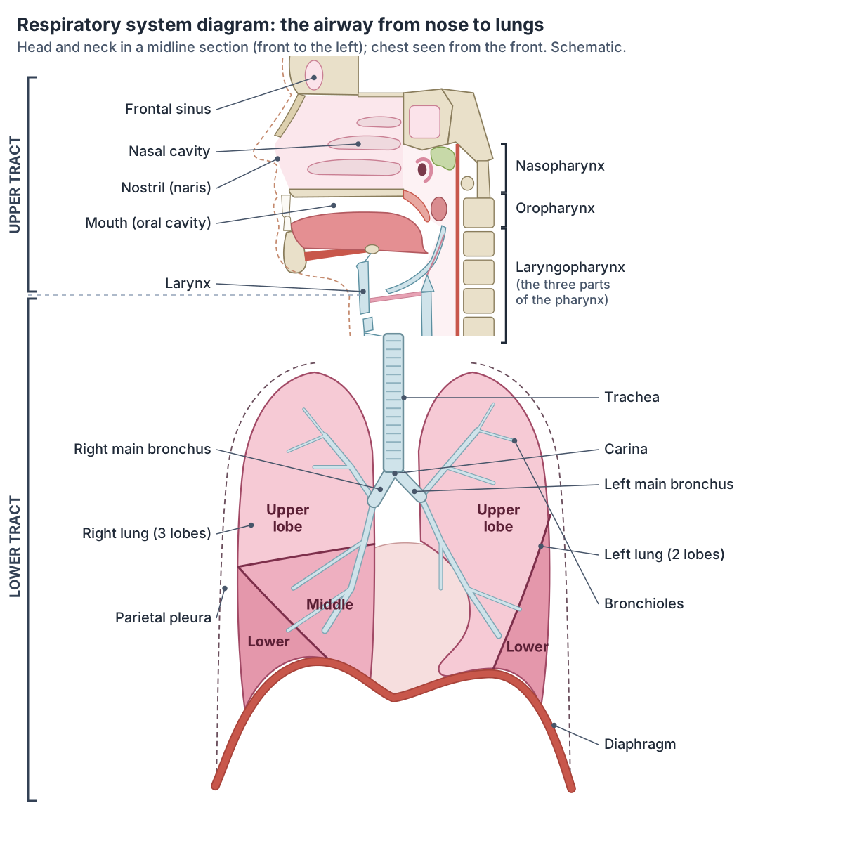
Upper vs lower respiratory tract
| Upper respiratory tract | Lower respiratory tract | |
|---|---|---|
| Parts | Nose and nasal cavity, paranasal sinuses, pharynx, larynx (all of it, or the part above the vocal folds) | Trachea, bronchi, bronchioles, alveoli and lungs (plus the larynx below the vocal folds in some schemes) |
| Main jobs | Conduct, filter, warm and moisten air; smell; voice | Conduct air and exchange gases |
| Lining | Respiratory epithelium in the nose and nasopharynx; stratified squamous in the oropharynx | Pseudostratified ciliated columnar, becoming cuboidal in the bronchioles and squamous in the alveoli |
| Typical infections | Common cold, rhinitis, sinusitis, pharyngitis, laryngitis, epiglottitis | Bronchitis, bronchiolitis, pneumonia |
From Kenhub, Cleveland Clinic and NCI SEER Training. Sources draw the boundary at the cricoid cartilage, at the vocal folds or, in one Kenhub article, at the carina.
Conducting zone vs respiratory zone
Physiologists divide the airway by what it does. The conducting zone includes the organs and structures not directly involved in gas exchange, from the nose to the terminal bronchioles; it moves air, removes debris and warms and moistens incoming air. The respiratory zone is where gas exchange happens, beginning where the terminal bronchioles join the respiratory bronchioles. (OpenStax Anatomy and Physiology 2e, 22.1: Organs and Structures of the Respiratory System.)
LITFL describes the lower airway as dividing 23 times from trachea to alveolus. The first 16 divisions form the conducting zone and the last 7 the respiratory zone. The bronchi make up the first four divisions, giving 5 lobar and 18 segmental bronchi. The total cross-sectional area of the airways is smallest at the third division and grows rapidly in the respiratory zone. (LITFL Part One: Airway and Alveolar Anatomy.)
Duke University's histology notes, after Weibel, number the trachea as generation 0 and the alveolar ducts and sacs as generations 20 to 23, with the first 16 generations purely conducting. Each airway splits into two daughter branches that usually differ in diameter or length, a pattern called irregular dichotomous branching. (Duke University Microscopic Anatomy: The Respiratory System (course notes, 2015).)
OpenAnesthesia adds a transitional zone between the two: the respiratory bronchioles and the first alveoli, where some gas exchange takes place. (OpenAnesthesia: Respiratory anatomy.)
Air in the conducting zone never reaches the alveoli, so it takes no part in gas exchange. This anatomical dead space is usually about 150 mL. (TeachMePhysiology: Lung Volumes and Capacities.)
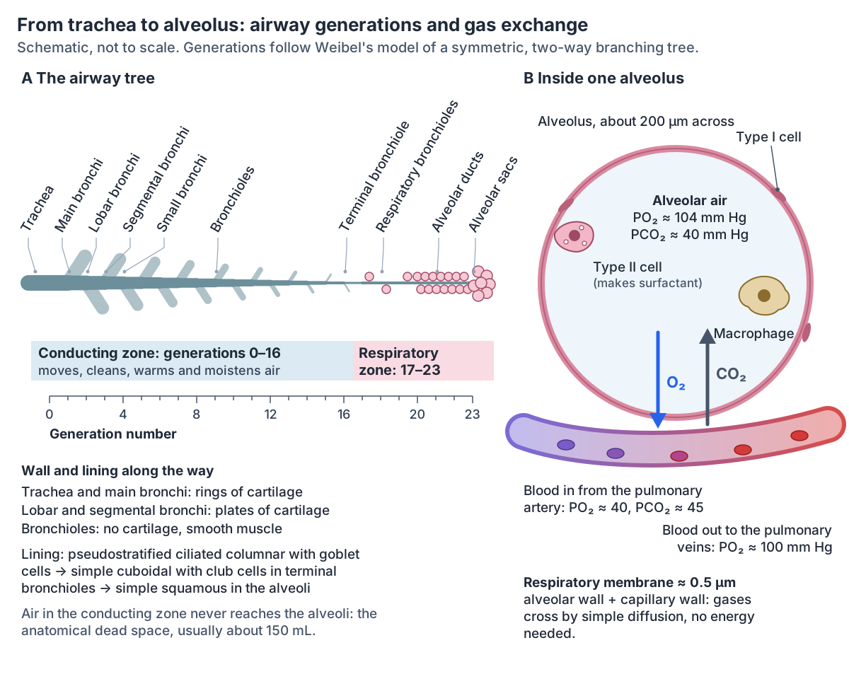
Airway generations from trachea to alveolus
| Generation | Airway | Wall | Zone |
|---|---|---|---|
| 0 | Trachea | C-shaped rings of hyaline cartilage, trachealis muscle | Conducting |
| 1 | Main (primary) bronchi | Rings of cartilage | Conducting |
| 2 | Lobar (secondary) bronchi: 3 right, 2 left | Plates of cartilage | Conducting |
| 3 | Segmental (tertiary) bronchi | Plates of cartilage | Conducting |
| 4 | Smaller bronchi | Plates of cartilage | Conducting |
| 5 to 15 | Bronchioles, about 1 mm across | No cartilage; smooth muscle | Conducting |
| 16 | Terminal bronchioles | Smooth muscle; club cells | Conducting (last generation) |
| 17 to 19 | Respiratory bronchioles | Alveoli start to bud from the wall | Respiratory |
| 20 to 23 | Alveolar ducts and alveolar sacs | Walls made of alveoli | Respiratory |
Generation numbers follow Weibel's symmetric model as given by Duke University and LITFL (16 conducting and 7 respiratory generations). Duke's notes describe about 16 generations to the terminal bronchioles and a further three to seven to the farthest alveoli, so real paths differ in length.
Nose and nasal cavity
The nose is the main entrance and exit of the respiratory system. Each nostril (naris) opens into the nasal cavity, which the nasal septum divides into left and right halves. The conchae on the side walls increase the surface area and disturb the airflow, so the air is cleaned and warmed, and olfactory epithelium for smell lies deep in the roof of the cavity. (OpenStax Anatomy and Physiology 2e, 22.1: Organs and Structures of the Respiratory System.)
The nasal lining is moist and warms and humidifies incoming air. Hairs and mucus in the front of the nose trap large particles, and the respiratory epithelium contains mucus-secreting goblet cells and cilia that sweep mucus and debris back towards the throat. (OpenStax Anatomy and Physiology 2e, 22.1: Organs and Structures of the Respiratory System.)
Kenhub describes the nasal cavity as opening in front through the two nares and behind through the two choanae, with the hard palate as its floor and three conchae (superior, middle and inferior) on each side wall that create turbulent airflow. (Kenhub: Respiratory system.)
The external nose, nasal cavity, septum and conchae in detail. Nose anatomy.
Paranasal sinuses
The four paranasal sinuses, the maxillary, frontal, sphenoidal and ethmoidal sinuses, are air-filled spaces connected to the nasal cavity. They are lined with respiratory epithelium that traps particles. (Kenhub: Respiratory system.)
OpenStax adds that the sinuses help warm and humidify the air and make the skull lighter. (OpenStax Anatomy and Physiology 2e, 22.1: Organs and Structures of the Respiratory System.)
Where each sinus lies and where it drains into the nose. Paranasal sinuses.
Pharynx: the throat
The pharynx is a muscular tube that runs from the base of the skull to the lower border of the cricoid cartilage at the level of the sixth cervical vertebra (C6). It has three parts: the nasopharynx behind the nose, the oropharynx behind the mouth and the laryngopharynx behind the larynx. (TeachMeAnatomy: The Pharynx.)
The nasopharynx serves only as an airway. The oropharynx carries both air and food. In the nasopharynx sits the pharyngeal tonsil (adenoid), which is large in children and tends to shrink with age; the palatine tonsils lie in the oropharynx and the lingual tonsil at the base of the tongue. (OpenStax Anatomy and Physiology 2e, 22.1: Organs and Structures of the Respiratory System.)
The lining changes along the way: respiratory epithelium in the nasopharynx, then non-keratinised stratified squamous epithelium in the oropharynx, which has to withstand food. The laryngopharynx divides into the larynx in front and the oesophagus behind. (Kenhub: Respiratory system.)
The three parts of the pharynx, the tonsils and swallowing. Throat anatomy.
Larynx: the voice box
The larynx lies in the front of the neck at the level of the third to sixth cervical vertebrae (C3 to C6). It begins at the laryngeal inlet and ends at the lower border of the cricoid cartilage, where it continues as the trachea. Its framework is nine cartilages: the unpaired cricoid, thyroid and epiglottis, and the paired arytenoid, corniculate and cuneiform cartilages. It conducts air into the lower respiratory tract, protects the airway by closing during swallowing, and produces sound. (Kenhub: Larynx.)
The thyroid cartilage is the largest. The epiglottis, a flexible flap of elastic cartilage attached to the thyroid cartilage, closes over the airway when you swallow. The glottis is made of the vestibular folds (false vocal cords), the true vocal cords and the space between them, and the free edges of the true vocal cords vibrate to make sound. (OpenStax Anatomy and Physiology 2e, 22.1: Organs and Structures of the Respiratory System.)
The larynx houses the vocal cords, and the epiglottis closes the laryngeal inlet during swallowing. Above the vocal cords the larynx is lined by stratified squamous epithelium and below them by pseudostratified ciliated columnar epithelium with goblet cells. (Kenhub: Respiratory system.)
The laryngeal cartilages, vocal folds and the muscles that move them. Larynx anatomy.
Trachea and bronchi: the lower airway
The trachea begins at the lower margin of the cricoid cartilage at C6 and is usually 10 to 13 cm long and 1.5 to 2 cm wide, wider in men. It is held open by 15 to 20 D-shaped rings of cartilage. It divides at the carina, at the level of T4 when lying down, shifting to T5 or even T6 when upright. (Radiopaedia: Trachea.)
LITFL gives a mean tracheal length of 11 cm and a mean diameter of 1.8 cm, inside Radiopaedia's ranges, and describes the trachea as D-shaped in cross-section. (LITFL Part One: Airway and Alveolar Anatomy.)
OpenStax counts 16 to 20 stacked, C-shaped pieces of hyaline cartilage. The cartilage stops the trachea from collapsing, and a fibroelastic membrane lets it stretch slightly. (OpenStax Anatomy and Physiology 2e, 22.1: Organs and Structures of the Respiratory System.)
The right main bronchus is about 2.5 cm long, shorter and more vertical than the left, which is about 5 cm long and more oblique, so inhaled material tends to enter the right side. Bronchi contain cartilage and bronchioles do not, which makes the smaller airways easier to compress. (OpenAnesthesia: Respiratory anatomy.)
The carina is the most sensitive area for triggering a cough. The trachea and bronchi are lined by ciliated pseudostratified columnar epithelium with goblet cells, which together form the mucociliary escalator that carries trapped particles up to the throat. (TeachMeAnatomy: The Tracheobronchial Tree.)
The windpipe, the carina and the main, lobar and segmental bronchi. Trachea and bronchial tree.
Lungs and pleura
The lungs are paired, pyramid-shaped organs joined to the trachea by the main bronchi, with the diaphragm beneath them. The right lung is shorter and wider and has three lobes; the left lung is smaller, has two lobes and carries a cardiac notch for the heart. (OpenStax Anatomy and Physiology 2e, 22.2: The Lungs.)
Each lung is wrapped in pleura. The visceral pleura covers the lung, the parietal pleura lines the chest wall, and a thin film of fluid between them lets the lung slide and holds it against the chest wall, so it expands when the chest expands. (TeachMeAnatomy: The Pleurae.)
Together the lungs contain about 1500 miles of airways and 300 to 500 million alveoli, with a combined surface area of about 70 square metres. Each day about 10,000 litres of air and 7,000 litres of blood pass through the lungs of a resting adult. (Radiopaedia: Lungs.)
The lobes, fissures, hilum, segments, pleura and blood supply of the lungs. Lung anatomy.
Alveoli and gas exchange
An alveolus is a tiny air sac, about 200 µm across, at the end of the airway. Its wall is mostly type I alveolar cells, thin squamous cells that let gases pass; type II cells secrete surfactant, which lowers surface tension; and alveolar macrophages remove debris and microbes. Type I cells make up as much as 97% of the alveolar surface. Alveolar pores connect neighbouring alveoli. The alveolar and capillary walls together form the respiratory membrane, only about 0.5 µm thick. (OpenStax Anatomy and Physiology 2e, 22.1: Organs and Structures of the Respiratory System.)
Duke University's notes give 200 to 500 million alveoli in adult lungs, with a mean diameter of 200 µm, and say type I cells cover about 95% of the alveolar surface. The capillary bed of the lungs is the largest in the body and receives the entire output of the heart. (Duke University Microscopic Anatomy: The Respiratory System (course notes, 2015).)
LITFL puts type I cells at about 90% and type II cells at about 10% of the alveolar surface. Because alveoli have no cilia, alveolar macrophages clear inhaled particles by engulfing them. (LITFL Part One: Airway and Alveolar Anatomy.)
OpenAnesthesia gives 300 to 480 million alveoli with 50 to 100 square metres of surface for gas exchange, and an air-blood barrier less than 1 µm thick. (OpenAnesthesia: Respiratory anatomy.)
Gases cross the respiratory membrane by simple diffusion, which needs no energy. Alveolar air has an oxygen partial pressure of about 104 mm Hg and carbon dioxide of about 40 mm Hg. Blood arriving from the pulmonary arteries has about 40 mm Hg of oxygen and 45 mm Hg of carbon dioxide, so oxygen moves into the blood and carbon dioxide out. Carbon dioxide's gradient is small, but it is about 20 times more soluble than oxygen. (OpenStax Anatomy and Physiology 2e, 22.4: Gas Exchange.)
Ventilation and blood flow are matched: arterioles serving poorly ventilated alveoli constrict and those serving well-ventilated alveoli dilate, and bronchioles widen when carbon dioxide in the alveoli rises. (OpenStax Anatomy and Physiology 2e, 22.4: Gas Exchange.)
How the right heart pumps blood to the lungs and the left heart receives it back. Blood flow through the heart.
Muscles of breathing: how breathing works
Air flows from higher to lower pressure. In quiet breathing in, the diaphragm and external intercostal muscles contract, the chest gets bigger, the pressure in the alveoli falls and air flows in. Quiet breathing out is passive, driven by elastic recoil. In forced breathing in, neck muscles such as the scalenes lift the chest wall; in forced breathing out, the abdominal muscles push the abdominal organs up against the diaphragm and the internal intercostals compress the rib cage. (OpenStax Anatomy and Physiology 2e, 22.3: The Process of Breathing.)
The diaphragm is a double-domed sheet of muscle and tendon that forms the floor of the chest. It attaches to the lumbar vertebrae and arcuate ligaments, the costal cartilages of ribs 7 to 10 and ribs 11 and 12, and the xiphoid process, and its fibres converge on a central tendon. It is supplied by the phrenic nerve from C3 to C5. When it contracts it flattens and makes the chest taller; the right dome sits slightly higher than the left. (TeachMeAnatomy: The Diaphragm.)
TeachMeAnatomy describes the external intercostals as stabilising the ribs in normal breathing and raising them only in forced inspiration, while the internal and innermost intercostals lower the ribs in forced expiration; all are supplied by the intercostal nerves T1 to T11. It lists pectoralis major and minor, serratus anterior and the scalenes as accessory muscles of respiration. So sources differ on whether the external intercostals work in every quiet breath. (TeachMeAnatomy: Muscles of the Thoracic Cage.)
Duke University's notes add that the lungs can hold up to about 7 litres of air at maximum inspiration and as little as about 1 litre after a forced breath out. (Duke University Microscopic Anatomy: The Respiratory System (course notes, 2015).)
The main muscle of breathing, its openings and its nerve supply. Diaphragm.
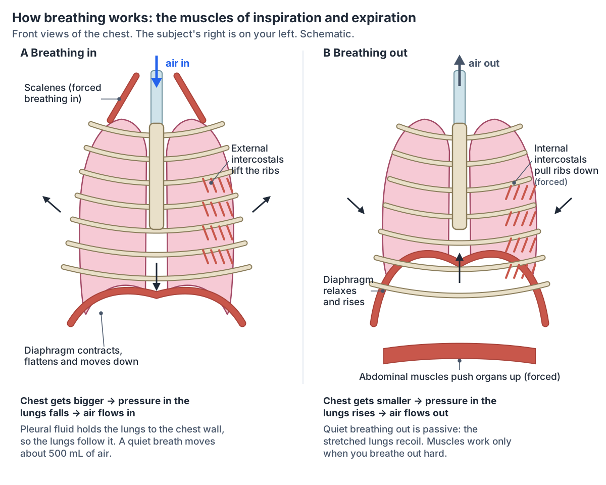
Muscles of breathing
| Muscle | When it works | What it does |
|---|---|---|
| Diaphragm | Every breath in, quiet or forced | Contracts and flattens, making the chest taller; relaxes back into a dome when you breathe out |
| External intercostals | Breathing in (quiet breathing in OpenStax; forced breathing in TeachMeAnatomy) | Raise the ribs and widen the chest |
| Scalenes | Forced breathing in | Lift the chest wall from the neck |
| Pectoralis major and minor, serratus anterior | Forced breathing in | Accessory muscles of respiration that attach to the chest wall |
| Internal and innermost intercostals | Forced breathing out | Pull the ribs down |
| Abdominal muscles, including the obliques | Forced breathing out | Push the abdominal organs up against the diaphragm |
| None | Quiet breathing out | Passive elastic recoil of the lungs and chest wall |
From OpenStax and TeachMeAnatomy. The diaphragm is supplied by the phrenic nerve (C3 to C5); the intercostal muscles by the intercostal nerves (T1 to T11).
Lung volumes and the control of breathing
A quiet breath, the tidal volume, moves about 500 mL of air. The vital capacity, the most air you can move in one breath, is 4000 to 5000 mL, and the total lung capacity is about 6000 mL in men and 4200 mL in women. OpenStax gives the expiratory reserve volume as up to 1200 mL in men, and a breathing rate of 12 to 18 breaths a minute in adolescents, similar to adults, against 30 to 60 in babies under one year. (OpenStax Anatomy and Physiology 2e, 22.3: The Process of Breathing.)
TeachMePhysiology gives similar average values: tidal volume 0.5 L, vital capacity 4.5 L and total lung capacity 6 L, with an inspiratory reserve of 2.5 L, an expiratory reserve of 1.5 L (a little more than OpenStax's figure) and a residual volume of 1.5 L, the air that stays in the lungs after the hardest breath out. (TeachMePhysiology: Lung Volumes and Capacities.)
Breathing is driven by the brainstem. In the medulla oblongata the ventral respiratory group sets the rhythm and the dorsal respiratory group integrates sensory input; centres in the pons adjust the depth and rate. Carbon dioxide, acting through the acidity of the blood and the fluid around the brain, is the main chemical stimulus; peripheral chemoreceptors also respond when blood oxygen falls to about 60 mm Hg or below. (OpenStax Anatomy and Physiology 2e, 22.3: The Process of Breathing.)
The ribs and sternum that move with every breath. Rib cage.
How the airway lining changes
From the larynx to the segmental bronchi the airway is lined by pseudostratified ciliated columnar epithelium with goblet cells. Smaller bronchi have simple columnar ciliated cells, the terminal bronchioles simple cuboidal cells with club cells (formerly Clara cells) that contribute to surfactant, and the alveoli simple squamous cells. (Kenhub: Respiratory system.)
Cartilage also fades out along the tree. The main bronchi have rings of cartilage, the lobar and segmental bronchi only crescent-shaped plates, and the bronchioles neither cartilage nor goblet cells. Club cells in the bronchioles secrete a surfactant-like lipoprotein that keeps the walls of the small airways from sticking together when you breathe out. (TeachMeAnatomy: The Tracheobronchial Tree.)
Common respiratory conditions by tract
Upper respiratory tract infections are usually caused by influenza viruses, rhinoviruses and streptococcus bacteria; they include the common cold, the most frequent of all, and rhinitis, sinusitis, pharyngitis, epiglottitis and laryngitis. Lower tract infections include bronchitis, bronchiolitis and pneumonia, which involves the alveoli. (Kenhub: Respiratory system.)
Other common conditions include asthma, chronic obstructive pulmonary disease (COPD), cystic fibrosis, interstitial lung disease and blockages by foreign objects or tumours. Pulse oximetry, lung function tests, chest X-rays and arterial blood gases are the usual tests. (Cleveland Clinic: Respiratory System.)
Respiratory system in the 3D model
- Arytenoid cartilage
- The arytenoid cartilages are a pair of pyramid-shaped cartilages at the back of the larynx.
- Cricoid cartilage
- The cricoid cartilage is a complete signet-ring-shaped cartilage forming the lower part of the larynx.
- Diaphragm
- The dome-shaped muscle separating the thoracic and abdominal cavities and the main muscle of inspiration.
- Epiglottis
- The epiglottis is a leaf-shaped elastic cartilage that covers the laryngeal inlet during swallowing.
- External intercostal muscle
- The outermost layer of muscles between the ribs.
- Inferior nasal concha
- A thin curved bone on the lateral wall of the nose.
- Inferior pharyngeal constrictor
- The lowest of the three constrictors of the pharynx.
- Internal intercostal muscle
- The middle layer of intercostal muscles.
- Lateral nasal cartilage
- The lateral nasal cartilages are paired triangular cartilages forming the upper lateral part of the external nose.
- Lower lobe of left lung
- The lower lobe of the left lung lies below and behind the oblique fissure. It forms the back and the base of the left lung, resting on the left dome of the diaphragm.
- Lower lobe of right lung
- The lower lobe of the right lung lies below and behind the oblique fissure. It forms the back and the base of the right lung, resting on the right dome of the diaphragm above the liver.
- Main bronchus
- The left main (primary) bronchus, one of the two airways formed by the division of the trachea.
- Main bronchus proper
- The right main (primary) bronchus, the extrapulmonary airway from the carina to the hilum of the right lung.
- Major alar cartilage
- The major alar cartilage is a U-shaped cartilage forming the tip and sides of the nostril.
- Middle lobe of right lung
- The middle lobe is the wedge of the right lung that lies between the horizontal and oblique fissures, at the front and side of the lower chest. Only the right lung has one.
- Middle pharyngeal constrictor
- The middle of the three pharyngeal constrictors.
- Nasal bone
- A small, paired rectangular bone forming the bridge of the nose.
- Scalenus anterior
- A deep neck muscle and an important landmark in the root of the neck.
- Scalenus medius
- The largest and longest of the scalene muscles.
- Scalenus posterior
- The smallest and deepest of the scalene muscles.
- Septal nasal cartilage
- The septal nasal cartilage is the cartilage that forms the front part of the nasal septum.
- Soft palate
- The soft palate is the muscular back part of the roof of the mouth. Its free back edge carries the uvula.
- Superior pharyngeal constrictor
- The uppermost of the three constrictors forming the pharyngeal wall.
- Thyroid cartilage
- The thyroid cartilage is the largest cartilage of the larynx, forming the laryngeal prominence.
- Trachea
- The trachea (windpipe) is a flexible air tube extending from the larynx to the bronchi.
- Upper lobe of left lung
- The upper lobe of the left lung is one of the left lung's two lobes. It lies above and in front of the oblique fissure, forms the apex of the lung and includes the lingula.
- Upper lobe of right lung
- The upper lobe of the right lung is the top of the right lung's three lobes. It lies above the horizontal fissure and above the upper part of the oblique fissure, and forms the apex of the lung.
Frequently asked questions
What are the main parts of the respiratory system?
The nose and nasal cavity, the paranasal sinuses, the pharynx (throat), the larynx (voice box), the trachea (windpipe), the bronchi and bronchioles, and the lungs with their alveoli. The diaphragm and the intercostal muscles move air in and out, and the pleura around each lung lets it slide against the chest wall.
What is the difference between the upper and lower respiratory tract?
The upper tract is the nose, nasal cavity, sinuses, pharynx and larynx; it filters, warms and moistens air. The lower tract is the trachea, bronchi, bronchioles and lungs, where air is conducted to the alveoli for gas exchange. Sources disagree on the larynx: some put all of it in the upper tract, others split it at the vocal cords.
What is the conducting zone of the respiratory system?
The conducting zone is every airway that carries air but does not exchange gases: the nose, pharynx, larynx, trachea, bronchi and bronchioles down to the terminal bronchioles, the first 16 airway generations. Its air, about 150 mL, is called anatomical dead space. Gas exchange starts in the respiratory zone, at the respiratory bronchioles.
How many times does the airway branch?
In Weibel's model the airway divides 23 times from the trachea (generation 0) to the alveolar sacs (generation 23). Generations 0 to 16 conduct air and generations 17 to 23 take part in gas exchange. Real branching is uneven, so the number of generations differs from one path to another.
Where does gas exchange take place?
In the alveoli and the respiratory bronchioles, across the respiratory membrane between alveolar air and the pulmonary capillaries. The membrane is only about 0.5 µm thick. Oxygen diffuses from alveolar air (about 104 mm Hg) into blood arriving at about 40 mm Hg, and carbon dioxide diffuses the other way.
What is the main muscle of breathing?
The diaphragm. When it contracts it flattens and makes the chest taller, so the pressure in the lungs falls and air flows in. It is supplied by the phrenic nerve from C3 to C5. The external intercostals help to raise the ribs, and in forced breathing the scalenes and other accessory muscles join in.
How many alveoli are in the lungs?
Several hundred million. Radiopaedia gives 300 to 500 million, Duke University's notes 200 to 500 million and OpenAnesthesia 300 to 480 million. Together they give a surface for gas exchange of about 50 to 100 square metres, often compared to half a tennis court. Each alveolus is about 200 µm across.
References
- Betts JG, et al. Anatomy and Physiology 2e, §22.1 Organs and Structures of the Respiratory System. OpenStax, Rice University, 2022.
- Betts JG, et al. Anatomy and Physiology 2e, §22.2 The Lungs. OpenStax, Rice University, 2022.
- Betts JG, et al. Anatomy and Physiology 2e, §22.3 The Process of Breathing. OpenStax, Rice University, 2022.
- Betts JG, et al. Anatomy and Physiology 2e, §22.4 Gas Exchange. OpenStax, Rice University, 2022.
- Cleveland Clinic. Respiratory System: Functions, Facts and Organs.
- Cleveland Clinic. Lungs: Function, Location and Anatomy.
- Kenhub. Respiratory system: Anatomy and functions.
- Kenhub. Lung: Anatomy, blood supply, innervation, functions.
- Kenhub. Larynx.
- National Cancer Institute SEER Training Modules. Conducting Passages.
- LITFL Part One. Airway and Alveolar Anatomy.
- OpenAnesthesia. Respiratory anatomy.
- Duke University. Microscopic Anatomy: The Respiratory System (course notes), 2015.
- TeachMePhysiology. Lung Volumes and Capacities.
- TeachMeAnatomy. The Pharynx.
- TeachMeAnatomy. The Tracheobronchial Tree.
- TeachMeAnatomy. The Pleurae.
- TeachMeAnatomy. The Diaphragm.
- TeachMeAnatomy. Muscles of the Thoracic Cage.
- Radiopaedia. Trachea (reference article).
- Radiopaedia. Lungs (reference article).
- Mitsuhashi N. et al. (2009). BodyParts3D: 3D structure database for anatomical concepts. Nucleic Acids Research 37, D782–D785. Data © The Database Center for Life Science, CC BY 4.0.
- Browne K., Schlehlein H. (2026). 3D Reference Organ Set for Male, v1.10 (Visible Human Male). Human Reference Atlas 3D Reference Object Library, HuBMAP / NIH. CC BY 4.0.
- Radiopaedia. Oblique fissure (surface anatomy used to place the oblique fissures in the 3D lungs).
- Radiopaedia. Horizontal fissure (surface anatomy used to place the horizontal fissure in the 3D right lung).
Related anatomy
- LungsLung anatomy with labelled diagrams and a 3D model of all five lobes: fissures, hilum, segments, pleura, blood supply…
- TracheaTrachea anatomy explained: where the windpipe runs, C-shaped rings, the carina, right vs left main bronchi and the…
- LarynxLarynx anatomy with 3D model and diagrams: thyroid, cricoid, arytenoid and epiglottis cartilages, vocal folds…
- Nose anatomyNose anatomy with a 3D model and labelled diagrams: nasal bones and cartilages, nostrils, nasal cavity, conchae…
- Throat anatomyThroat anatomy with a 3D model and labelled diagrams: the three parts of the pharynx, constrictor muscles, tonsils…
- DiaphragmDiaphragm muscle anatomy with a labelled diagram and 3D model: attachments, crura, central tendon, openings at T8, T10…
Explore the whole body in the interactive 3D human anatomy model, or browse the A–Z list of human structures.