

What is the back?
The back (dorsal trunk) is the posterior surface of the trunk, running from the lower edge of the neck down to the gluteal region. The upper back is the posterior wall of the thorax, and the lower back is the lumbar region, behind the abdominal cavity. (Kenhub: Back: Anatomy, parts, diagrams and function.)
From the surface inwards it is built of skin, subcutaneous tissue, the superficial and deep back muscles, the posterior parts of the ribs and the vertebral column with the spinal cord and its meninges. Its jobs are to protect the spinal cord, hold the head and trunk upright, carry the weight of the upper body and help move the arms and legs. (Kenhub: Back: Anatomy, parts, diagrams and function.)
Regions of the back
Kenhub divides the surface of the back into vertebral, scapular, interscapular, suprascapular, infrascapular, lumbar and sacral regions. The area beside the scapula belongs to the lateral thoracic region of the chest wall, and the posterior trunk as a whole is divided into the back and the buttocks. (Kenhub: Back: Anatomy, parts, diagrams and function.)
The shoulder blade lies on the back but is a bone of the upper limb: Kenhub's guide to the upper limb notes that the scapula is classified with the limb, not with the back. (Kenhub: Regions of the upper limb.)
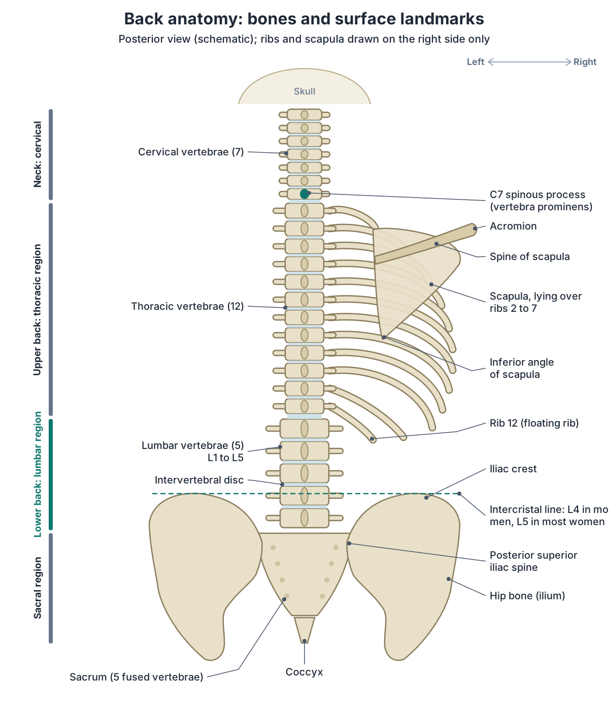
Regions of the back
| Region | Where it is |
|---|---|
| Vertebral region | Over the thoracic and lumbar vertebral column, in the midline |
| Scapular region | Over the scapula |
| Interscapular region | Between the medial borders of the two scapulae |
| Suprascapular region | Above and medial to the spine of the scapula |
| Infrascapular region | Below and medial to the scapular region |
| Lumbar region (lower back) | On either side of the lumbar vertebrae |
| Sacral region | Over the sacrum |
Source: Kenhub, Back: Anatomy, parts, diagrams and function.
The spine: the backbone of the back
The adult vertebral column (spine) has 24 vertebrae plus the sacrum and coccyx: 7 cervical, 12 thoracic and 5 lumbar vertebrae, a sacrum made of 5 fused vertebrae and a coccyx made of 4. It starts as 33 vertebrae, and the sacral and coccygeal ones fuse from after the age of 20 into middle age. (OpenStax Anatomy and Physiology 2e, 7.3: The Vertebral Column.)
Kenhub gives the coccyx 3 to 5 fused vertebrae, so the total count varies slightly between books. It describes four curvatures: cervical lordosis, thoracic kyphosis, lumbar lordosis and sacral kyphosis. A lordosis is concave towards the back and a kyphosis is concave towards the front. (Kenhub: Anatomy of the back: Spine and back muscles.)
The thoracic and sacrococcygeal curves are primary curves kept from the fetus. The cervical and lumbar curves are secondary curves that appear after birth as a child learns to hold up the head, sit, stand and walk. Each typical vertebra has a weight-bearing body in front and a vertebral arch of pedicles and laminae behind; together the vertebral foramina form the vertebral canal for the spinal cord, and each spinal nerve leaves through an intervertebral foramen between two vertebrae. (OpenStax Anatomy and Physiology 2e, 7.3: The Vertebral Column.)
Vertebrae, regions, curves, discs and ligaments of the vertebral column. Spine anatomy.
Regions of the vertebral column under the back
| Region | Vertebrae | Key features | Part of the back |
|---|---|---|---|
| Cervical | 7 (C1–C7) | Small bodies, bifid spinous processes, transverse foramina; C7 spinous process easy to feel | Back of the neck |
| Thoracic | 12 (T1–T12) | Long, downward-pointing spinous processes; facets for the ribs | Upper and middle back |
| Lumbar | 5 (L1–L5) | Largest, thickest bodies; short, blunt spinous processes | Lower back |
| Sacral | 5 fused into the sacrum | Sacral canal and foramina; sacroiliac joints with the hip bones | Base of the back, between the hip bones |
| Coccygeal | 4 fused into the coccyx (3–5 in some sources) | Small fused segments below the sacrum | Below the sacrum |
Sources: OpenStax Anatomy and Physiology 2e, 7.3; Kenhub.
Discs, facet joints and ligaments of the back
Each intervertebral disc has a tough outer ring, the anulus fibrosus, around a gel-like centre, the nucleus pulposus, whose high water content resists compression and falls with age. In a herniated disc, the nucleus pushes backwards through a weakened anulus; this happens most often at the L4/L5 and L5/S1 discs. (OpenStax Anatomy and Physiology 2e, 7.3: The Vertebral Column.)
Vertebrae are joined by the discs between their bodies and by facet (zygapophyseal) joints between their articular processes. The sacroiliac joints connect the spine to the pelvis, and costovertebral joints connect the ribs to the thoracic vertebrae. (Kenhub: Anatomy of the back: Spine and back muscles.)
Ligaments bind the column together. The anterior longitudinal ligament runs down the front of the vertebral bodies and resists bending backwards; the posterior longitudinal ligament lies on their back surface, in front of the spinal cord; the elastic ligamenta flava join the laminae behind the cord; and the supraspinous ligament joins the spinous processes and continues in the neck as the nuchal ligament. (OpenStax Anatomy and Physiology 2e, 7.3: The Vertebral Column.)
Sacrum and coccyx: the base of the back
The sacrum forms from five fused sacral vertebrae and contains the sacral canal, the lower end of the vertebral canal, with sacral foramina on each side. The coccyx below it forms from fused coccygeal vertebrae. (OpenStax Anatomy and Physiology 2e, 7.3: The Vertebral Column.)
The sacroiliac joints between the sacrum and the hip bones connect the vertebral column to the pelvis. On the surface, the sacral region of the back lies over the sacrum, just above the buttocks. (Kenhub: Anatomy of the back: Spine and back muscles.)
Parts, foramina, sacroiliac joints and sacrum vs coccyx. Sacrum.
Ribs and shoulder blades in the upper back
The thoracic cage has 12 pairs of ribs, and all of them articulate behind with the thoracic vertebrae T1 to T12. The head of each rib meets the body of its vertebra and the tubercle meets the transverse process. Ribs 11 and 12 are floating ribs with no attachment in front, and the angles of the ribs form the most posterior part of the thoracic cage. (OpenStax Anatomy and Physiology 2e, 7.4: The Thoracic Cage.)
Most rib heads touch two vertebral bodies and the disc between them: ribs 2 to 9 do this, while ribs 1, 10, 11 and 12 meet a single vertebra. Costotransverse joints between rib necks and tubercles and the transverse processes are found from T1 to T10. (Kenhub: Anatomy of the back: Spine and back muscles.)
The scapula lies over the posterior surface of ribs two to seven. Its acromion, a lateral extension of the spine of the scapula, can be felt at the top of the shoulder. (Kenhub: Scapula.)
True, false and floating ribs, rib joints and the sternum. Rib cage.
Back muscles: extrinsic and intrinsic layers
The back muscles form two groups. The extrinsic (superficial) muscles attach the spine to the shoulder girdle and arm, so they move the shoulder, and some help breathing. The intrinsic (deep) muscles act on the joints of the vertebral column, are all supplied by posterior rami of spinal nerves, and are separated from the extrinsic group by the thoracolumbar fascia. (Kenhub: Anatomy of the back: Spine and back muscles.)
Kenhub splits the extrinsic muscles into a superficial and an intermediate layer, and the intrinsic muscles, which run from the skull to the pelvis beside the vertebral column, into superficial, intermediate, deep and deepest layers. The intrinsic group keeps the posture of the trunk and moves the vertebral column. (Kenhub: Back: Anatomy, parts, diagrams and function.)
The erector spinae group forms most of the muscle mass of the back. It extends the vertebral column, controls bending and twisting, and maintains the lumbar curve. (OpenStax Anatomy and Physiology 2e, 11.3: Axial Muscles of the Head, Neck, and Back.)
Every back muscle with origin, insertion, nerve and action. Back muscles.
Layers of the back muscles
| Layer | Muscles | Nerve supply | Main job |
|---|---|---|---|
| Extrinsic, superficial | Trapezius, latissimus dorsi, levator scapulae, rhomboid major and minor | Accessory nerve (trapezius), thoracodorsal nerve (latissimus dorsi), dorsal scapular nerve (levator scapulae, rhomboids) | Move the scapula and arm |
| Extrinsic, intermediate | Serratus posterior superior and inferior | Intercostal nerves | Move the ribs; slight role in breathing |
| Intrinsic, superficial | Splenius capitis and cervicis | Posterior rami of spinal nerves | Extend and rotate the head and neck |
| Intrinsic, intermediate | Erector spinae: iliocostalis, longissimus, spinalis | Posterior rami of spinal nerves | Extend and laterally flex the spine; maintain the lumbar curve |
| Intrinsic, deep | Transversospinales: semispinalis, multifidus, rotatores | Posterior rami of spinal nerves | Extend and rotate the spine; stabilise it |
| Intrinsic, deepest | Interspinales, intertransversarii | Posterior rami of spinal nerves | Stabilise neighbouring vertebrae |
Sources: Kenhub; TeachMeAnatomy; OpenStax Anatomy and Physiology 2e, 11.3.
Trapezius, latissimus dorsi and the other superficial muscles
Trapezius arises from the skull, the ligamentum nuchae and the spinous processes of C7 to T12 and inserts on the clavicle, acromion and spine of the scapula. The accessory nerve (cranial nerve XI) supplies it. Its upper fibres elevate and rotate the scapula, its middle fibres pull it back and its lower fibres pull it down. (TeachMeAnatomy: The Superficial Back Muscles.)
Latissimus dorsi arises from the spinous processes of T6 to T12, the thoracolumbar fascia, the iliac crest and the lowest three ribs and inserts into the intertubercular sulcus of the humerus. The thoracodorsal nerve supplies it, and it extends, adducts and medially rotates the arm. Levator scapulae elevates the scapula and the rhomboids retract and rotate it; all three are supplied by the dorsal scapular nerve. (TeachMeAnatomy: The Superficial Back Muscles.)
The intermediate layer is two thin muscles, serratus posterior superior and inferior, which attach the spine to the upper and lower ribs. They are supplied by intercostal nerves and are thought to have only a slight role in breathing. (TeachMeAnatomy: The Intermediate Back Muscles.)
Descending, transverse and ascending parts, accessory nerve and actions. Trapezius anatomy.
Erector spinae and the deep back muscles
The deep muscles of the back run from the sacrum to the base of the skull. In the intermediate layer, iliocostalis, longissimus and spinalis form a column on each side, the erector spinae, which shares a common tendinous origin from the lower thoracic and lumbar vertebrae, the sacrum, the back of the iliac crest and the sacroiliac and supraspinous ligaments. (TeachMeAnatomy: The Deep (Intrinsic) Back Muscles.)
Iliocostalis is the lateral column, longissimus the intermediate one and spinalis the medial one. Higher up, the splenius muscles run from the midline upwards and outwards to extend, bend and rotate the head. Deeper still, the transversospinales run from transverse processes to spinous processes; in the lumbar region multifidus helps extend and bend the spine sideways, and small segmental muscles between neighbouring vertebrae help stabilise it. (OpenStax Anatomy and Physiology 2e, 11.3: Axial Muscles of the Head, Neck, and Back.)
Of the transversospinales, semispinalis is the most superficial, multifidus lies beneath it and mainly stabilises the vertebral column, and the rotatores are the deepest, most prominent in the thoracic region and thought to have a proprioceptive role. (TeachMeAnatomy: The Deep (Intrinsic) Back Muscles.)
The superficial intrinsic back muscle of the neck and head. Splenius capitis.
Spinal cord and spinal nerves in the back
The spinal cord does not run the whole length of the back. Its tapered end, the conus medullaris, lies most often at the level of the L1/L2 disc in children and adults, with a reported range from mid-T11 to mid-L3. Below it, the lumbar and sacral nerve roots continue down the vertebral canal as the cauda equina, and a thin fibrous strand, the filum terminale, runs on to the sacrum. (Radiopaedia: Conus medullaris.)
There are 31 pairs of spinal nerves: 8 cervical, 12 thoracic, 5 lumbar, 5 sacral and 1 coccygeal. Each carries both sensory and motor fibres. The thoracic nerves T2 to T11 do not join a plexus but run between the ribs as the intercostal nerves. (OpenStax Anatomy and Physiology 2e, 13.4: The Peripheral Nervous System.)
Spinal nerves leave the vertebral canal through the intervertebral foramina. Their posterior rami supply the skin of the back, the spine itself and the intrinsic back muscles, while the extrinsic muscles are supplied by the plexuses, the accessory nerve and the intercostal and subcostal nerves. Spinal nerves also form four plexuses: cervical (C1 to C4), brachial (C5 to T1), lumbar (L1 to L4) and sacral (L4 to S4). (Kenhub: Anatomy of the back: Spine and back muscles.)
Regions, conus medullaris, cauda equina, tracts and spinal nerves. Spinal cord.
Lower back anatomy: lumbar vertebrae and discs
The lower back is the lumbar region of the back, the part that lies behind the abdominal cavity, between the thorax above and the sacral region and buttocks below. On the surface, the lumbar regions lie on either side of the lumbar vertebrae. (Kenhub: Back: Anatomy, parts, diagrams and function.)
Its skeleton is the five lumbar vertebrae, L1 to L5, which have the largest and thickest bodies of the whole spine and short, blunt spinous processes. The discs between them, especially L4/L5 and L5/S1, are the commonest sites of disc herniation. (OpenStax Anatomy and Physiology 2e, 7.3: The Vertebral Column.)
Kenhub links the shape of lumbar vertebrae to their load: they have the largest bodies and a relatively narrow vertebral canal, whereas cervical vertebrae have the smallest bodies and the widest canal. (Kenhub: Anatomy of the back: Spine and back muscles.)
The lowest lumbar vertebra and the lumbosacral junction. L5 vertebra.
Muscles and fascia of the lower back
In the lower back, the deep muscles are wrapped by the thoracolumbar fascia, a girdle of aponeurotic and fascial layers that separates the paraspinal muscles from the muscles of the posterior abdominal wall. Its superficial layer is made largely of the aponeuroses of latissimus dorsi and serratus posterior inferior, and at the base of the spine its layers merge and attach firmly to the posterior superior iliac spine and the sacrotuberous ligament, where gluteus maximus also attaches. It plays a part in posture, load transfer and breathing. (Willard FH et al. The thoracolumbar fascia: anatomy, function and clinical considerations. J Anat. 2012.)
Quadratus lumborum is a muscle of the posterior abdominal wall. It runs from the iliac crest and iliolumbar ligament to the transverse processes of L1 to L4 and the lower border of the 12th rib, is supplied by the anterior rami of T12 to L4 and extends and laterally flexes the vertebral column. (TeachMeAnatomy: Quadratus Lumborum.)
Kenhub calls quadratus lumborum the deepest muscle of the posterior abdominal wall and notes that its overuse and strain are among the major causes of chronic lower back pain. Contracting both sides together extends the lumbar spine. (Kenhub: Quadratus lumborum muscle.)
The lumbar plexus forms within the substance of psoas major, in front of the lumbar transverse processes, from the anterior rami of L1 to L4 with a contribution from T12. (TeachMeAnatomy: The Lumbar Plexus.)
The hip flexor beside the lumbar spine that contains the lumbar plexus. Psoas major.
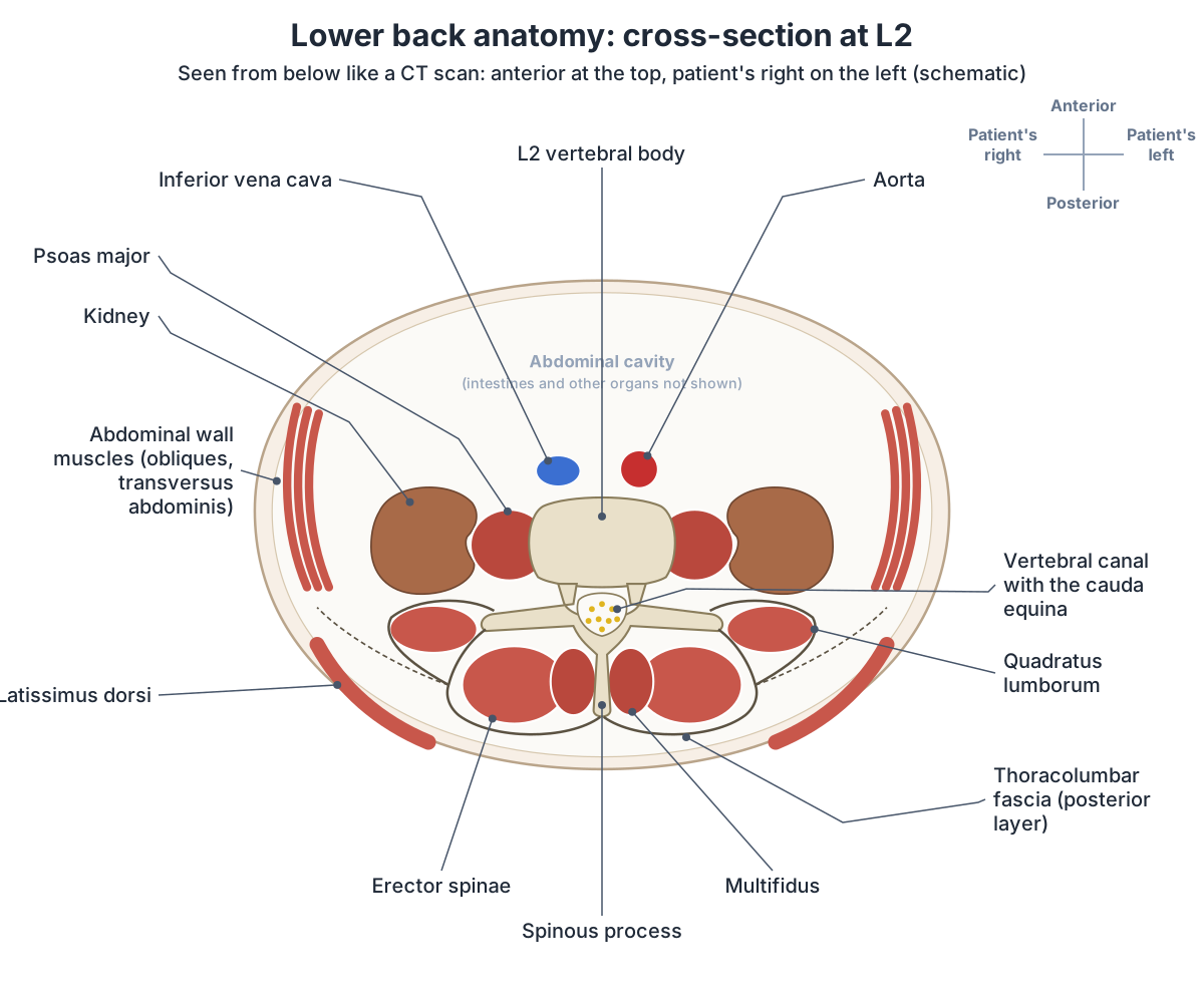
What organs are in the lower back? The kidneys
The kidneys lie behind the peritoneum (retroperitoneally) on either side of the vertebral column, typically from T12 to L3, with the right kidney often slightly lower because of the liver. Behind them lie the diaphragm, the 11th and 12th ribs (only the 12th on the right), psoas major, quadratus lumborum and transversus abdominis, with the subcostal, iliohypogastric and ilioinguinal nerves crossing that wall. (TeachMeAnatomy: The Kidneys.)
Where the kidneys are, their coverings, relations and inner anatomy. Kidney.
Blood supply of the back
The back is supplied by posterior branches of the vertebral, ascending cervical, deep cervical, occipital, posterior intercostal, subcostal, lumbar and lateral sacral arteries. Its veins drain into the internal and external vertebral venous plexuses, which empty into the vertebral and segmental veins, the azygos system and the inferior vena cava. (Kenhub: Anatomy of the back: Spine and back muscles.)
Kenhub names the aorta as the main arterial supply to much of the posterior thorax and abdomen, with the subclavian arteries supplying the neck and scapular regions. (Kenhub: Back: Anatomy, parts, diagrams and function.)
Thoracic and abdominal aorta and their intercostal and lumbar branches. Aorta.
Surface landmarks of the back
The spinous process of C7 is the prominent bump at the base of the neck, found by running a finger down the midline of the back of the neck. (OpenStax Anatomy and Physiology 2e, 7.3: The Vertebral Column.)
A horizontal line across the highest points of both iliac crests, the intercristal (Tuffier's or Jacoby's) line, is used to number the lumbar vertebrae. On radiographs it most often crosses the body of L4 or its lower endplate in men and the body of L5 or its upper endplate in women; across the population it falls anywhere between these levels. (Radiopaedia: Intercristal line.)
The triangle of auscultation is a small gap bounded by trapezius, latissimus dorsi and rhomboid major (or the medial border of the scapula). Lower down, the superior lumbar triangle lies between the 12th rib, internal oblique and quadratus lumborum, and the inferior lumbar triangle (of Petit) between the iliac crest, latissimus dorsi and external oblique. (Kenhub: Back: Anatomy, parts, diagrams and function.)
C1 atlas, C2 axis and C7, the vertebra prominens. Cervical vertebrae.
Female back anatomy: what is different?
Women and men have the same back bones, muscles and nerves, but the lower curve differs in detail. A CT study of 158 adults (77 women) found that the female lumbar curve has a different shape, with its upper part positioned further back, yet the overall amount of inward curving (lordosis) was similar in both sexes. (Hay O et al. The Lumbar Lordosis in Males and Females, Revisited. PLoS One. 2015.)
An MRI study of 40 girls and 40 boys aged 9 to 13 found a greater lumbar lordosis angle in the girls, whose lumbar vertebrae had about 15.5% smaller cross-sectional area. Smaller vertebrae accounted for the sex difference, and the authors note it is unknown whether the result applies to adults. (Wren TAL et al. Association between vertebral cross-sectional area and lumbar lordosis angle in adolescents. PLoS One. 2017.)
A 2007 study in Nature by Whitcome, Shapiro and Lieberman reported that the female lumbar curve spans three vertebrae against two in men, and that female lumbar joints are relatively larger. This lets a pregnant woman increase her lordosis to keep her balance; in 19 pregnant women, lordosis increased by up to 60% by term. (Harvard Gazette: Female lower back has evolved to accommodate strain of pregnancy (2007).)
Use the Male / Female switch in the 3D viewer to compare. The female body shares the skeleton and back muscles of the male model; its pelvis, sacrum and coccyx come from the Human Reference Atlas female model.
Hip bones, sacrum and the differences between the male and female pelvis. Pelvis.
Why the lower back is prone to pain
The World Health Organization defines low back pain as pain between the lower edge of the ribs and the buttock. In 2020 it affected 619 million people worldwide, and it is the single leading cause of disability in the world. About 90% of cases are classed as non-specific low back pain. (World Health Organization: Low back pain fact sheet.)
The lower back is also where the nerve roots of the sciatic nerve (L4 to S3) leave the spine, so the anatomy of this region matters for the whole leg. The sciatic nerve is the largest nerve in the body. (TeachMeAnatomy: The Sciatic Nerve.)
Lumbar and sacral plexuses, sciatic nerve and cluneal nerves. Nerves of the leg.
Exploring back anatomy in the 3D model
The 3D view opens from behind on the BodyParts3D adult male. It highlights the twelve thoracic and five lumbar vertebrae, the two lowest lumbar discs and the sacrum; the three parts of trapezius, the rhomboids, levator scapulae and serratus posterior inferior; and the erector spinae columns (iliocostalis, longissimus and spinalis). The model does not contain latissimus dorsi, multifidus, quadratus lumborum, the thoracolumbar fascia, the spinal cord or spinal nerves, and the male body has no coccyx. Switch on the urinary system to see the kidneys in front of the lower ribs.
To find your way, start at the C7 spinous process at the base of the neck and count down the spinous processes; in the lumbar region they are short and blunt. (OpenStax Anatomy and Physiology 2e, 7.3: The Vertebral Column.)
Back anatomy in the 3D model
- Ascending part of trapezius
- The lower fibres of trapezius, running upward and laterally to the scapular spine.
- Descending part of trapezius
- The upper fibres of trapezius, running down from the skull and neck.
- Eighth thoracic vertebra
- The eighth of the twelve thoracic vertebrae, labelled T8, which articulate with ribs.
- Eleventh thoracic vertebra
- The eleventh of the twelve thoracic vertebrae, labelled T11, which articulate with ribs.
- Fifth lumbar vertebra
- The fifth of the five lumbar vertebrae, labelled L5, in the lower back.
- Fifth thoracic vertebra
- The fifth of the twelve thoracic vertebrae, labelled T5, which articulate with ribs.
- First lumbar vertebra
- The first of the five lumbar vertebrae, labelled L1, in the lower back.
- First thoracic vertebra
- The first of the twelve thoracic vertebrae, labelled T1, which articulate with ribs.
- Fourth lumbar vertebra
- The fourth of the five lumbar vertebrae, labelled L4, in the lower back.
- Fourth thoracic vertebra
- The fourth of the twelve thoracic vertebrae, labelled T4, which articulate with ribs.
- Iliocostalis lumborum
- The lumbar part of the iliocostalis column of erector spinae.
- Iliocostalis thoracis
- The thoracic part of the iliocostalis column of erector spinae.
- Intervertebral disk of fifth lumbar vertebra
- The intervertebral disc between L5 and S1 (the L5–S1 disc), a fibrocartilage pad with a gel-like nucleus pulposus inside a ring-like anulus fibrosus.
- Intervertebral disk of fourth lumbar vertebra
- The intervertebral disc between L4 and L5 (the L4–L5 disc), a fibrocartilage pad with a gel-like nucleus pulposus inside a ring-like anulus fibrosus.
- Levator scapulae
- A muscle of the posterolateral neck that raises the scapula.
- Longissimus thoracis
- The largest part of the longissimus column of erector spinae.
- Ninth thoracic vertebra
- The ninth of the twelve thoracic vertebrae, labelled T9, which articulate with ribs.
- Rhomboid major
- A flat muscle deep to trapezius that connects the spine to the scapula.
- Rhomboid minor
- A small muscle lying above rhomboid major.
- Sacrum
- A triangular bone of five fused sacral vertebrae.
- Second lumbar vertebra
- The second of the five lumbar vertebrae, labelled L2, in the lower back.
- Second thoracic vertebra
- The second of the twelve thoracic vertebrae, labelled T2, which articulate with ribs.
- Serratus posterior inferior
- A thin muscle of the lower back deep to latissimus dorsi.
- Seventh thoracic vertebra
- The seventh of the twelve thoracic vertebrae, labelled T7, which articulate with ribs.
- Sixth thoracic vertebra
- The sixth of the twelve thoracic vertebrae, labelled T6, which articulate with ribs.
- Spinalis thoracis
- The thoracic part of spinalis, the medial column of the erector spinae.
- Tenth thoracic vertebra
- The tenth of the twelve thoracic vertebrae, labelled T10, which articulate with ribs.
- Third lumbar vertebra
- The third of the five lumbar vertebrae, labelled L3, in the lower back.
- Third thoracic vertebra
- The third of the twelve thoracic vertebrae, labelled T3, which articulate with ribs.
- Transverse part of trapezius
- The middle fibers of trapezius, running horizontally.
- Twelfth thoracic vertebra
- The twelfth of the twelve thoracic vertebrae, labelled T12, which articulate with ribs.
Frequently asked questions
What are the main parts of the back?
The back runs from the base of the neck to the buttocks. The upper back is the posterior wall of the chest, with the thoracic vertebrae, ribs and shoulder blades; the lower back is the lumbar region behind the abdomen, with the five lumbar vertebrae. Below them the sacrum joins the spine to the pelvis. Muscles in several layers cover the spine on each side.
How many bones are in the back?
The adult spine has 24 separate vertebrae (7 cervical, 12 thoracic and 5 lumbar) plus the sacrum and the coccyx, which form from fused vertebrae. The back also contains the posterior parts of the 12 pairs of ribs, which all articulate with the thoracic vertebrae, and the two shoulder blades, which belong to the upper limb skeleton.
What is the lower back called in anatomy?
The lower back is the lumbar region. Its skeleton is the five lumbar vertebrae, L1 to L5, which have the largest vertebral bodies in the spine. The lumbar spine curves inwards (lumbar lordosis), and the discs at L4/L5 and L5/S1 at its lower end are the commonest sites of disc herniation.
What organs are in the lower back?
The kidneys. They lie behind the peritoneum on either side of the spine, from about T12 to L3, with the right kidney slightly lower because of the liver. Behind them are the diaphragm, the lowest ribs, psoas major and quadratus lumborum.
What are the main muscles of the back?
Superficially, trapezius, latissimus dorsi, the rhomboids and levator scapulae, which move the shoulder and arm. Deep to the thoracolumbar fascia are the intrinsic muscles that move the spine: splenius in the neck, the erector spinae (iliocostalis, longissimus and spinalis), which form most of the back's muscle mass, and the deep transversospinales such as multifidus.
Where does the spinal cord end?
Most often at the level of the disc between the first and second lumbar vertebrae (L1/L2), with a normal range from mid-T11 to mid-L3. Below that the vertebral canal contains the cauda equina, a bundle of lumbar and sacral nerve roots, and the thin filum terminale, which anchors the cord to the sacrum.
Is female back anatomy different from male?
The bones, muscles and nerves are the same. Studies differ on the lumbar curve: one CT study found a different curve shape in women but a similar amount of lordosis, while an MRI study of children found a greater lordosis angle in girls linked to smaller vertebrae. Research on pregnancy suggests the female lumbar curve spans three vertebrae rather than two.
References
- Kenhub. Back: Anatomy, parts, diagrams and function.
- Kenhub. Anatomy of the back: Spine and back muscles.
- OpenStax. Anatomy and Physiology 2e, §7.3 The Vertebral Column. Rice University, 2022.
- OpenStax. Anatomy and Physiology 2e, §7.4 The Thoracic Cage. Rice University, 2022.
- OpenStax. Anatomy and Physiology 2e, §11.3 Axial Muscles of the Head, Neck, and Back. Rice University, 2022.
- OpenStax. Anatomy and Physiology 2e, §13.4 The Peripheral Nervous System. Rice University, 2022.
- TeachMeAnatomy. The Superficial Back Muscles.
- TeachMeAnatomy. The Intermediate Back Muscles.
- TeachMeAnatomy. The Deep (Intrinsic) Back Muscles.
- TeachMeAnatomy. Quadratus Lumborum.
- Kenhub. Quadratus lumborum muscle.
- TeachMeAnatomy. The Lumbar Plexus.
- TeachMeAnatomy. The Sciatic Nerve.
- TeachMeAnatomy. The Kidneys.
- Kenhub. Scapula.
- Kenhub. Regions of the upper limb.
- Willard FH, Vleeming A, Schuenke MD, Danneels L, Schleip R. The thoracolumbar fascia: anatomy, function and clinical considerations. J Anat. 2012;221(6):507–536.
- Radiopaedia. Conus medullaris.
- Radiopaedia. Intercristal line.
- World Health Organization. Low back pain (fact sheet).
- Hay O, Dar G, Abbas J, et al. The Lumbar Lordosis in Males and Females, Revisited. PLoS One. 2015;10(8):e0133685.
- Wren TAL, Aggabao PC, Poorghasamians E, et al. Association between vertebral cross-sectional area and lumbar lordosis angle in adolescents. PLoS One. 2017;12(2):e0172844.
- Harvard Gazette. Female lower back has evolved to accommodate strain of pregnancy. 12 December 2007 (reporting Whitcome KK, Shapiro LJ, Lieberman DE, Nature 2007).
- Mitsuhashi N. et al. (2009). BodyParts3D: 3D structure database for anatomical concepts. Nucleic Acids Research 37, D782–D785. Data © The Database Center for Life Science, CC BY 4.0.
Related anatomy
- Back musclesBack muscles anatomy with a labeled diagram and 3D model: the lats (latissimus dorsi), trapezius, rhomboids, erector…
- Vertebral columnSpine anatomy with a 3D model and labelled diagrams: cervical, thoracic and lumbar vertebrae, parts of a vertebra…
- Spinal cordSpinal cord anatomy with labelled diagrams: cross-section, grey matter horns, spinothalamic and dorsal column tracts…
- Rib cageRib cage anatomy: 12 pairs of ribs, true, false and floating ribs, parts of a rib, sternum and jugular notch, costal…
- KidneyWhere are your kidneys? Kidney location on the back (T12 to L3), coverings, relations, cortex, medulla, calyces, renal…
- SacrumSacrum anatomy explained: promontory, ala, sacral foramina, crests and hiatus, joints and ligaments, sacrum vs coccyx…
Explore the whole body in the interactive 3D human anatomy model, or browse the A–Z list of human structures.