

What are the tonsils?
The tonsils are collections of lymphoid tissue in the lining of the pharynx. They are mucosa-associated lymphoid tissue (MALT) containing T cells, B cells and macrophages, and they act as a first line of defence against microbes entering through the nose and mouth. (TeachMeAnatomy: The Tonsils (Waldeyer's Ring).)
Kenhub describes the tonsils as lymphoid nodules arranged in a ring around the oropharynx and nasopharynx. Their crypts, deep pits in the surface, greatly increase the area where the immune system can sample what we breathe in and swallow. (Kenhub: Tonsils.)
OpenStax compares a tonsil to a lymph node: the pharyngeal tonsil is an aggregate of lymphoid reticular tissue similar to a lymph node, and the palatine and lingual tonsils are made of the same lymphoid tissue. (OpenStax Anatomy and Physiology 2e, 22.1: Organs and Structures of the Respiratory System.)
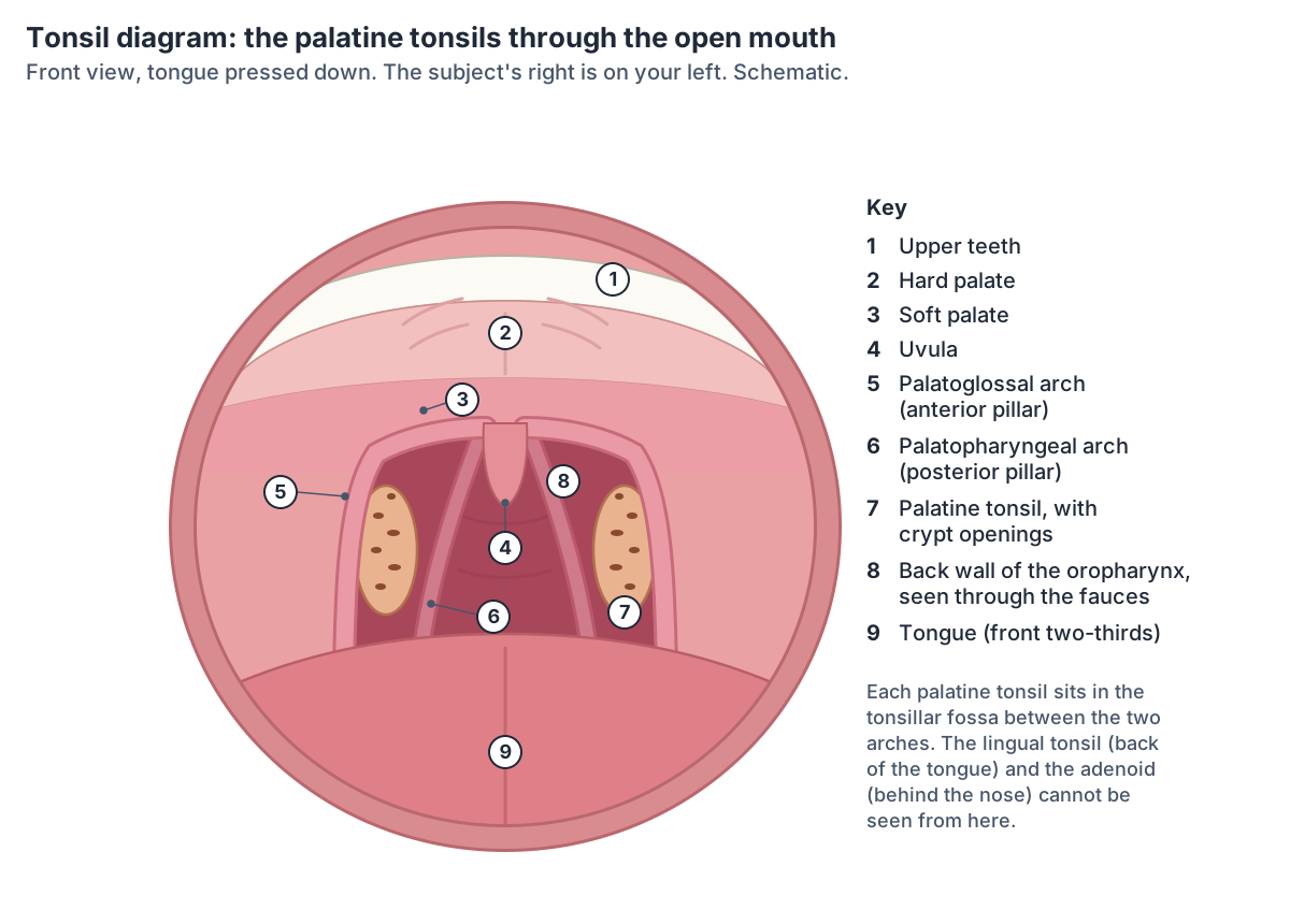
Healthy palatine tonsils look like oval, pinkish mounds on each side of the back of the throat, just behind the soft palate. They turn red and swollen when they are inflamed or infected. (Cleveland Clinic: Tonsils.)
Waldeyer's ring: the four types of tonsil
Waldeyer's ring is a ring of lymphoid tissue in the nasopharynx and oropharynx at the entrance to the airway and food passage. It is made of the palatine (faucial) tonsils, the adenoid (nasopharyngeal) tonsil, the tubal tonsils, also called Gerlach tonsils, near the openings of the auditory tubes, and the lingual tonsil at the back of the tongue. (Radiopaedia: Waldeyer ring.)
Counting each tonsil, the ring has six: one pharyngeal tonsil at the top, two tubal tonsils and two palatine tonsils at the sides, and one lingual tonsil at the bottom. (TeachMeAnatomy: The Tonsils (Waldeyer's Ring).)
The ring is named after the German anatomist Heinrich Wilhelm Gottfried von Waldeyer-Hartz, who lived from 1836 to 1921. (Radiopaedia: Waldeyer ring.)
The nasopharynx, oropharynx and laryngopharynx around the tonsils. Throat anatomy.
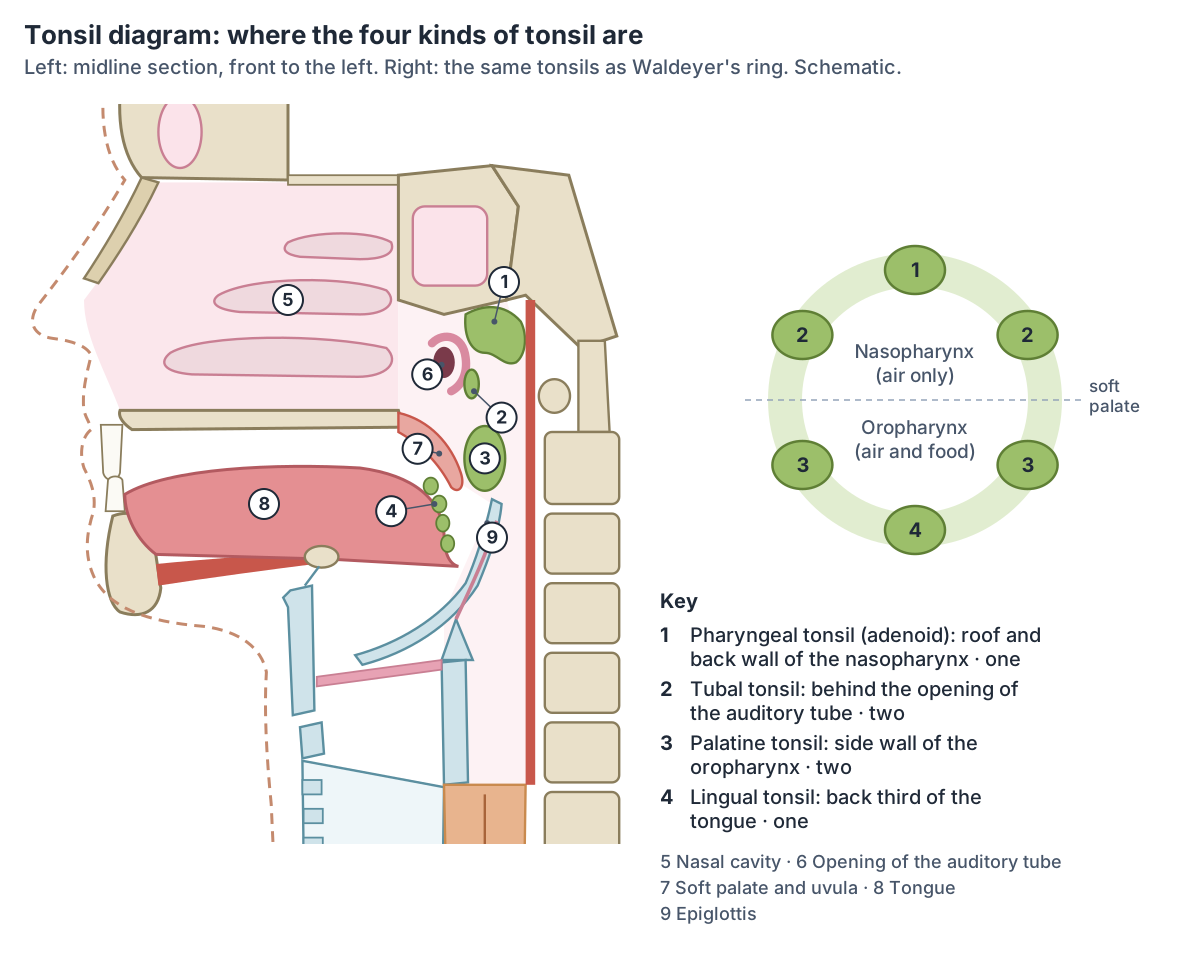
The tonsils of Waldeyer's ring
| Tonsil | Number | Where | Surface lining | Crypts | Lymph drains to |
|---|---|---|---|---|---|
| Pharyngeal tonsil (adenoid) | 1, midline | Roof and back wall of the nasopharynx | Ciliated pseudostratified (respiratory) epithelium, thrown into folds | None | Retropharyngeal and deep cervical nodes (Kenhub: retropharyngeal and pharyngomaxillary) |
| Tubal tonsils | 2 | Side wall of the nasopharynx, around and just behind the opening of each auditory (Eustachian) tube | Ciliated pseudostratified epithelium | Not described | Retropharyngeal and deep cervical nodes |
| Palatine tonsils | 2 | Side walls of the oropharynx, between the palatoglossal and palatopharyngeal arches | Non-keratinised stratified squamous epithelium | 15 to 20 (Kenhub) or 10 to 30 (Samara et al.) | Jugulodigastric and upper deep cervical nodes |
| Lingual tonsil | 1, midline | Back (pharyngeal) third of the tongue | Non-keratinised stratified squamous epithelium | Each nodule has a single crypt | Jugulodigastric and deep cervical nodes |
Compiled from TeachMeAnatomy and Kenhub, with crypt counts from Kenhub and Samara et al. (2023).
Palatine tonsils: where are the tonsils and what do they look like?
The palatine tonsils are what most people call the tonsils. Each sits in the tonsillar bed on the side wall of the oropharynx, between the palatoglossal arch in front and the palatopharyngeal arch behind. Its inner surface is free and bulges into the throat; its outer surface has a fibrous capsule, separated from the superior pharyngeal constrictor by loose connective tissue. (TeachMeAnatomy: The Tonsils (Waldeyer's Ring).)
Radiopaedia places the palatine tonsils in the oropharyngeal isthmus (the fauces). Each has two borders, two poles and two surfaces: the upper pole reaches the soft palate and the lower pole the back of the tongue. The tonsil is only partly encapsulated and is covered by non-keratinised stratified squamous epithelium that dips into long, branched crypts. (Radiopaedia: Palatine tonsil.)
Kenhub counts 15 to 20 tonsillar crypts. Their walls hold many lymphoid nodules, and their spaces contain lymphocytes, bacteria and shed epithelial cells. (Kenhub: Tonsils.)
A 2023 review by Samara and colleagues gives a wider range, about 10 to 30 crypts per palatine tonsil after birth, and describes the crypts as lined by stratified squamous epithelium and lymphoepithelium. (Samara P, Athanasopoulos M, Athanasopoulos I. Unveiling the enigmatic adenoids and tonsils. Microorganisms. 2023.)
The tonsil fills most of its fossa but leaves a small space above it, the supratonsillar fossa. Laterally it is enclosed by its fibrous capsule, and the loose tissue between the capsule and the superior constrictor forms a surgical plane of major importance in tonsillectomy. (IMAIOS e-Anatomy: Palatine tonsil.)
The palatine tonsil's own page in the 3D model, with its neighbours. Palatine tonsil in 3D.
Blood supply of the tonsils
The main artery of the palatine tonsil is the tonsillar branch of the facial artery, which pierces the superior constrictor to reach it. Blood leaves through the external palatine vein, which drains to the facial vein, and through smaller veins to the pharyngeal plexus. (TeachMeAnatomy: The Tonsils (Waldeyer's Ring).)
Kenhub lists five arteries for the palatine tonsil: the ascending palatine and tonsillar branches of the facial artery, the ascending pharyngeal artery, the dorsal lingual branch of the lingual artery and the lesser palatine branch of the descending palatine artery. Its veins form a peritonsillar plexus that drains to the internal jugular vein. (Kenhub: Tonsils.)
IMAIOS names the greater palatine artery instead of the lesser, alongside the tonsillar, ascending palatine, ascending pharyngeal and dorsal lingual arteries, and describes drainage through the peritonsillar venous plexus to the pharyngeal plexus and facial vein. (IMAIOS e-Anatomy: Palatine tonsil.)
The adenoid has its own supply: the ascending palatine and ascending pharyngeal arteries, the pharyngeal branch of the maxillary artery, the artery of the pterygoid canal, the basisphenoid artery and the tonsillar branch of the facial artery. Its veins pierce the superior constrictor to join the pharyngeal plexus. The tubal tonsils are supplied mainly by the ascending pharyngeal artery, and the lingual tonsil by the lingual artery with help from the facial and ascending pharyngeal arteries. (TeachMeAnatomy: The Tonsils (Waldeyer's Ring).)
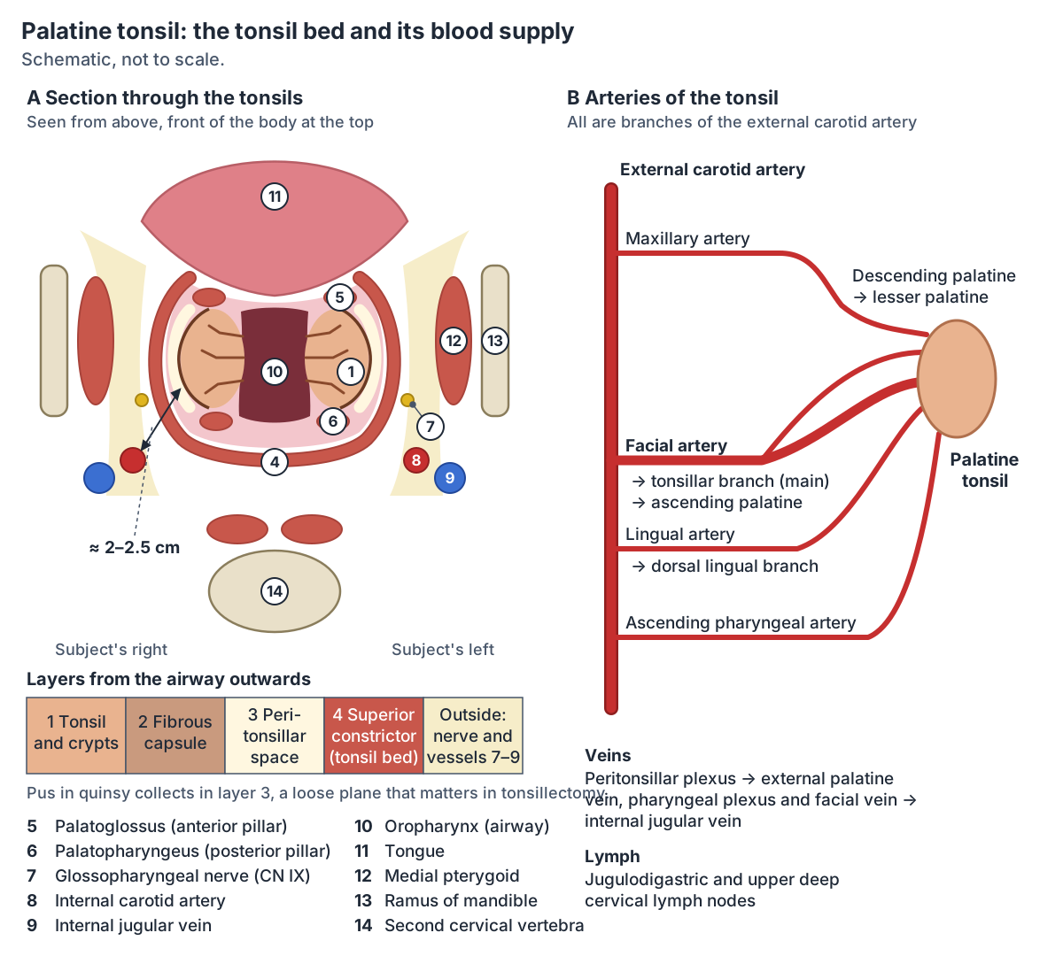
Arteries of the palatine tonsil
| Artery | Comes from | Notes |
|---|---|---|
| Tonsillar branch | Facial artery | The main supply; pierces the superior constrictor to reach the tonsil |
| Ascending palatine artery | Facial artery | The suspected bleeding vessel in one of Opatowsky's cases, near the junction of the tonsil bed and soft palate |
| Dorsal lingual branch | Lingual artery | The lingual artery also supplies the lingual tonsil |
| Ascending pharyngeal artery | External carotid artery | Also supplies the adenoid and tubal tonsils |
| Lesser (or greater) palatine artery | Descending palatine branch of the maxillary artery | Kenhub and Opatowsky et al. name the descending and lesser palatine arteries; IMAIOS names the greater palatine artery |
All five arise from branches of the external carotid artery. From Kenhub, TeachMeAnatomy, Radiopaedia, IMAIOS and Opatowsky et al. (2001).
Lymph drainage and nerves of the tonsils
The palatine and lingual tonsils drain to the jugulodigastric and upper deep cervical lymph nodes. The adenoid and tubal tonsils drain to the retropharyngeal nodes, which empty into the deep cervical chain. (TeachMeAnatomy: The Tonsils (Waldeyer's Ring).)
IMAIOS notes that the palatine tonsil drains mainly to the jugulodigastric node, which commonly becomes enlarged in tonsillitis. (IMAIOS e-Anatomy: Palatine tonsil.)
Sensation from the palatine tonsil is carried mainly by the glossopharyngeal nerve (CN IX). Because this nerve also supplies the middle ear, a sore tonsil can cause pain felt in the ear (referred otalgia). (IMAIOS e-Anatomy: Palatine tonsil.)
Radiopaedia lists the glossopharyngeal nerve and the lesser palatine nerves as the nerves of the palatine tonsil, and places the jugulodigastric nodes just below the angle of the mandible. (Radiopaedia: Palatine tonsil.)
TeachMeAnatomy gives the maxillary and glossopharyngeal nerves for the palatine and tubal tonsils, the glossopharyngeal nerve for the lingual tonsil, and the vagus and glossopharyngeal nerves for the adenoid. (TeachMeAnatomy: The Tonsils (Waldeyer's Ring).)
The jugulodigastric node and the deep cervical chain that drain the tonsils. Lymph nodes of the head and neck.
Adenoids (pharyngeal tonsil)
The pharyngeal tonsil is lymphoid tissue in the lining of the roof of the nasopharynx, in the midline, forming the top of Waldeyer's ring. When it is enlarged it is called the adenoids. It is covered by ciliated pseudostratified epithelium. (TeachMeAnatomy: The Tonsils (Waldeyer's Ring).)
Kenhub adds that the adenoid is attached to the periosteum of the sphenoid bone, that its surface is thrown into folds rather than crypts, and that its capsule is thinner than that of the palatine tonsil. It drains to the retropharyngeal and pharyngomaxillary nodes. (Kenhub: Tonsils.)
Samara and colleagues place the adenoid where the sphenoid and occipital bones meet, along the roof and back wall of the nasopharynx, and describe connective tissue septa that divide it into 4 to 6 segments. (Samara P, Athanasopoulos M, Athanasopoulos I. Unveiling the enigmatic adenoids and tonsils. Microorganisms. 2023.)
Enlarged adenoids are common in children and much less common in adults. They can cause nasal blockage, repeated middle ear infections, swallowing difficulties and a blocked-nose (hyponasal) voice. (Radiopaedia: Adenoids.)
The nasal cavity that opens into the nasopharynx in front of the adenoid. Nose anatomy.
Tubal tonsils and the lingual tonsil
The tubal tonsils are lymphoid tissue around the opening of the auditory (Eustachian) tube in the side wall of the nasopharynx, lined by ciliated pseudostratified epithelium. The lingual tonsil is a collection of lymphoid nodules in the submucosa of the back third of the tongue; it forms the bottom of the ring and gives the back of the tongue its bumpy surface. (TeachMeAnatomy: The Tonsils (Waldeyer's Ring).)
Kenhub places the tubal tonsils just behind the opening of the auditory tube and gives their arteries as branches of the sphenopalatine and ascending pharyngeal arteries. It describes the lingual tonsil as small, round elevations whose stratified squamous epithelium dips in to form a single crypt each, varying greatly in size and shape between people. (Kenhub: Tonsils.)
The tongue, whose back third carries the lingual tonsil. Tongue.
Do tonsils shrink with age?
Yes, mostly. OpenStax notes that the pharyngeal tonsils are large in children but tend to regress with age and may even disappear. (OpenStax Anatomy and Physiology 2e, 22.1: Organs and Structures of the Respiratory System.)
TeachMeAnatomy gives the usual period of enlargement of the adenoids as about 3 to 8 years of age, after which they regress. (TeachMeAnatomy: The Pharynx.)
An MRI study of the adenoid in 270 people aged 3 months to 34 years by Perry and colleagues reviews earlier work: adenoid tissue is present by about 6 weeks of age, grows fast in the first 2 years and can fill up to half of the nasopharynx by age 2, with maximal growth reported between 7 and 10 years and a fall in volume after about 9. The study's own peak ages differ for each measurement: about 8 to 9 years for height and thickness, and between under 2 and 4 years for depth, a figure the paper reports differently in different places. (Perry JL et al. A midsagittal-view MRI study of the growth and involution of the adenoid mass. J Speech Lang Hear Res. 2022.)
The palatine tonsils shrink from puberty onwards. In tonsils removed at surgery, Pastor and colleagues found the share of germinal centre B cells fell from about 33% in children aged 2 to 4 to about 17% at 12 to 18, and dated the start of functional decline to about 10 years. Enlarged tonsils were the main reason for tonsillectomy under 10, and recurrent tonsillitis after puberty. (Pastor R et al. Role of germinal center and CD39highCD73+ B cells in the age-related tonsillar involution. Immun Ageing. 2024.)
Tonsillitis, quinsy and tonsil stones
Tonsillitis is inflammation of the palatine tonsils. It is usually viral; bacteria cause about a third of cases. It causes painful swallowing, often with fever and bad breath, and the tonsils look red and swollen, sometimes with pus, with enlarged neck lymph nodes. (TeachMeAnatomy: The Tonsils (Waldeyer's Ring).)
A peritonsillar abscess, or quinsy, is the most common deep neck infection and almost always follows acute or recurrent tonsillitis. Infection breaks through the tonsil's capsule into the peritonsillar space, between the capsule and the superior constrictor. It is commonest at 20 to 40 years, in males and in smokers, and causes one-sided pain on swallowing, a muffled hot-potato voice, difficulty opening the mouth (trismus) and drooling. Beta-haemolytic streptococci are the commonest cause, and it usually needs aspiration or incision and drainage. (Radiopaedia: Peritonsillar abscess.)
Repeated inflammation can leave calcified debris of bacteria and mucus in the crypts, forming tonsil stones (tonsilloliths), which cause bad breath, a sore throat and a cough. (Kenhub: Tonsils.)
Enlarged tonsils can narrow the airway and cause snoring or sleep apnoea. Tonsil cancer is the most common form of oropharyngeal cancer and is often linked to human papillomavirus (HPV). (Cleveland Clinic: Tonsils.)
Tonsillectomy: the anatomy behind the operation
Tonsillectomy removes both palatine tonsils from the sides of the oropharynx. It is done for airway obstruction by enlarged tonsils, sleep apnoea, snoring and recurrent tonsillitis. Because the tonsils have a rich blood supply, bleeding is the main complication. (Kenhub: Tonsils.)
A 2023 review by Samara and colleagues gives the usual thresholds for recurrent tonsillitis: seven or more episodes in one year, five a year for two years, or three a year for three years. Other indications are repeated peritonsillar abscess, suspected cancer and sleep-disordered breathing. The review found no clinically significant long-term harm to the immune system from removing the tonsils. (Samara P, Athanasopoulos M, Athanasopoulos I. Unveiling the enigmatic adenoids and tonsils. Microorganisms. 2023.)
The internal carotid artery lies behind and to the side of the tonsil bed, about 2 to 2.5 cm from the tonsillar fossa, so close neurovascular relations make tonsillectomy an operation where anatomy matters. (IMAIOS e-Anatomy: Palatine tonsil.)
Gill and colleagues cite earlier work showing that in children this distance increases with age and weight until it reaches about 25 mm in adults. Their case reports show that an internal carotid artery running an unusual course behind the pharynx can lie only a few millimetres from the pharyngeal wall. (Gill JK et al. Clinically significant anatomical variation of the retropharyngeal internal carotid arteries. Radiol Case Rep. 2017.)
Bleeding within 24 hours of surgery is called primary haemorrhage and bleeding after 24 hours secondary, most often 5 to 10 days after the operation. Opatowsky and colleagues quote rates from under 1% to over 10%, and more recent figures of about 2 to 3%. Their cases bled from the ascending palatine and lingual arteries. (Opatowsky MJ et al. Endovascular treatment of hemorrhage after tonsillectomy in children. AJNR Am J Neuroradiol. 2001.)
Rates vary with the patients studied: in 778 adults treated at a national ENT centre in Georgia, 14.7% had some bleeding, 8.1% within 24 hours and 6.6% later, with most episodes on the first day and a second peak around day 7. (Besiashvili N et al. Evaluation and risk factor analysis of post-tonsillectomy hemorrhage in an adult population. Cureus. 2024.)
The palate, uvula and arches that frame the tonsils. Mouth anatomy.
Tonsils in the 3D model
- Internal carotid artery
- The internal carotid artery supplies the brain and the eye.
- Middle pharyngeal constrictor
- The middle of the three pharyngeal constrictors.
- Palatine tonsil
- The palatine tonsils are the two masses of lymphoid tissue at the back of the mouth, one on each side of the oropharynx. They are what most people mean by the tonsils.
- Palatopharyngeus
- A muscle of the soft palate and pharynx that forms the posterior pillar of the fauces.
- Soft palate
- The soft palate is the muscular back part of the roof of the mouth. Its free back edge carries the uvula.
- Stylopharyngeus
- A slender pharyngeal muscle arising from the styloid process.
- Superior pharyngeal constrictor
- The uppermost of the three constrictors forming the pharyngeal wall.
- Tongue
- The tongue is a muscular organ of the mouth involved in taste, chewing, swallowing and speech.
- Uvular muscle
- A small paired muscle forming the core of the uvula.
Frequently asked questions
What are the four types of tonsils?
The pharyngeal tonsil (adenoid) in the roof of the nasopharynx, the two tubal tonsils beside the openings of the auditory tubes, the two palatine tonsils on the side walls of the oropharynx, and the lingual tonsil on the back third of the tongue. Together these six masses of lymphoid tissue form Waldeyer's ring around the upper throat.
Where are your tonsils located?
The palatine tonsils, the ones you can see, sit on each side of the back of the mouth, behind the soft palate, in a hollow between two folds: the palatoglossal arch in front and the palatopharyngeal arch behind. The adenoid is hidden behind the nose, and the lingual tonsil lies further back on the tongue, out of view in the open mouth.
What is the difference between tonsils and adenoids?
Both are lymphoid tissue in Waldeyer's ring. The palatine tonsils are a pair in the oropharynx, covered by stratified squamous epithelium with deep crypts. The adenoid is a single pharyngeal tonsil in the roof of the nasopharynx, covered by ciliated respiratory epithelium thrown into folds, without crypts. Enlarged adenoids block the nose and are linked to repeated ear infections; enlarged palatine tonsils narrow the throat.
Why do tonsils have holes in them?
The holes are the openings of tonsillar crypts, deep pits in the surface of the palatine tonsil. Kenhub counts 15 to 20 and a 2023 review 10 to 30 per tonsil. The crypts greatly increase the surface where immune cells meet swallowed and inhaled microbes, but trapped debris can also harden into tonsil stones.
Do tonsils shrink as you get older?
Yes. The adenoid grows fastest in early childhood, is largest at around 3 to 10 years depending on the study, and then regresses, sometimes disappearing in adults. The palatine tonsils begin to involute from puberty, and their germinal centres become less active from about 10 years of age.
What artery supplies the tonsil?
Mainly the tonsillar branch of the facial artery, which pierces the superior pharyngeal constrictor to reach the tonsil. The ascending palatine, ascending pharyngeal, dorsal lingual and descending palatine branches also contribute, all ultimately from the external carotid artery. The external palatine vein is a main route of venous drainage.
What is quinsy?
Quinsy is a peritonsillar abscess: pus that has broken through the tonsil's capsule into the loose peritonsillar space between the capsule and the superior constrictor muscle. It follows tonsillitis, is commonest in young adults, causes one-sided throat pain, a muffled voice and difficulty opening the mouth, and usually needs drainage.
References
- TeachMeAnatomy. The Tonsils (Waldeyer's Ring): Lingual, Pharyngeal, Palatine, Tubal.
- TeachMeAnatomy. The Pharynx.
- Kenhub. Tonsils: Anatomy and histology of the types of tonsils.
- Betts JG, et al. Anatomy and Physiology 2e, §22.1 Organs and Structures of the Respiratory System. OpenStax, Rice University, 2022.
- Radiopaedia. Palatine tonsil (reference article).
- Radiopaedia. Adenoids (reference article).
- Radiopaedia. Waldeyer ring (reference article).
- Radiopaedia. Peritonsillar abscess (reference article).
- IMAIOS e-Anatomy. Palatine tonsil (tonsilla palatina).
- Cleveland Clinic. Tonsils: Anatomy, Definition and Function.
- Perry JL, Haenssler AE, Kotlarek KJ, et al. A midsagittal-view magnetic resonance imaging study of the growth and involution of the adenoid mass and related changes in selected velopharyngeal structures. J Speech Lang Hear Res. 2022;65(4):1282-1293.
- Samara P, Athanasopoulos M, Athanasopoulos I. Unveiling the enigmatic adenoids and tonsils: exploring immunology, physiology, microbiome dynamics, and the transformative power of surgery. Microorganisms. 2023;11(7):1624.
- Pastor R, Puyssegur J, de la Guardia MP, et al. Role of germinal center and CD39highCD73+ B cells in the age-related tonsillar involution. Immun Ageing. 2024;21:24.
- Opatowsky MJ, Browne JD, McGuirt WF Jr, Morris PP. Endovascular treatment of hemorrhage after tonsillectomy in children. AJNR Am J Neuroradiol. 2001;22(4):713-716.
- Besiashvili N, Datikashvili-David IG, Gakharia T. Evaluation and risk factor analysis of post-tonsillectomy hemorrhage in an adult population. Cureus. 2024;16(9):e68371.
- Gill JK, Sadiq M, Badar Z, Ezhapilli S. Clinically significant anatomical variation of the retropharyngeal internal carotid arteries. Radiol Case Rep. 2017;12(3):514-518.
- Mitsuhashi N. et al. (2009). BodyParts3D: 3D structure database for anatomical concepts. Nucleic Acids Research 37, D782–D785. Data © The Database Center for Life Science, CC BY 4.0.
- Browne K., Schlehlein H. (2026). 3D Reference Organ Set for Male, v1.10 (Visible Human Male). Human Reference Atlas 3D Reference Object Library, HuBMAP / NIH. CC BY 4.0.
Related anatomy
- Palatine tonsilPalatine tonsil: The palatine tonsils are the two masses of lymphoid tissue at the back of the mouth, one on each side…
- Throat anatomyThroat anatomy with a 3D model and labelled diagrams: the three parts of the pharynx, constrictor muscles, tonsils…
- Lymph nodes of the head and neckLymph node map of the head and neck: where neck lymph nodes are, submental and submandibular nodes, levels I to VI…
- Mouth anatomyMouth anatomy with a 3D model and labelled diagrams: lips, cheeks, hard and soft palate, uvula, tongue, teeth, gums…
- Nose anatomyNose anatomy with a 3D model and labelled diagrams: nasal bones and cartilages, nostrils, nasal cavity, conchae…
- Respiratory systemAirway and respiratory system anatomy with labelled diagrams and a 3D model: nose, pharynx, larynx, trachea, bronchi…
Explore the whole body in the interactive 3D human anatomy model, or browse the A–Z list of human structures.