

What is in the neck? Compartments and contents
The neck attaches the head to the trunk, holds the head in position and lets it turn. Kenhub divides its interior into four compartments: a vertebral compartment with the cervical vertebrae and postural muscles; a visceral compartment with the thyroid, parathyroid and thymus glands, larynx, pharynx and trachea; and two vascular compartments, one on each side, each holding the common carotid artery, internal jugular vein and vagus nerve. (Kenhub: Neck anatomy.)
The compartments are wrapped in deep cervical fascia. Its outermost layer, the investing layer, forms a collar around the whole neck; the pretracheal layer wraps the viscera at the front; and the prevertebral layer forms a tube around the vertebral column. Between the skin and the investing layer lies the thin superficial fascia with the platysma, cutaneous nerves and vessels and superficial lymph nodes. (Kenhub: Cervical fasciae.)
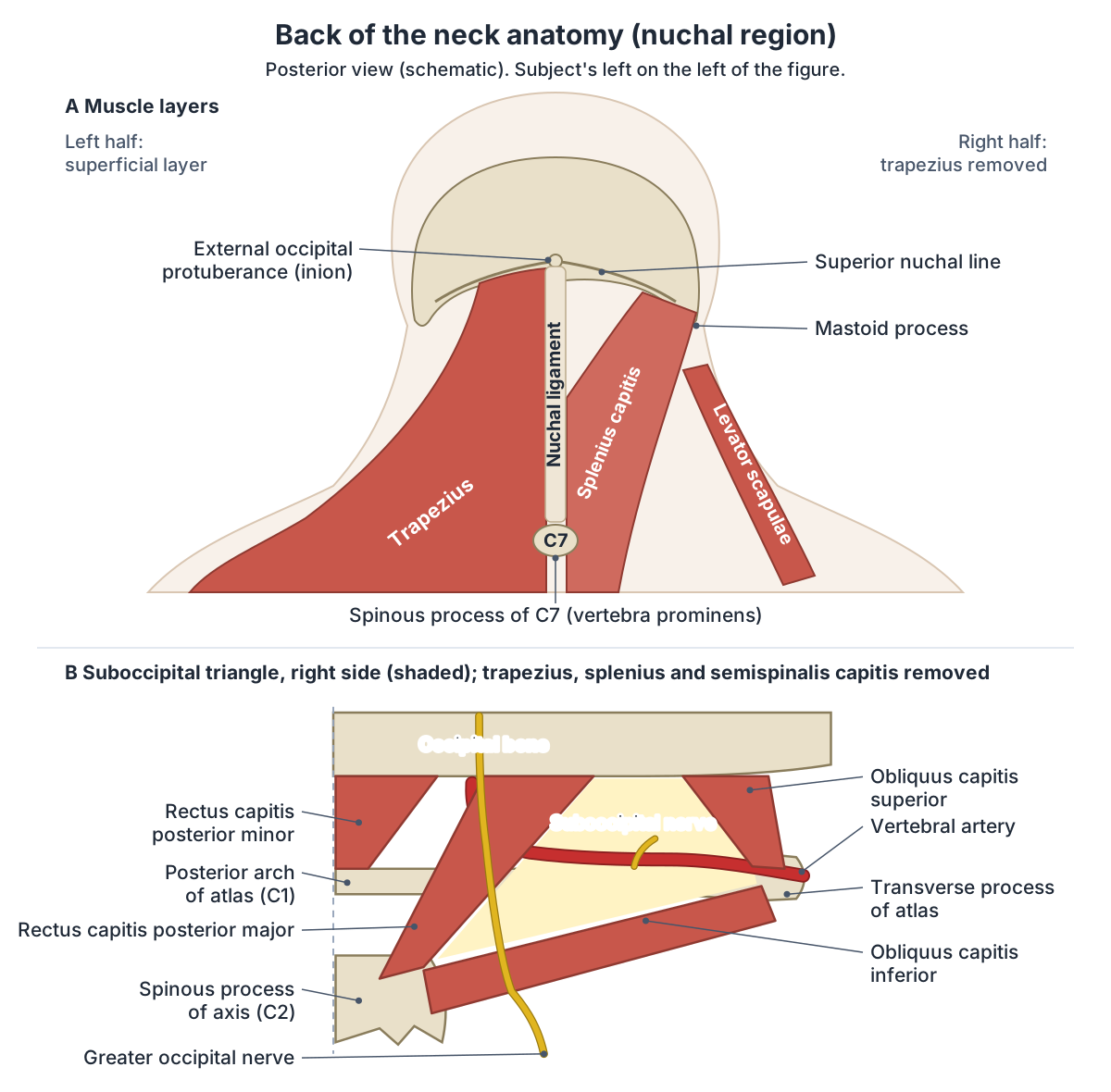
The back of the neck is described as its own region, the nuchal (posterior cervical) region. It lies deep to the trapezius, and its limits are the superior nuchal line and external occipital protuberance of the skull above and a line through the seventh cervical vertebra below. (Kenhub: Nuchal region.)
The head and neck hub: skull, brain, face, sense organs and ENT structures. Head anatomy.
Neck landmarks: hyoid (C3), thyroid cartilage (C4) and cricoid (C6)
Three firm landmarks can be felt in the front of the neck. The highest is the hyoid bone, which lies between the mandible and the thyroid cartilage at about the level of C3/C4. Apart from sesamoid bones, it is the only bone that does not articulate directly with another bone. (Radiopaedia: Hyoid bone.)
Below the hyoid is the thyroid cartilage. Its two flat laminae fuse in the midline to form the laryngeal prominence (Adam's apple), and its superior border sits at about the level of C4. Its lower horns form the cricothyroid joints with the cricoid cartilage. (Radiopaedia: Thyroid cartilage.)
The cricoid cartilage, a ring with a thin arch in front and a broad lamina behind, lies at the level of C6. It is the only complete ring of cartilage in the whole airway, and the tracheal rings start just below it. (Radiopaedia: Cricoid cartilage.)
These levels are textbook values, and textbooks themselves differ: some place the top of the thyroid cartilage at C4, Gray's at the C3/C4 disc. A CT study of 78 adults found the hyoid most often at C4 (47.5%), the top of the thyroid cartilage at C5 (35.9%) and the lower border of the cricoid at C7 (42.3%), with lower levels in men than in women. Even the most common level matched in fewer than half of patients, so the authors advise confirming levels with imaging rather than by touch. (Ko TS et al. The Inaccuracy of Surface Landmarks for the Anterior Approach to the Cervical Spine in Southern Chinese Patients. Asian Spine J 2018.)
At the back, the spinous process of C7 is much longer than those of C3 to C6. You can feel this prominent spine at the base of the neck by running a finger down the midline. (OpenStax Anatomy and Physiology 2e, 7.3: The Vertebral Column.)
The thyroid, cricoid and arytenoid cartilages, vocal folds and laryngeal muscles. Larynx anatomy.
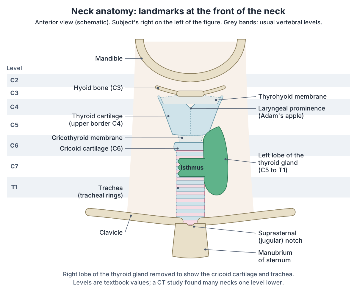
Neck landmarks and their vertebral levels
| Landmark | Usual teaching level | What it marks | CT study (Ko et al. 2018) |
|---|---|---|---|
| Hyoid bone | C3 (Radiopaedia: C3/C4) | Anchor of the suprahyoid and infrahyoid muscles; border between node levels I and VI in front and II and III at the side | Most often C4 (47.5%) |
| Thyroid cartilage, upper border | C4 (some textbooks: C3/C4 disc) | Laryngeal prominence (Adam's apple); the common carotid artery divides at about this level | Most often C5 (35.9%); C4 in women |
| Cricoid cartilage | C6 | End of the larynx and pharynx; start of the trachea and oesophagus; border between node levels III and IV | Lower border most often C7 (42.3%); C6 in women |
| Thyroid gland | C5 to T1 | Isthmus on the 2nd to 4th tracheal rings (Radiopaedia) or the 2nd and 3rd (Kenhub) | Not measured |
| C7 spinous process | C7 | Vertebra prominens, the bump at the base of the back of the neck; lower limit of the nuchal region | Not measured |
Teaching levels from Radiopaedia, TeachMeAnatomy and Kenhub. The last column is the most common level in a CT study of 78 adults in Hong Kong, where even the commonest level matched fewer than half of patients.
Triangles of the neck
Sternocleidomastoid crosses each side of the neck obliquely and splits it into two large triangles. The anterior triangle is bounded by the inferior border of the mandible, the midline of the neck and the anterior border of sternocleidomastoid. The posterior triangle lies behind it, between the posterior border of sternocleidomastoid, the anterior border of trapezius and the middle third of the clavicle. (Kenhub: Triangles of the neck.)
The hyoid bone and the suprahyoid and infrahyoid muscles divide the anterior triangle into four smaller triangles: submental, submandibular, carotid and muscular. The carotid triangle holds the carotid sinus, a dilated part of the artery with pressure receptors; in people with a hypersensitive sinus, pressing on it can slow the heart and cause fainting. (TeachMeAnatomy: The Anterior Triangle of the Neck.)
The inferior belly of omohyoid crosses the posterior triangle and divides it into an occipital triangle above and a subclavian triangle below. Its roof is the investing fascia and its floor the prevertebral fascia over splenius capitis, levator scapulae and the scalene muscles. The accessory nerve crosses it inside the investing fascia, close to the surface, which leaves it vulnerable to injury. (TeachMeAnatomy: The Posterior Triangle of the Neck.)
Sternocleidomastoid, suprahyoid, infrahyoid, scalene and suboccipital muscles, with a triangles diagram. Neck muscles.
Triangles of the neck: borders and contents
| Triangle | Borders | Main contents |
|---|---|---|
| Anterior triangle (whole) | Inferior border of the mandible; midline of the neck; anterior border of sternocleidomastoid | Suprahyoid and infrahyoid muscles, larynx, trachea, oesophagus, thyroid and parathyroid glands, submandibular gland, carotid arteries, internal jugular vein, vagus and hypoglossal nerves, lymph nodes |
| Submental | Body of the hyoid (base); anterior bellies of the two digastric muscles; midline | Submental lymph nodes, tributaries of the anterior jugular vein; floor: mylohyoid |
| Submandibular (digastric) | Inferior border of the mandible; anterior and posterior bellies of digastric | Submandibular gland and nodes, facial artery and vein, hypoglossal and mylohyoid nerves |
| Carotid | Posterior belly of digastric and stylohyoid; superior belly of omohyoid; anterior border of sternocleidomastoid | Common carotid artery and its division, internal and external carotid arteries, internal jugular vein, vagus and hypoglossal nerves, part of the sympathetic trunk |
| Muscular (omotracheal) | Hyoid bone; superior belly of omohyoid and sternocleidomastoid; midline | Infrahyoid (strap) muscles, thyroid and parathyroid glands, larynx, trachea, oesophagus |
| Posterior triangle (whole) | Posterior border of sternocleidomastoid; anterior border of trapezius; middle third of the clavicle | Accessory nerve, cervical plexus branches, brachial plexus trunks, external jugular vein, third part of the subclavian artery, transverse cervical and suprascapular vessels, lymph nodes |
| Occipital | Sternocleidomastoid; trapezius; inferior belly of omohyoid | Accessory nerve, cervical plexus branches, upper part of the brachial plexus |
| Subclavian (omoclavicular, supraclavicular) | Inferior belly of omohyoid; sternocleidomastoid; clavicle | Third part of the subclavian artery, brachial plexus trunks, nerve to subclavius, lymph nodes |
Compiled from Kenhub and TeachMeAnatomy, which agree on all borders.
Layers of the neck: fascia, spaces and the carotid sheath
The investing layer of deep cervical fascia splits to enclose trapezius and sternocleidomastoid. It is attached above to the external occipital protuberance and superior nuchal line, in front to the hyoid bone, below to the spine and acromion of the scapula, the clavicle and the manubrium, and behind to the nuchal ligament. (TeachMeAnatomy: Fascial Layers of the Neck.)
The pretracheal layer runs from the hyoid bone into the thorax, where it fuses with the pericardium. Its muscular part encloses the infrahyoid muscles and its visceral part the thyroid gland, trachea and oesophagus. The prevertebral layer surrounds the vertebral column with the scalene, prevertebral and deep back muscles; it forms the floor of the posterior triangle and continues around the brachial plexus and subclavian artery as the axillary sheath. (TeachMeAnatomy: Fascial Layers of the Neck.)
The carotid sheath is a fascial tube on each side that runs from the base of the skull to the root of the neck. It contains the common and internal carotid arteries, the internal jugular vein, the vagus nerve, some deep cervical lymph nodes, the carotid sinus nerve and sympathetic fibres. (Kenhub: Cervical fasciae.)
The fascial layers create spaces. The retropharyngeal space lies between the buccopharyngeal and prevertebral fascia and reaches from the skull base to the posterior mediastinum, and the visceral space runs from the hyoid to the superior mediastinum. The tough layers can stop a superficial abscess spreading deeper, but the carotid sheath and these spaces can carry infection down into the chest. (TeachMeAnatomy: Fascial Layers of the Neck.)
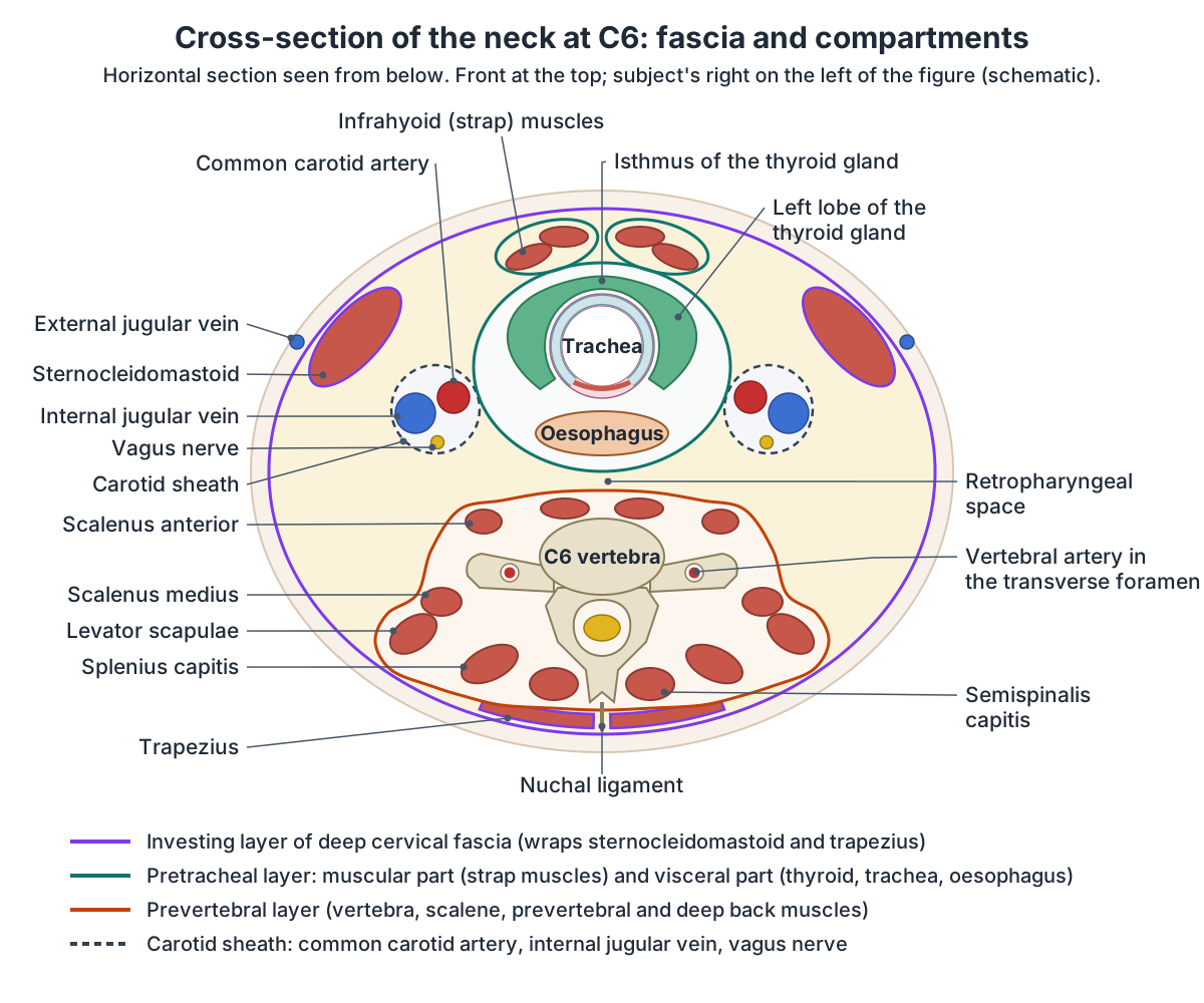
Layers of deep cervical fascia
| Layer | What it encloses | Extent and notes |
|---|---|---|
| Investing layer | Every structure of the neck; splits around sternocleidomastoid and trapezius; also the submandibular and parotid glands | From the external occipital protuberance, superior nuchal line and hyoid to the scapular spine, acromion, clavicle and manubrium; roof of both triangles |
| Pretracheal layer | Muscular part: infrahyoid muscles. Visceral part: thyroid gland, trachea and oesophagus | From the hyoid down into the thorax, where it blends with the pericardium |
| Prevertebral layer | Vertebral column with the prevertebral, scalene and deep back muscles; cervical sympathetic trunks | Skull base down to the thorax, where it blends with the endothoracic fascia; floor of the posterior triangle; continues around the brachial plexus and subclavian artery as the axillary sheath |
| Carotid sheath | Common and internal carotid arteries, internal jugular vein, vagus nerve, some deep cervical lymph nodes | Built from all three layers; runs from the skull base to the root of the neck and communicates with the mediastinum |
From TeachMeAnatomy and Kenhub.
Bones of the neck: cervical vertebrae and the hyoid
The neck is supported by seven cervical vertebrae, C1 to C7. They have small bodies, usually bifid (forked) spinous processes and a transverse foramen in each transverse process, through which an artery to the brain climbs the neck. The atlas (C1) is a ring with no body or spinous process that supports the skull, and the axis (C2) carries the upward-pointing dens, the pivot for turning the head. (OpenStax Anatomy and Physiology 2e, 7.3: The Vertebral Column.)
The hyoid bone has a body, two greater horns and two lesser horns. The suprahyoid muscles attach above it and the infrahyoid muscles below it, and the middle pharyngeal constrictor arises from its greater horn, so it acts as the anchor between the floor of the mouth, the larynx and the pharynx. (Radiopaedia: Hyoid bone.)
The atlas, axis and C3 to C7, the transverse foramen and the upper neck joints. Cervical vertebrae.
Muscles of the neck
Cleveland Clinic counts about 30 neck muscles and sorts them into anterior, lateral and posterior groups. Together they lift the upper ribs when you breathe in, help with chewing, swallowing and speaking, move the head, neck and shoulder blades, and stabilise the head and cervical spine. (Cleveland Clinic: Neck Muscles.)
Many front-of-neck muscles attach to the hyoid bone. The suprahyoid group (stylohyoid, digastric, mylohyoid and geniohyoid) raises the hyoid when you swallow, and the infrahyoid group (omohyoid, sternohyoid, thyrohyoid and sternothyroid) pulls it down. The muscles in the floor of the posterior triangle include splenius capitis, levator scapulae and the anterior, middle and posterior scalenes. (Kenhub: Neck anatomy.)
Platysma lies in the superficial fascia, above the deep fascia that wraps the other neck muscles. Its two heads arise from the fascia over pectoralis major and deltoid, cross the clavicle and insert on the lower border of the mandible. It is supplied by the cervical branch of the facial nerve. (TeachMeAnatomy: Fascial Layers of the Neck.)
Every neck muscle group with attachments, nerve supply and actions. Neck muscles.
Back of the neck anatomy (nuchal region)
The back of the neck, or nape, is the nuchal region. Its landmarks can all be felt through the skin: the external occipital protuberance and superior nuchal line of the skull above, the spinous process of C7 (vertebra prominens) below, and between them the nuchal ligament in the midline. (Kenhub: Nuchal region.)
The nuchal ligament (ligamentum nuchae) continues the supraspinous ligament up the neck, where the cervical spinous processes are short. It is attached to the cervical spinous processes, reaches the base of the skull and helps support the head. (OpenStax Anatomy and Physiology 2e, 7.3: The Vertebral Column.)
Under the skin, the muscles lie in layers. The descending part of trapezius is the most superficial. Beneath it are splenius capitis and splenius cervicis, then the erector spinae, then the transversospinalis group: semispinalis capitis and cervicis, multifidus and, deepest, the rotatores. Under the skull lie four small suboccipital muscles: rectus capitis posterior major and minor and obliquus capitis superior and inferior. (Kenhub: Nuchal region.)
Three of the suboccipital muscles frame the suboccipital triangle, which contains the third part of the vertebral artery, the suboccipital nerve (posterior ramus of C1) and a venous plexus. The greater occipital nerve, the posterior ramus of C2, is the largest skin nerve of the region and pierces trapezius near the external occipital protuberance; the occipital artery, a branch of the external carotid, supplies the deeper muscles. (Kenhub: Nuchal region.)
Trapezius, splenius, erector spinae and the deep back muscles in 3D. Back muscles.
The throat (pharynx) in the neck
The pharynx begins at the base of the skull and ends at the inferior border of the cricoid cartilage (C6). Its lowest part, the laryngopharynx, runs from the top of the epiglottis to the cricoid and continues below as the oesophagus. Three circular constrictor muscles, superior, middle and inferior, form its wall, and almost all pharyngeal muscles are supplied by the vagus nerve. (TeachMeAnatomy: The Pharynx.)
The nasopharynx, oropharynx and laryngopharynx, constrictors and tonsils. Throat anatomy.
The larynx (voice box)
The larynx is continuous with the pharynx above and the trachea below. It conducts air and, with the epiglottis, keeps food out of the airway. It is built from nine cartilages: three unpaired (thyroid, cricoid and epiglottis) and three paired (arytenoid, corniculate and cuneiform). (Kenhub: Neck anatomy.)
Its nerves are branches of the vagus. The external branch of the superior laryngeal nerve supplies cricothyroid, the internal branch carries sensation from the laryngopharynx and upper larynx, and the recurrent laryngeal nerve supplies most of the other intrinsic muscles. Damage to one recurrent laryngeal nerve causes a hoarse voice; damage to both causes loss of voice and noisy breathing (stridor). (TeachMeAnatomy: The Vagus Nerve (CN X).)
Cartilages, vocal folds, laryngeal muscles and their nerves. Larynx.
The trachea in the neck
The trachea (windpipe) begins at the lower margin of the cricoid cartilage at C6 and is 10 to 13 cm long, held open by 15 to 20 D-shaped rings of cartilage. In the neck the oesophagus lies directly behind it and the recurrent laryngeal nerves run at its sides; it ends at the carina, at about T4 lying down and lower when upright. (Radiopaedia: Trachea.)
Tracheal rings, the carina and the bronchial tree in 3D. Trachea.
The cervical oesophagus (esophagus)
The oesophagus (esophagus) begins at the lower border of the cricoid cartilage, at about C5/C6, where the cricopharyngeus muscle forms the upper oesophageal sphincter. It is on average 22 to 24 cm long, and in the neck the recurrent laryngeal nerves lie in the groove between it and the trachea. (Radiopaedia: Oesophagus.)
Course, sphincters, constrictions and blood supply of the food pipe. Esophagus.
Thyroid and parathyroid glands
The thyroid gland lies in the visceral compartment of the front of the neck and spans the C5 to T1 vertebrae. Its two lobes, joined in front by an isthmus, wrap around the cricoid cartilage and the upper tracheal rings inside the pretracheal fascia. The carotid sheath lies beside each lobe, and the recurrent laryngeal nerves run along its deep (medial) side. (TeachMeAnatomy: The Thyroid Gland.)
Behind the thyroid lie the parathyroid glands. About 80% of people have four, two superior and two inferior, each about 5 to 6 mm long and weighing 40 to 60 mg. They make parathyroid hormone, which controls the calcium level of the blood. (Radiopaedia: Parathyroid glands.)
Lobes, isthmus, blood supply, recurrent laryngeal nerve, parathyroids and histology. Thyroid gland anatomy.
Arteries of the neck: carotid, vertebral and subclavian
The right common carotid artery arises from the brachiocephalic trunk and the left directly from the arch of the aorta. Each divides at about C4 into an internal carotid artery, which climbs to the skull without giving any branches in the neck, and an external carotid artery, whose branches are the superior thyroid, ascending pharyngeal, lingual, facial, occipital and posterior auricular arteries, ending as the maxillary and superficial temporal arteries. (TeachMeAnatomy: Arterial Supply to the Head and Neck.)
Inside the carotid sheath, the internal jugular vein lies lateral to the common carotid artery with the vagus nerve between them. The two common carotids are separated low in the neck by the trachea and higher up by the thyroid gland, larynx and pharynx, and the level at which each divides varies between people. (Radiopaedia: Common carotid artery.)
The subclavian arteries supply the lower neck. Each gives off a vertebral artery, which ascends through the transverse foramina of C6 to C1 to reach the brain, and a thyrocervical trunk, whose branches are the inferior thyroid, ascending cervical, transverse cervical and suprascapular arteries. (TeachMeAnatomy: Arterial Supply to the Head and Neck.)
Common, internal and external carotids, the eight external carotid branches and the vertebral artery. Carotid arteries.
Veins of the neck: internal, external and anterior jugular veins
The internal jugular vein begins at the jugular foramen as the continuation of the sigmoid sinus. It descends in the carotid sheath, deep to sternocleidomastoid and lateral to the common carotid artery, and joins the subclavian vein behind the sternal end of the clavicle to form the brachiocephalic vein. Its tributaries include the facial, lingual, occipital and superior and middle thyroid veins. (TeachMeAnatomy: Venous Drainage of the Head and Neck.)
The external jugular vein forms just behind the angle of the mandible, crosses sternocleidomastoid in the superficial fascia and drains into the subclavian vein. The paired anterior jugular veins run near the midline, are often joined by a jugular venous arch and also end in the subclavian vein. (TeachMeAnatomy: Venous Drainage of the Head and Neck.)
Course, carotid sheath relations, tributaries and central line anatomy. Internal jugular vein.
Nerves of the neck: cervical plexus, phrenic nerve and brachial plexus
The cervical plexus is formed by the anterior rami of C1 to C4 and lies within the prevertebral fascia, about halfway up sternocleidomastoid. Its skin branches (lesser occipital, great auricular, transverse cervical and supraclavicular nerves) emerge together at the middle of the muscle's posterior border, the nerve point of the neck. The ansa cervicalis (C1 to C3) supplies the infrahyoid muscles, and the phrenic nerve (C3 to C5) supplies the diaphragm. (TeachMeAnatomy: The Cervical Plexus.)
The roots of the brachial plexus, the anterior rami of C5 to T1, pass between the anterior and middle scalene muscles into the base of the neck. There they join to form the superior, middle and inferior trunks, which cross the posterior triangle and divide before entering the axilla. (TeachMeAnatomy: The Brachial Plexus.)
The cervical sympathetic trunk lies within the prevertebral fascia. Kenhub lists three cervical ganglia: the superior, the largest, at C1 to C3; the middle, most often at C6; and the inferior at C7 to T1. (Kenhub: Thyroid gland.)
The brachial plexus from roots to terminal branches. Upper limb nerves.
Nerves of the neck
| Nerve | Origin | Course in the neck | Supplies |
|---|---|---|---|
| Cervical plexus | Anterior rami of C1 to C4 | Within the prevertebral fascia about halfway up sternocleidomastoid; skin branches emerge at the middle of its posterior border | Skin of the scalp, ear, neck and upper chest; ansa cervicalis to the infrahyoid muscles |
| Phrenic nerve | C3 to C5 | Descends within the prevertebral fascia into the chest | Diaphragm |
| Brachial plexus (roots and trunks) | Anterior rami of C5 to T1 | Between scalenus anterior and medius, then across the posterior triangle | Upper limb |
| Vagus nerve (CN X) | Jugular foramen | Inside the carotid sheath with the internal jugular vein and carotid artery | Pharynx and soft palate, larynx (superior and recurrent laryngeal nerves), heart |
| Accessory nerve (CN XI) | Spinal roots of C1 to C5/C6; leaves the skull through the jugular foramen | To sternocleidomastoid, then superficially across the posterior triangle | Sternocleidomastoid and trapezius |
| Hypoglossal nerve (CN XII) | Hypoglossal canal | Below the angle of the mandible, crossing the internal and external carotid arteries | Muscles of the tongue except palatoglossus |
| Cervical sympathetic trunk | Superior (C1 to C3), middle (usually C6) and inferior (C7 to T1) cervical ganglia | Within the prevertebral fascia | Sympathetic fibres to the neck viscera, for example the thyroid gland and the pharyngeal plexus |
From TeachMeAnatomy (cervical plexus, brachial plexus, cranial nerves, posterior triangle) and Kenhub (cervical fascia, thyroid gland).
Vagus, accessory and hypoglossal nerves in the neck
The vagus nerve (CN X) leaves the skull through the jugular foramen and runs down the neck inside the carotid sheath with the internal jugular vein and carotid artery. Its pharyngeal branches supply most of the pharynx and soft palate. On the right, the recurrent laryngeal nerve hooks under the right subclavian artery; on the left, it hooks under the arch of the aorta. (TeachMeAnatomy: The Vagus Nerve (CN X).)
The spinal part of the accessory nerve (CN XI) arises from the C1 to C5/C6 segments, enters the skull through the foramen magnum and leaves through the jugular foramen. It descends to sternocleidomastoid and crosses the posterior triangle to trapezius. Because it lies so near the surface, operations such as cervical lymph node biopsy can injure it, weakening head turning and shoulder shrugging. (TeachMeAnatomy: The Accessory Nerve (CN XI).)
The hypoglossal nerve (CN XII) leaves the skull through the hypoglossal canal, runs below the angle of the mandible across the internal and external carotid arteries and supplies all the tongue muscles except palatoglossus. Fibres from C1 travel in its sheath to geniohyoid and thyrohyoid. (TeachMeAnatomy: The Hypoglossal Nerve (CN XII).)
All 12 cranial nerves, their exits from the skull and what they supply. Cranial nerves.
Lymph nodes of the neck
Superficial lymph nodes form a ring where the head meets the neck, from under the chin to the back of the head. All lymph of the head and neck then reaches the deep cervical nodes, a vertical chain beside the internal jugular vein in the carotid sheath. The jugular lymph trunk joins the thoracic duct on the left and forms the right lymphatic duct on the right. (TeachMeAnatomy: Lymphatic Drainage of the Head and Neck.)
For imaging and surgery the neck nodes are grouped into levels: I submental and submandibular, II to IV the upper, middle and lower internal jugular chain (divided at the hyoid and the cricoid), V the posterior triangle and VI the central compartment in front of the trachea. Level IV includes the medial supraclavicular nodes, among them Virchow's node. (Radiopaedia: Cervical lymph node levels.)
Node groups, what they drain, and neck levels I to VI with diagrams. Lymph nodes of the head and neck.
Neck anatomy in the 3D model
The 3D viewer opens on the left side of the neck. It highlights the seven cervical vertebrae, the hyoid bone, the thyroid and cricoid cartilages, the trachea, the common and internal carotid, vertebral and subclavian arteries, the internal jugular veins, and the sternocleidomastoid, the three scalenes, omohyoid, sternohyoid, longus capitis and two suboccipital muscles (rectus capitis posterior major and obliquus capitis inferior). Open the back muscles guide to see trapezius, splenius capitis and semispinalis capitis in 3D.
The model is BodyParts3D, built from a whole-body set of MRI images of an adult male volunteer. It has no thyroid or parathyroid glands, lymph nodes, neck nerves, external carotid artery or external jugular vein, so the diagrams on this page and the linked guides show those structures. (Mitsuhashi N et al. BodyParts3D: 3D structure database for anatomical concepts. Nucleic Acids Res 2009.)
Neck anatomy in the 3D model
- Atlas
- The first cervical vertebra (C1), a ring-shaped bone that supports the skull.
- Axis
- The second cervical vertebra (C2), distinguished by a tooth-like upward projection called the dens.
- Common carotid artery
- The common carotid artery is the main artery of the neck and splits into the external and internal carotid arteries.
- Cricoid cartilage
- The cricoid cartilage is a complete signet-ring-shaped cartilage forming the lower part of the larynx.
- Fifth cervical vertebra
- The fifth of the seven cervical (neck) vertebrae, labelled C5.
- Fourth cervical vertebra
- The fourth of the seven cervical (neck) vertebrae, labelled C4.
- Hyoid bone
- A U-shaped bone in the neck.
- Internal carotid artery
- The internal carotid artery supplies the brain and the eye.
- Internal jugular vein
- The internal jugular vein is the main vein draining the head and neck.
- Longus capitis
- A prevertebral muscle in front of the upper cervical spine.
- Obliquus capitis inferior
- A suboccipital muscle below the atlas.
- Omohyoid
- A thin infrahyoid strap muscle with two bellies joined by an intermediate tendon.
- Rectus capitis posterior major
- A suboccipital muscle forming the superomedial border of the suboccipital triangle.
- Scalenus anterior
- A deep neck muscle and an important landmark in the root of the neck.
- Scalenus medius
- The largest and longest of the scalene muscles.
- Scalenus posterior
- The smallest and deepest of the scalene muscles.
- Seventh cervical vertebra
- The seventh of the seven cervical (neck) vertebrae, labelled C7.
- Sixth cervical vertebra
- The sixth of the seven cervical (neck) vertebrae, labelled C6.
- Sternocleidomastoid
- A thick superficial neck muscle forming the main landmark dividing the anterior and posterior triangles.
- Sternohyoid
- A thin infrahyoid strap muscle at the front of the neck.
- Subclavian artery
- The major artery to the upper limb, neck and part of the brain.
- Third cervical vertebra
- The third of the seven cervical (neck) vertebrae, labelled C3.
- Thyroid cartilage
- The thyroid cartilage is the largest cartilage of the larynx, forming the laryngeal prominence.
- Trachea
- The trachea (windpipe) is a flexible air tube extending from the larynx to the bronchi.
- Vertebral artery
- A branch of the subclavian artery supplying the brain and spinal cord.
Frequently asked questions
What are the main structures of the neck?
The neck contains the seven cervical vertebrae and the spinal cord, about 30 muscles, the hyoid bone, the larynx and trachea (airway), the pharynx and oesophagus (food passage), the thyroid and parathyroid glands, the carotid and vertebral arteries, the jugular veins, the vagus, accessory and hypoglossal nerves, the cervical and brachial plexuses and many lymph nodes.
What organs are in the neck?
The organs of the neck lie mainly in its visceral compartment at the front: the larynx (voice box), the trachea (windpipe), the pharynx and oesophagus, and the thyroid and parathyroid glands. The upper part of the thymus can also reach the root of the neck. Lymph nodes lie along the jugular veins and around these organs.
What are the triangles of the neck?
Sternocleidomastoid divides each side of the neck into an anterior and a posterior triangle. The anterior triangle, between the mandible, the midline and sternocleidomastoid, is split into submental, submandibular, carotid and muscular triangles. The posterior triangle, between sternocleidomastoid, trapezius and the clavicle, is split by omohyoid into occipital and subclavian triangles.
How many bones are in the neck?
The neck has seven cervical vertebrae, C1 (the atlas) to C7, plus the hyoid bone at the front, which is unusual because it does not articulate directly with any other bone. The thyroid and cricoid cartilages of the larynx are cartilage, not bone.
What is the bump at the back of the neck?
The bump you feel at the base of the back of the neck is usually the spinous process of the seventh cervical vertebra (C7), called the vertebra prominens because its spine is much longer than those above it. The nuchal ligament runs from it up to the external occipital protuberance, the bump at the back of the skull.
What is inside the carotid sheath?
The carotid sheath is a tube of deep fascia on each side of the neck. It contains the common carotid artery and, above its division, the internal carotid artery, the internal jugular vein lateral to the artery, the vagus nerve between them, and some deep cervical lymph nodes and sympathetic fibres. It runs from the skull base to the root of the neck.
What level is the cricoid cartilage?
The cricoid cartilage is usually taught at the level of the sixth cervical vertebra (C6). It marks the end of the larynx and pharynx and the start of the trachea and oesophagus. A CT study found that in many adults, especially men, its lower border lies lower, often at C7, so the level is a guide rather than a fixed rule.
References
- Betts JG, et al. Anatomy and Physiology 2e, §7.3 The Vertebral Column. OpenStax, Rice University, 2022.
- Kenhub. Neck anatomy. Accessed 2026-10-24.
- Kenhub. Triangles of the neck. Accessed 2026-10-24.
- Kenhub. Cervical fasciae. Accessed 2026-10-24.
- Kenhub. Nuchal region. Accessed 2026-10-24.
- Kenhub. Thyroid gland. Accessed 2026-10-24.
- TeachMeAnatomy. The Anterior Triangle of the Neck. Accessed 2026-10-24.
- TeachMeAnatomy. The Posterior Triangle of the Neck. Accessed 2026-10-24.
- TeachMeAnatomy. Fascial Layers of the Neck. Accessed 2026-10-24.
- TeachMeAnatomy. The Pharynx. Accessed 2026-10-24.
- TeachMeAnatomy. The Thyroid Gland. Accessed 2026-10-24.
- TeachMeAnatomy. Arterial Supply to the Head and Neck. Accessed 2026-10-24.
- TeachMeAnatomy. Venous Drainage of the Head and Neck. Accessed 2026-10-24.
- TeachMeAnatomy. Lymphatic Drainage of the Head and Neck. Accessed 2026-10-24.
- TeachMeAnatomy. The Cervical Plexus. Accessed 2026-10-24.
- TeachMeAnatomy. The Brachial Plexus. Accessed 2026-10-24.
- TeachMeAnatomy. The Vagus Nerve (CN X). Accessed 2026-10-24.
- TeachMeAnatomy. The Accessory Nerve (CN XI). Accessed 2026-10-24.
- TeachMeAnatomy. The Hypoglossal Nerve (CN XII). Accessed 2026-10-24.
- Radiopaedia. Hyoid bone. Accessed 2026-10-24.
- Radiopaedia. Thyroid cartilage. Accessed 2026-10-24.
- Radiopaedia. Cricoid cartilage. Accessed 2026-10-24.
- Radiopaedia. Trachea. Accessed 2026-10-24.
- Radiopaedia. Oesophagus. Accessed 2026-10-24.
- Radiopaedia. Common carotid artery. Accessed 2026-10-24.
- Radiopaedia. Cervical lymph node levels. Accessed 2026-10-24.
- Radiopaedia. Parathyroid glands. Accessed 2026-10-24.
- Cleveland Clinic. Neck Muscles: What They Are, Types, Anatomy & Function. Accessed 2026-10-24.
- Ko TS, Tse MSH, Wong KK, Wong WC. The Inaccuracy of Surface Landmarks for the Anterior Approach to the Cervical Spine in Southern Chinese Patients. Asian Spine J. 2018;12(6):1123-1126.
- Mitsuhashi N, et al. BodyParts3D: 3D structure database for anatomical concepts. Nucleic Acids Res. 2009;37:D782-D785.
- Mitsuhashi N. et al. (2009). BodyParts3D: 3D structure database for anatomical concepts. Nucleic Acids Research 37, D782–D785. Data © The Database Center for Life Science, CC BY 4.0.
Related anatomy
- Neck musclesNeck muscles explained: sternocleidomastoid, suprahyoid, infrahyoid, scalene, prevertebral and suboccipital groups…
- Cervical vertebraeCervical vertebrae explained: typical C3 to C6 features, the atlas (C1), axis and dens (C2), C7 vertebra prominens, AO…
- Thyroid glandThyroid gland anatomy with labelled diagrams: lobes, isthmus, pyramidal lobe, arteries and veins, recurrent laryngeal…
- Carotid arteriesCarotid arteries explained: common carotid, bifurcation, internal carotid, the 8 external carotid branches with a…
- Lymph nodes of the head and neckLymph node map of the head and neck: where neck lymph nodes are, submental and submandibular nodes, levels I to VI…
- Throat anatomyThroat anatomy with a 3D model and labelled diagrams: the three parts of the pharynx, constrictor muscles, tonsils…
Explore the whole body in the interactive 3D human anatomy model, or browse the A–Z list of human structures.