

What is the cardiovascular system?
OpenStax describes eleven organ systems in the human body. One of them is the cardiovascular system, whose organs are the heart and the blood vessels. (OpenStax Anatomy and Physiology 2e, 1.2: Structural Organization of the Human Body.)
Circulatory system and cardiovascular system are two names for the same system. Its four main parts are the heart, arteries, veins and capillaries. Blood passes through the lungs, but the lungs themselves belong to the respiratory system. (Cleveland Clinic: Circulatory System.)
The fluid it moves is blood: about 5 to 6 litres in adult males and 4 to 5 litres in females, roughly 8 percent of body weight. Blood carries oxygen, nutrients, hormones and wastes, helps defend the body against infection, and helps control body temperature and pH. (OpenStax Anatomy and Physiology 2e, 18.1: An Overview of Blood.)
The lymphatic system is a separate but connected system of vessels, cells and organs, such as lymph nodes, the spleen and the thymus. It carries excess fluid from the tissues back into the bloodstream. (OpenStax Anatomy and Physiology 2e, 21.1: Anatomy of the Lymphatic and Immune Systems.)
Lymph vessels, lymph nodes and the thoracic duct are described in the guide to the Lymphatic system.
Pulmonary vs systemic circulation
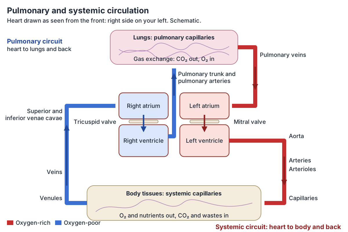
The pulmonary circuit takes blood to the lungs and back. Blood flows from the right atrium to the right ventricle, then through the pulmonary trunk, which splits into the right and left pulmonary arteries. These branch into arterioles and then the pulmonary capillaries around the alveoli, where gas exchange happens. Blood returns through pulmonary venules and four pulmonary veins, two on each side, to the left atrium. (OpenStax Anatomy and Physiology 2e, 20.5: Circulatory Pathways.)
The systemic circuit takes blood to the rest of the body. Blood moves from the left atrium to the left ventricle and into the aorta, whose branches supply nearly every organ. It comes back through the systemic veins: the superior vena cava drains everything above the diaphragm and the inferior vena cava everything below it, and both empty into the right atrium. (OpenStax Anatomy and Physiology 2e, 20.5: Circulatory Pathways.)
Whether a vessel is an artery or a vein depends on direction, not oxygen. An artery carries blood away from the heart. In the systemic circuit arteries carry oxygen-rich blood, but in the pulmonary circuit they carry blood low in oxygen to the lungs. (OpenStax Anatomy and Physiology 2e, 20.1: Structure and Function of Blood Vessels.)
The heart muscle has its own branch of the systemic circuit, the coronary circulation. The coronary arteries arise from the first part of the aorta and encircle the heart. (OpenStax Anatomy and Physiology 2e, 20.5: Circulatory Pathways.)
Before birth the lungs are not used, so three shunts let most blood bypass them and the liver: the foramen ovale between the atria, the ductus arteriosus between the pulmonary trunk and aorta, and the ductus venosus past the liver. After birth they become the fossa ovalis, the ligamentum arteriosum and the ligamentum venosum. (OpenStax Anatomy and Physiology 2e, 20.6: Development of Blood Vessels and Fetal Circulation.)
Each step of the path through the chambers and valves is shown on an interactive 3D heart in Blood flow through the heart.
The heart: the pump at the centre
The heart lies in the mediastinum between the lungs. It has four chambers: the right atrium receives the superior and inferior venae cavae and the coronary sinus, the left atrium receives four pulmonary veins, and the two ventricles pump blood into the pulmonary trunk and the aorta. The left ventricle's wall is much thicker than the right's because it drives the whole systemic circuit. (OpenStax Anatomy and Physiology 2e, 19.1: Heart Anatomy.)
Each ventricle ejects about 70 mL of blood per beat at rest. At 75 beats a minute that is about 5.25 litres a minute, pumped through roughly 60,000 miles of blood vessels. (OpenStax Anatomy and Physiology 2e, 19.1: Heart Anatomy.)
Cleveland Clinic gives matching figures: more than 7,500 litres (about 2,000 gallons) of blood pumped a day through more than 60,000 miles of blood vessels. (Cleveland Clinic: Circulatory System.)
The chambers, valves, wall layers, great vessels and conduction system are covered in detail in Heart anatomy.
Types of blood vessels: arteries, arterioles, capillaries, venules and veins
Arteries carry blood away from the heart and branch into smaller and smaller vessels. The smallest, the arterioles, have a lumen of about 30 micrometres or less and lead into capillaries, microscopic channels with a lumen of 5 to 10 micrometres. Capillaries drain into venules, about 8 to 100 micrometres across, which join to form veins, thin-walled vessels with large, irregular lumens that carry blood back towards the heart. (OpenStax Anatomy and Physiology 2e, 20.1: Structure and Function of Blood Vessels.)
Arteries come in two main kinds. Elastic (conducting) arteries, mostly more than 10 mm across, have plenty of elastic fibres so they stretch and recoil with each heartbeat. Muscular (distributing) arteries, about 0.1 to 10 mm across, have a thick layer of smooth muscle and do most of the narrowing (vasoconstriction). (OpenStax Anatomy and Physiology 2e, 20.1: Structure and Function of Blood Vessels.)
The large elastic arteries are the aorta and the pulmonary arteries, closest to the heart; their middle layer holds 40 to 70 fenestrated elastic membranes. Muscular arteries have about 40 layers of smooth muscle, and arterioles, less than 0.1 mm across, usually about three. (TeachMeAnatomy: Blood Vessels.)
Blood pressure falls along the way from arteries to arterioles, capillaries, venules and veins. The biggest drop is in the arterioles, which offer the most resistance and so control blood pressure. Blood also slows sharply in the capillaries, which gives time for exchange, and speeds up again in the veins. (OpenStax Anatomy and Physiology 2e, 20.2: Blood Flow, Blood Pressure, and Resistance.)
There are three types of capillary. Continuous capillaries are the commonest; in the brain they have no gaps and form the blood-brain barrier. Fenestrated capillaries have pores and are found in the small intestine, kidneys, choroid plexus and many endocrine glands. Sinusoid capillaries, the least common, have large gaps and are found in the liver, spleen, bone marrow and lymph nodes. (OpenStax Anatomy and Physiology 2e, 20.1: Structure and Function of Blood Vessels.)
Each capillary bed of about 10 to 100 capillaries is supplied by a metarteriole. Precapillary sphincters at the entrance to the capillaries open when the tissue needs more oxygen or has waste to clear. (OpenStax Anatomy and Physiology 2e, 20.1: Structure and Function of Blood Vessels.)
Artery vs vein: the structure of the vessel wall
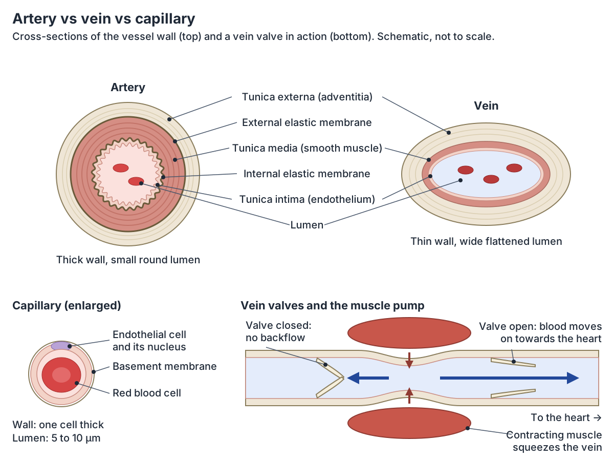
Arteries and veins have the same three layers, called tunics. The tunica intima, the innermost, is lined by endothelium. The tunica media is smooth muscle and elastic fibres and is usually the thickest layer of an artery. The tunica externa (adventitia) is connective tissue and is usually the thickest layer of a vein. (OpenStax Anatomy and Physiology 2e, 20.1: Structure and Function of Blood Vessels.)
In cross-section an artery has a thick wall, a small lumen and a round shape, and its endothelium looks wavy. A vein has a thin wall, a large lumen and a flattened shape. Larger arteries have internal and external elastic membranes, which veins lack. Arterial pressure is high and venous pressure low, and only veins have valves. (OpenStax Anatomy and Physiology 2e, 20.1: Structure and Function of Blood Vessels.)
Compared with the artery it travels with, a vein is usually wider with a thinner wall, more connective tissue and fewer elastic and muscle fibres. (TeachMeAnatomy: Blood Vessels.)
The walls of large arteries and veins are too thick to be fed from the lumen alone, so they have their own small vessels, the vasa vasorum. In arteries these are limited to the outer layers, because the high pressure inside would squeeze them shut. (OpenStax Anatomy and Physiology 2e, 20.1: Structure and Function of Blood Vessels.)
Artery vs vein vs capillary
| Feature | Artery | Vein | Capillary |
|---|---|---|---|
| Direction of flow | Away from the heart | Towards the heart | Links arterioles to venules |
| Wall | Thick; tunica media thickest | Thin; tunica externa thickest | One layer of endothelium on a basement membrane |
| Lumen and shape | Small, round | Large, flattened or irregular | Lumen 5 to 10 micrometres |
| Elastic membranes | Internal and external in larger arteries | Absent | Absent |
| Valves | None | Present, mostly in the limbs | None |
| Pressure | High | Low | Low; the site of exchange |
| Share of blood at rest | About 13 percent (systemic) | About 64 percent (systemic) | About 7 percent (systemic) |
From OpenStax Anatomy and Physiology 2e, section 20.1, and TeachMeAnatomy.
Vein anatomy: valves, deep and superficial veins
Veins start as postcapillary venules, 10 to 30 micrometres across and leakier than capillaries. TeachMeAnatomy describes venules up to 1 mm wide with very thin walls and valves, and large veins more than 10 mm across, with a thicker intima and longitudinal smooth muscle in the adventitia. (TeachMeAnatomy: Blood Vessels.)
Venous valves are made of thickened endothelium reinforced with connective tissue. They let blood flow only towards the heart and stop it flowing back. They are commonest in the veins of the limbs. (OpenStax Anatomy and Physiology 2e, 20.1: Structure and Function of Blood Vessels.)
Veins are capacitance vessels: they stretch easily at low pressure and hold most of the blood. At rest about 64 percent of the blood is in the systemic veins, 13 percent in the systemic arteries, 7 percent in the systemic capillaries, 9 percent in the pulmonary circuit and 7 percent in the heart. About 21 percent of venous blood sits in the liver, bone marrow and skin as a reserve. (OpenStax Anatomy and Physiology 2e, 20.1: Structure and Function of Blood Vessels.)
Pressure in the veins is low, so two pumps help return blood. In the skeletal muscle pump, contracting muscles squeeze nearby veins; the valve above opens and the valve below closes, so blood can only move towards the heart. In the respiratory pump, breathing in lowers the pressure in the chest and draws blood up from the abdominal veins. (OpenStax Anatomy and Physiology 2e, 20.2: Blood Flow, Blood Pressure, and Resistance.)
Deep veins usually travel with an artery of the same name. Many deep arteries have a pair of veins, the venae comitantes, wrapped with them in one sheath, and the pulse of the artery helps push venous blood along. (TeachMeAnatomy: Blood Vessels.)
Superficial veins run in the fat under the skin and have no matching artery: examples are the cephalic and basilic veins of the arm and the great saphenous vein of the leg. Veins are also more variable from person to person than arteries. (OpenStax Anatomy and Physiology 2e, 20.5: Circulatory Pathways.)
When valves fail, blood pools in the veins and they become stretched and twisted: these are varicose veins. (OpenStax Anatomy and Physiology 2e, 20.1: Structure and Function of Blood Vessels.)
The longest superficial vein, its valves and its role in varicose veins are covered on the page about the Great saphenous vein.
Where the blood is at rest
| Part of the circulation | Share of blood volume |
|---|---|
| Systemic veins and venules | About 64 percent |
| Systemic arteries | About 13 percent |
| Pulmonary circuit | About 9 percent |
| Systemic capillaries | About 7 percent |
| Heart | About 7 percent |
Figures from OpenStax Anatomy and Physiology 2e, section 20.1.
Major arteries of the body: the aorta and its branches
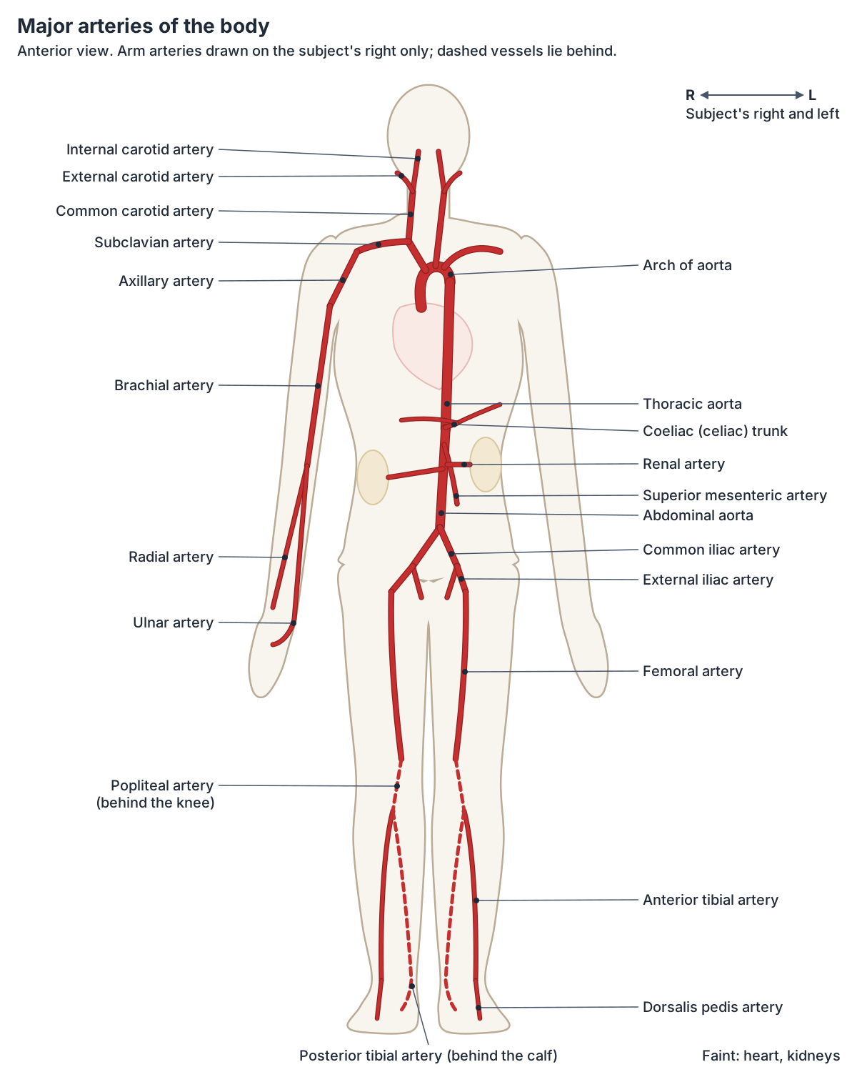
The aorta is the largest artery in the body. The ascending aorta rises about 5 cm from the left ventricle and gives off the coronary arteries. The aortic arch curves to the left and gives three branches: the brachiocephalic trunk (on the right only), the left common carotid artery and the left subclavian artery. (OpenStax Anatomy and Physiology 2e, 20.5: Circulatory Pathways.)
The thoracic aorta gives small branches to the bronchi, pericardium, oesophagus and mediastinum, and intercostal and superior phrenic arteries to the chest wall and diaphragm. The abdominal aorta gives the coeliac (celiac) trunk, which divides into the left gastric, splenic and common hepatic arteries, the superior and inferior mesenteric arteries, and the inferior phrenic, adrenal, renal, gonadal and lumbar arteries. It ends at the level of L4 by dividing into the two common iliac arteries. (OpenStax Anatomy and Physiology 2e, 20.5: Circulatory Pathways.)
Every part of the aorta, its branches and their vertebral levels are set out in the guide to The aorta.
Arteries of the head and neck
Each common carotid artery divides into an external carotid artery, which supplies the face, lower jaw and neck, and an internal carotid artery, which with the vertebral artery is one of the two main suppliers of blood to the brain. The vertebral arteries arise from the subclavian arteries and, at the base of the brain, join the internal carotids in a ring of linked arteries called the arterial circle (circle of Willis). (OpenStax Anatomy and Physiology 2e, 20.5: Circulatory Pathways.)
The carotid and vertebral arteries in the neck are described in the guide to the Carotid arteries.
Arteries of the arm
One artery runs the length of the arm but changes its name as it goes: the subclavian artery becomes the axillary artery in the armpit and the brachial artery in the arm. At the elbow the brachial artery divides into the radial and ulnar arteries, which join in the hand to form the palmar arches that supply the digital arteries of the fingers. (OpenStax Anatomy and Physiology 2e, 20.5: Circulatory Pathways.)
Each artery and vein of the upper limb, with its branches, is traced in Arteries and veins of the arm.
Arteries of the leg
In the lower limb the external iliac artery becomes the femoral artery in the thigh and the popliteal artery behind the knee. The popliteal artery divides into the anterior tibial artery, which continues on the top of the foot as the dorsalis pedis artery, and the posterior tibial artery, which gives off the fibular artery and ends as the medial and lateral plantar arteries in the sole. (OpenStax Anatomy and Physiology 2e, 20.5: Circulatory Pathways.)
The iliac, femoral, popliteal, tibial and foot arteries and the leg pulses are covered in Arteries of the lower limb.
Major arteries of the body
| Artery | Arises from | Main area supplied |
|---|---|---|
| Ascending aorta | Left ventricle | Heart (coronary arteries) |
| Brachiocephalic trunk | Aortic arch (right side only) | Right side of the head, neck and right arm |
| Common carotid artery | Brachiocephalic trunk (right), aortic arch (left) | Head and neck, via the internal and external carotids |
| Vertebral artery | Subclavian artery | Brain (back), spinal cord |
| Subclavian, axillary and brachial arteries | Brachiocephalic trunk (right), aortic arch (left) | Shoulder and arm |
| Radial and ulnar arteries | Brachial artery at the elbow | Forearm and hand |
| Coeliac trunk | Abdominal aorta | Stomach, liver, spleen |
| Superior and inferior mesenteric arteries | Abdominal aorta | Intestines |
| Renal arteries | Abdominal aorta | Kidneys |
| Common iliac arteries | End of the abdominal aorta at L4 | Pelvis and legs |
| Femoral and popliteal arteries | External iliac artery | Thigh and knee |
| Anterior and posterior tibial arteries | Popliteal artery | Leg and foot |
From OpenStax Anatomy and Physiology 2e, section 20.5.
Major veins of the body: the venae cavae and their tributaries
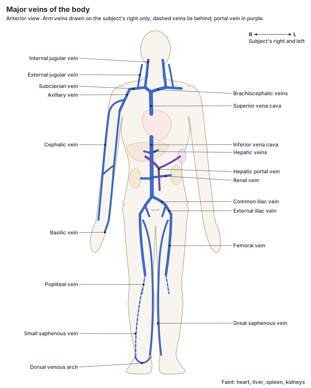
All systemic veins end in one of two great veins. The superior vena cava drains structures above the diaphragm; it is formed by the two brachiocephalic veins, which are fed by the subclavian and jugular veins, and it also receives the azygos vein. The inferior vena cava drains structures below the diaphragm. It forms at about L5 from the common iliac veins and receives the renal, hepatic, gonadal and lumbar veins; the renal veins are normally the largest veins entering it. (OpenStax Anatomy and Physiology 2e, 20.5: Circulatory Pathways.)
The inferior vena cava runs up the right side of the vertebral column with the aorta on its left, passes through the diaphragm at the caval opening at the level of T8 and drains into the right atrium. (Radiopaedia: Inferior vena cava.)
The superior vena cava enters the upper right atrium at about the level of the third rib. (TeachMeAnatomy: Great Vessels of the Heart.)
The course, tributaries and variants of both venae cavae are described on the page about the Inferior and superior vena cava.
Veins of the head, neck, arms and legs
The internal jugular veins drain the brain. The external jugular veins drain the superficial parts of the head and empty into the subclavian veins. (OpenStax Anatomy and Physiology 2e, 20.5: Circulatory Pathways.)
In the arm, digital veins drain into the palmar venous arches and then into the radial, ulnar and median antebrachial veins, which lead to the brachial vein. The superficial cephalic and basilic veins run up the outer and inner sides of the arm, linked at the elbow by the median cubital vein, the usual site for taking blood. The axillary vein becomes the subclavian vein. (OpenStax Anatomy and Physiology 2e, 20.5: Circulatory Pathways.)
In the leg, the dorsal venous arch of the foot drains into the anterior and posterior tibial and fibular veins, which form the popliteal vein behind the knee. This becomes the femoral vein, which receives the great saphenous vein and continues as the external iliac and then the common iliac vein into the inferior vena cava. (OpenStax Anatomy and Physiology 2e, 20.5: Circulatory Pathways.)
The main vein of the neck and its relations are described on the page about the Internal jugular vein.
Major veins of the body
| Vein | Drains | Empties into |
|---|---|---|
| Superior vena cava | Head, neck, arms and chest (above the diaphragm) | Right atrium |
| Inferior vena cava | Abdomen, pelvis and legs (below the diaphragm) | Right atrium |
| Brachiocephalic veins | Head, neck and arms | Superior vena cava |
| Internal jugular vein | Brain | Brachiocephalic vein |
| External jugular vein | Superficial head | Subclavian vein |
| Subclavian and axillary veins | Arm | Brachiocephalic vein |
| Cephalic and basilic veins | Superficial arm | Axillary vein |
| Hepatic portal vein | Stomach, intestines, spleen, pancreas | Liver, then hepatic veins to the inferior vena cava |
| Renal veins | Kidneys | Inferior vena cava |
| Common iliac veins | Pelvis and legs | Inferior vena cava (at L5) |
| Femoral and popliteal veins | Thigh, knee and leg | External iliac vein |
| Great saphenous vein | Superficial leg and foot | Femoral vein |
From OpenStax Anatomy and Physiology 2e, section 20.5.
Hepatic portal system
Blood from the gut does not go straight back to the heart. The hepatic portal vein, formed at about the level of L2 by the superior mesenteric and splenic veins, carries blood from the stomach, intestines, spleen and pancreas to the liver. After passing through the liver sinusoids it leaves through the hepatic veins into the inferior vena cava. (OpenStax Anatomy and Physiology 2e, 20.5: Circulatory Pathways.)
The tributaries, branches and clinical anatomy of the hepatic portal vein are covered on the page about the Portal vein.
Pulse points and blood pressure
The pulse is the expansion and recoil of an artery after each heartbeat. It is usually felt at the radial artery in the wrist or the common carotid artery in the neck. Other pulse points are the temporal and facial arteries of the head, the brachial artery in the upper arm, the femoral artery in the thigh, the popliteal artery behind the knee, the posterior tibial artery near the ankle and the dorsalis pedis artery on the foot. (OpenStax Anatomy and Physiology 2e, 20.2: Blood Flow, Blood Pressure, and Resistance.)
A normal adult blood pressure is about 120/80 mm Hg: 120 when the ventricles contract (systolic) and 80 when they relax (diastolic). The difference, about 40 mm Hg, is the pulse pressure, and the mean arterial pressure normally lies between 70 and 110 mm Hg. (OpenStax Anatomy and Physiology 2e, 20.2: Blood Flow, Blood Pressure, and Resistance.)
Where to feel the radial pulse, and the anatomy of the artery, are covered on the page about the Radial artery.
Common problems of the arteries and veins
An aneurysm is a widening of an artery to more than 50 percent above its normal diameter. It can follow weakness of the wall, as in Marfan syndrome, or a dissection, in which a tear in the inner wall lets blood run inside the wall itself. The main danger is rupture. (TeachMeAnatomy: Great Vessels of the Heart.)
Fluid can also leak out of the vessels into the tissues, causing swelling (oedema, US edema). It has many causes, including high blood pressure and heart failure. (OpenStax Anatomy and Physiology 2e, 20.1: Structure and Function of Blood Vessels.)
Coronary artery disease, angina and heart attacks are explained with the anatomy of the Coronary arteries.
The circulatory system in the 3D model
The 3D model comes from BodyParts3D, a whole-body model of an adult human male released as an anatomical reference database under a CC BY licence. (BodyParts3D, Database Center for Life Science.)
This page highlights the walls of the heart, the aorta and its main branches, the arteries of the neck, arms and legs, the pulmonary vessels, both venae cavae, the large veins of the neck and limbs, the renal veins and the hepatic portal vein. Switch off the skeleton to follow each vessel from the heart to the hands and feet, and click any vessel to open its own page. Capillaries and most small veins are too fine to be modelled, and the model has no external carotid artery or external jugular vein, so the diagrams show those.
Circulatory system in the 3D model
- Abdominal aorta
- The abdominal aorta is the part of the aorta that runs through the abdomen and supplies the abdominal organs, pelvis and lower limbs.
- Anterior tibial artery
- The anterior tibial artery is the artery of the front compartment of the leg and the main source of the dorsalis pedis artery.
- Arch of aorta
- The aortic arch is the curved part of the aorta between the ascending and descending aorta, giving the great vessels to the head and arms.
- Ascending aorta
- The ascending aorta is the first part of the aorta, rising from the left ventricle.
- Axillary artery
- The axillary artery is the main artery of the armpit, continuing the subclavian artery.
- Axillary vein
- The axillary vein is the main vein of the armpit and carries blood from the upper limb.
- Basilic vein
- The basilic vein is a large superficial vein on the medial side of the arm.
- Brachial artery
- The brachial artery is the main artery of the arm, continuing the axillary artery.
- Brachiocephalic artery
- The brachiocephalic artery (trunk) is the first branch of the aortic arch and supplies the right head, neck and arm.
- Brachiocephalic vein
- The brachiocephalic veins are two large veins that unite to form the superior vena cava.
- Celiac trunk
- The celiac trunk is the first major unpaired branch of the abdominal aorta and supplies the foregut.
- Cephalic vein
- The cephalic vein is a large superficial vein on the lateral side of the upper limb.
- Common carotid artery
- The common carotid artery is the main artery of the neck and splits into the external and internal carotid arteries.
- Common iliac artery
- The common iliac arteries are the two terminal branches of the abdominal aorta.
- Common iliac vein
- The common iliac veins are formed by the external and internal iliac veins and unite to form the inferior vena cava.
- Descending thoracic aorta
- The descending thoracic aorta is the part of the aorta in the posterior mediastinum.
- Dorsalis pedis artery
- The dorsalis pedis artery is the continuation of the anterior tibial artery onto the top of the foot.
- External iliac artery
- The external iliac artery is the main artery of the lower limb in the pelvis and becomes the femoral artery.
- External iliac vein
- The external iliac vein is the continuation of the femoral vein above the inguinal ligament.
- Femoral artery
- The femoral artery is the main artery of the thigh, continuing the external iliac artery.
- Femoral vein
- The femoral vein is the main deep vein of the thigh and continues the popliteal vein.
- Great saphenous vein
- The great saphenous vein is the longest vein in the body and runs up the medial side of the leg and thigh.
- Hepatic portal vein
- The hepatic portal vein carries nutrient-rich blood from the gut and spleen to the liver.
- Inferior pulmonary vein
- The inferior pulmonary veins (one per lung) carry oxygenated blood from the lower lobes of the lungs to the left atrium.
- Inferior vena cava
- The inferior vena cava is the largest vein in the body and returns blood from the lower body to the right atrium.
- Internal carotid artery
- The internal carotid artery supplies the brain and the eye.
- Internal iliac artery
- The internal iliac artery supplies the pelvis.
- Internal jugular vein
- The internal jugular vein is the main vein draining the head and neck.
- Median cubital vein
- A superficial vein across the front of the elbow linking the cephalic and basilic veins.
- Popliteal artery
- The continuation of the femoral artery behind the knee.
- Popliteal vein
- A deep vein behind the knee formed by the joining of the anterior and posterior tibial veins.
- Posterior tibial artery
- The larger terminal branch of the popliteal artery in the posterior leg.
- Pulmonary artery
- An artery carrying deoxygenated blood from the heart to a lung.
- Pulmonary trunk
- The large artery arising from the right ventricle that carries blood to the lungs.
- Radial artery
- One of the two terminal branches of the brachial artery, running on the lateral forearm.
- Renal artery
- A large paired branch of the abdominal aorta supplying the kidney.
- Renal vein
- A large vein draining the kidney into the inferior vena cava.
- Small saphenous vein
- A superficial vein of the posterior leg.
- Subclavian artery
- The major artery to the upper limb, neck and part of the brain.
- Subclavian vein
- The large vein draining the upper limb into the brachiocephalic vein.
- Superior mesenteric artery
- An unpaired artery of the midgut.
- Superior pulmonary vein
- One of the pulmonary veins draining the upper lung lobes into the left atrium.
- Superior vena cava
- The large vein draining the upper body into the right atrium.
- Ulnar artery
- The larger terminal branch of the brachial artery.
- Vertebral artery
- A branch of the subclavian artery supplying the brain and spinal cord.
- Wall of atrium
- The muscular walls of the right and left atria, the thin-walled receiving chambers of the heart.
- Wall of ventricle
- The muscular wall of a ventricle of the heart.
Frequently asked questions
What are the main parts of the cardiovascular system?
The cardiovascular system is the heart, the blood vessels and the blood inside them. The heart pumps; arteries carry blood away from it; capillaries are the tiny vessels where oxygen, nutrients and wastes are exchanged with the tissues; and veins carry blood back to the heart. An adult has about 4 to 6 litres of blood.
Is the circulatory system the same as the cardiovascular system?
Yes. Circulatory system and cardiovascular system are two names for the same system of the heart and blood vessels. The lymphatic system is separate, although it is linked: it collects excess fluid from the tissues and returns it to the bloodstream through the subclavian veins at the base of the neck.
What is the difference between arteries and veins?
Arteries carry blood away from the heart at high pressure, so they have thick, muscular and elastic walls and small, round lumens. Veins carry blood back to the heart at low pressure, so they have thin walls, wide flattened lumens and, especially in the limbs, valves that stop blood flowing backwards. About two-thirds of the blood is in the veins at rest.
Do arteries always carry oxygenated blood?
No. An artery is defined by carrying blood away from the heart, not by its oxygen content. Systemic arteries such as the aorta carry oxygen-rich blood, but the pulmonary trunk and pulmonary arteries carry oxygen-poor blood from the right ventricle to the lungs. In the same way, the pulmonary veins carry oxygen-rich blood back to the heart.
What are the three layers of a blood vessel wall?
Arteries and veins have three layers, or tunics. The tunica intima is the inner lining of endothelium. The tunica media is smooth muscle and elastic tissue and is the thickest layer in arteries. The tunica externa (adventitia) is the outer connective tissue coat and is the thickest layer in veins. Capillaries have only the endothelium and its basement membrane.
Why do veins have valves?
Blood pressure in the veins is low, especially in the legs, where blood has to return against gravity. Venous valves are folds of thickened endothelium that let blood flow only towards the heart. When nearby muscles contract and squeeze the veins, the valves stop blood being pushed backwards. Faulty valves let blood pool, which causes varicose veins.
What is the largest artery in the body?
The aorta is the largest artery. It leaves the left ventricle, rises as the ascending aorta, arches to the left over the heart, descends through the chest as the thoracic aorta and through the abdomen as the abdominal aorta, and divides into the two common iliac arteries at the level of the fourth lumbar vertebra.
References
- OpenStax. Anatomy and Physiology 2e, §1.2 Structural Organization of the Human Body. Rice University, 2022.
- OpenStax. Anatomy and Physiology 2e, §18.1 An Overview of Blood. Rice University, 2022.
- OpenStax. Anatomy and Physiology 2e, §19.1 Heart Anatomy. Rice University, 2022.
- OpenStax. Anatomy and Physiology 2e, §20.1 Structure and Function of Blood Vessels. Rice University, 2022.
- OpenStax. Anatomy and Physiology 2e, §20.2 Blood Flow, Blood Pressure, and Resistance. Rice University, 2022.
- OpenStax. Anatomy and Physiology 2e, §20.5 Circulatory Pathways. Rice University, 2022.
- OpenStax. Anatomy and Physiology 2e, §20.6 Development of Blood Vessels and Fetal Circulation. Rice University, 2022.
- OpenStax. Anatomy and Physiology 2e, §21.1 Anatomy of the Lymphatic and Immune Systems. Rice University, 2022.
- Jones O. Blood Vessels. TeachMeAnatomy.
- Jones O. Great Vessels of the Heart. TeachMeAnatomy.
- Inferior vena cava. Radiopaedia reference article.
- Circulatory System. Cleveland Clinic health library.
- BodyParts3D. Database Center for Life Science, Japan.
- Mitsuhashi N. et al. (2009). BodyParts3D: 3D structure database for anatomical concepts. Nucleic Acids Research 37, D782–D785. Data © The Database Center for Life Science, CC BY 4.0.
Related anatomy
- HeartHeart anatomy with labelled diagrams and a 3D model: location, pericardium, wall layers, four chambers, valves, great…
- AortaAorta anatomy explained: ascending aorta, aortic arch and its three branches, thoracic aorta, and every abdominal…
- Inferior vena cavaInferior vena cava anatomy: course, relations, tributaries by vertebral level, embryology, variants and IVC filters…
- Upper limb blood vesselsArteries and veins of the arm and hand: subclavian, axillary branches, brachial, radial and ulnar arteries, palmar…
- Lower limb arteriesArteries of the leg explained: common, internal and external iliac, femoral, deep femoral, popliteal, tibial, fibular…
- Coronary arteriesCoronary arteries explained: left main, LAD and diagonals, circumflex, RCA and posterior descending artery, dominance…
Explore the whole body in the interactive 3D human anatomy model, or browse the A–Z list of human structures.