

What are the parts of the nose?
The nose has an external part and an internal part. The external nose is pyramid-shaped: its root is at the top, continuous with the forehead, its dorsum runs down the middle, and it ends below in the apex, or tip. Just below the apex are the nostrils (nares), which open into the nasal vestibule. (TeachMeAnatomy: The External Nose.)
Behind the nostrils are two nasal cavities, one on each side of the nasal septum. Each opens to the face through a naris and to the nasopharynx at the back through a choana. Twelve skull bones help to build the nasal cavity: the paired nasal, maxillary, palatine and lacrimal bones and the unpaired ethmoid, sphenoid, frontal and vomer. The ethmoid makes the largest contribution. (Kenhub: Nasal cavity.)
Four pairs of paranasal sinuses, named after the frontal, maxillary, sphenoid and ethmoid bones that contain them, are air spaces in the bones around the nasal cavity. They are lined with the same respiratory mucosa and open into the nose. (OpenStax Anatomy and Physiology 2e, 22.1: Organs and Structures of the Respiratory System.)
The four pairs of air spaces around the nose and where each one drains. Paranasal sinuses.
External nose anatomy: root, dorsum, tip and alae
OpenStax names the surface landmarks of the external nose as follows. The root is the region between the eyebrows. The bridge joins the root to the rest of the nose, and the dorsum nasi is its length. The apex is the tip. Each ala is a cartilage-supported wing that forms the outer side of a nostril. OpenStax also describes the philtrum, the concave surface that connects the nose to the upper lip. (OpenStax Anatomy and Physiology 2e, 22.1: Organs and Structures of the Respiratory System.)
The upper part of the external nose is bony and the lower part is cartilage. Skin over the bony part is thin; skin over the cartilage is thicker and has many sebaceous glands. This skin continues through the nostrils into the vestibule, where hairs filter the air we breathe in. (TeachMeAnatomy: The External Nose.)
Small muscles of facial expression move the nose. Nasalis is the largest: its transverse part compresses the nasal opening and its alar part widens it. Depressor septi nasi pulls the septum down to help widen the nostril, and procerus pulls the eyebrows down and wrinkles the skin over the bridge. All are supplied by the facial nerve (CN VII). (TeachMeAnatomy: Muscles of Facial Expression.)
Nasalis, procerus and the other muscles of facial expression, with labelled diagrams. Facial muscles.
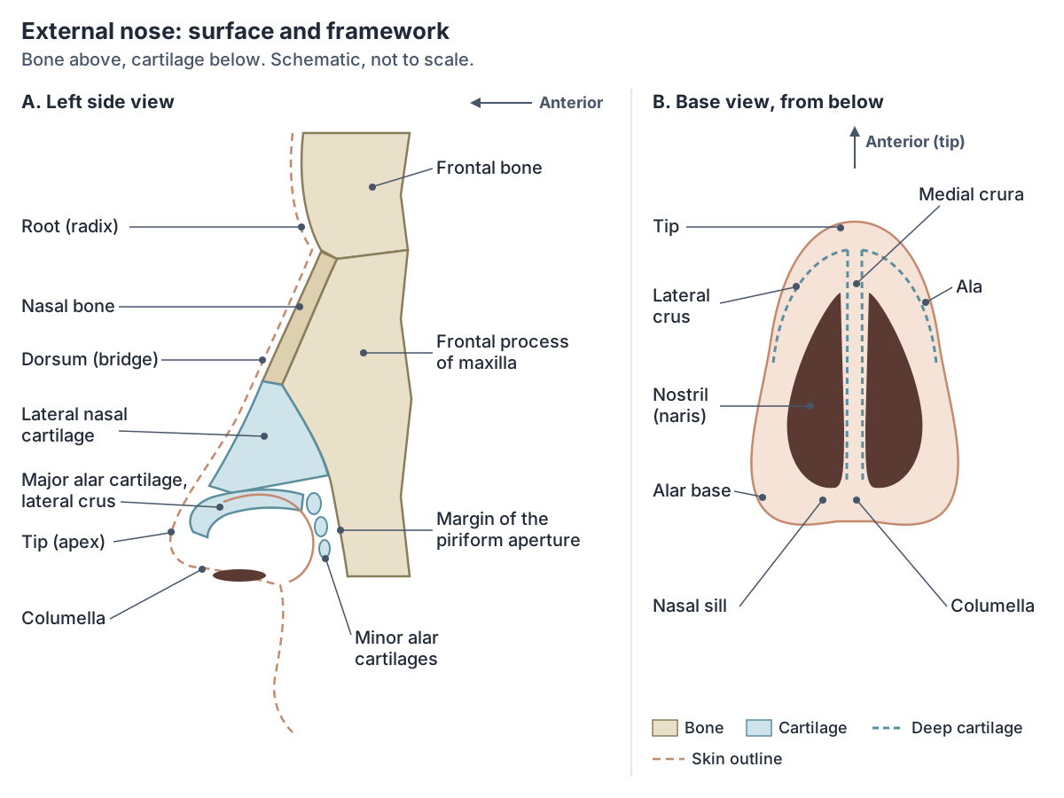
Is the nose bone or cartilage? Bones and cartilages of the nose
Both. The bony upper part is formed by the nasal bones, the maxillae and the frontal bone. The cartilaginous lower part is formed by two lateral cartilages, two major alar cartilages, one septal cartilage and some smaller (minor) alar cartilages. (TeachMeAnatomy: The External Nose.)
The nasal bones are small, paired, oblong bones. Each one meets the frontal bone above at the frontonasal suture, the frontal process of the maxilla at the side, its partner in the midline, and ends below in a free edge that continues into the lateral nasal cartilage. Procerus and nasalis attach to their outer surface, and a groove on the inner surface carries the anterior ethmoidal nerve. (Radiopaedia: Nasal bone.)
Kenhub lists the cartilages by what they shape: the major alar cartilage forms the apex, the minor alar cartilages support the ala, the lateral processes of the septal cartilage form the dorsum, and the septal cartilage bounds the nostrils medially. (Kenhub: Nasal cavity.)
Each major alar cartilage is a thin, flexible plate bent on itself. Its lateral crus forms the outer wall of the nostril and its medial crus forms the inner wall beside the septum. (AnatomyZone: Major alar cartilage.)
The major alar cartilage forms the front outer and front inner borders of each nostril and keeps it open. Its upper margin is attached to the lateral nasal cartilage, and behind it three or four minor alar cartilages complete the framework. Because the plate is thin and flexible, the lower nose is mobile. (Elsevier Complete Anatomy: Major alar cartilage.)
Bones and cartilages of the nose
| Part | Tissue | Where it is | What it does |
|---|---|---|---|
| Nasal bones (paired) | Bone | Upper bridge, meeting in the midline below the frontal bone | Form the bony bridge; the lateral nasal cartilages attach to their lower edge |
| Frontal processes of the maxillae | Bone | Each side of the nasal bones | Form the bony side walls of the upper nose |
| Lateral nasal (upper lateral) cartilages | Cartilage | Middle third, below the nasal bones | Shape the side walls; their lower edge bounds the internal nasal valve |
| Major alar cartilages (paired) | Cartilage | Tip and around each nostril; lateral and medial crura | Shape the tip and keep the nostrils open; the medial crura lie in the columella |
| Minor alar cartilages | Cartilage | Three or four small plates behind each major alar cartilage | Complete the framework of the ala |
| Septal (quadrangular) cartilage | Hyaline cartilage | Front of the nasal septum, in the midline | Divides the nose into two sides and supports the dorsum |
| Perpendicular plate of the ethmoid | Bone | Upper back part of the septum | Bony septum; hangs from the cribriform plate |
| Vomer | Bone | Lower back part of the septum | Bony septum; its free back edge separates the two choanae |
From TeachMeAnatomy, OpenStax, Radiopaedia, Kenhub and Elsevier Complete Anatomy (see references). BodyParts3D calls the upper lateral cartilage the lateral nasal cartilage; some texts call it the lateral process of the septal cartilage.
Nostril anatomy: nares, vestibule, columella and nasal valve
The nostrils (nares) are pear-shaped openings into the nasal vestibule. Each is bounded medially by the nasal septum and laterally by the ala nasi, the cartilage-supported wing of the nose. (TeachMeAnatomy: The External Nose.)
The nasal vestibule is the small, slightly widened space just inside each nostril, at the very front of the nasal cavity. The columella forms its medial wall, the ala its lateral and front wall, and the nasal sill its back edge. (Radiopaedia: Nasal vestibule.)
The columella is the strip of soft tissue between the two nostrils. It contains the septum mobile nasi, the lowest front part of the septum, which is formed by the medial crura of the two major alar cartilages rather than by the septal cartilage. (Radiopaedia: Nasal septum.)
The nares and front of the nasal cavity are lined with mucous membrane containing sebaceous glands and hair follicles, which block large particles. Deeper in, the lining changes to respiratory epithelium. (OpenStax Anatomy and Physiology 2e, 22.1: Organs and Structures of the Respiratory System.)
The internal nasal valve is the narrowest part of the nasal passage. It is bounded by the septum medially and by the upper and lower lateral cartilages above and to the side. Because its cross-section is so small, it accounts for between half and two-thirds of the total resistance to airflow through the nose. The angle at its apex is about 10 to 15 degrees. The external nasal valve is the part of the side wall that collapses when you breathe in hard. (Pirola F, Kim R, Douglas R. Nasal valve obstruction: a comprehensive analysis of the current literature. Front Surg 2025.)
Lee and Won place the internal valve about 1.5 cm inside the nostril, between the septum and the lower edge of the upper lateral cartilage, with the inferior concha below. They give a slightly wider normal range for its angle, about 10 to 20 degrees, and cite a study that measured about 22 degrees in Asian noses and 15.5 degrees in Caucasian noses. (Lee DY, Won TB. Management of Nasal Valve Dysfunction. Clin Exp Otorhinolaryngol 2024.)
Nasal cavity anatomy: walls, openings and regions
Each nasal cavity has four walls. The roof is the cribriform plate of the ethmoid. The floor is the hard palate, formed by the palatine processes of the maxillae and the horizontal plates of the palatine bones. The lateral wall carries the three conchae. The medial wall is the nasal septum. In front the cavity opens at the nostril; behind, it opens into the nasopharynx through the choana (posterior nasal aperture). (Radiopaedia: Nasal cavity.)
The nasal cavity is the uppermost part of the respiratory tract. It has three regions. The vestibule is the area around the front opening. The respiratory region, which is most of the cavity, is lined by ciliated pseudostratified epithelium with mucus-secreting goblet cells. The olfactory region at the top of the cavity carries the smell receptors. (TeachMeAnatomy: The Nasal Cavity.)
Several openings link the cavity to its neighbours. The cribriform plate lets olfactory nerve fibres through to the brain. The sphenopalatine foramen, at the level of the superior meatus, connects it to the pterygopalatine fossa and carries the sphenopalatine artery and nasal nerves. The incisive canal connects it to the roof of the mouth. (TeachMeAnatomy: The Nasal Cavity.)
The nasopharynx behind the nose and the rest of the pharynx and larynx. Throat anatomy.
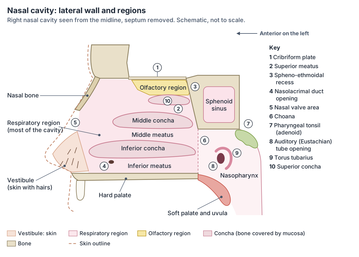
Nasal conchae (turbinates) and meatuses
Three curved shelves, the superior, middle and inferior nasal conchae, project from each lateral wall. The superior and middle conchae are parts of the ethmoid bone; the inferior conchae are separate bones. The conchae increase the surface area of the cavity and disturb the incoming air, and together with the meatuses they also trap water during exhalation, which helps to prevent dehydration. (OpenStax Anatomy and Physiology 2e, 22.1: Organs and Structures of the Respiratory System.)
The space under each concha is a meatus. The inferior meatus, between the inferior concha and the floor, receives the nasolacrimal duct. The middle meatus receives the frontal and maxillary sinuses and the anterior ethmoidal cells through the semilunar hiatus. The superior meatus receives the posterior ethmoidal cells. Above and behind the superior concha is the spheno-ethmoidal recess. (TeachMeAnatomy: The Nasal Cavity.)
The auditory (Eustachian) tube opens into the nasopharynx just behind the inferior concha. The tube links the middle ear to the nasopharynx, equalises pressure across the eardrum and drains secretions from the middle ear. (TeachMeAnatomy: The Eustachian Tube.)
Which sinus drains into which meatus, with front and side diagrams. Paranasal sinuses and drainage.
What opens into the nasal cavity
| Space | Where it is | What opens into it |
|---|---|---|
| Spheno-ethmoidal recess | Above and behind the superior concha | Sphenoid sinus |
| Superior meatus | Between the superior and middle conchae | Posterior ethmoidal air cells |
| Middle meatus | Between the middle and inferior conchae | Frontal and maxillary sinuses and the anterior ethmoidal air cells, through the semilunar hiatus |
| Inferior meatus | Between the inferior concha and the floor | Nasolacrimal duct (tears) |
| Nasopharynx, just behind the cavity | Lateral wall, level with the inferior meatus | Auditory (Eustachian) tube from the middle ear |
From TeachMeAnatomy and Radiopaedia. The paranasal sinuses page covers sinus drainage in detail.
Nasal septum: cartilage and bone
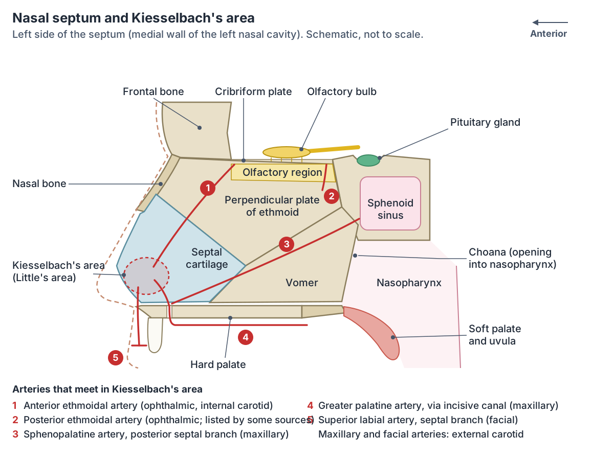
The nasal septum divides the nasal cavity into left and right halves. Its front part is septal cartilage, a flexible plate of hyaline cartilage. Its back part is bone: the perpendicular plate of the ethmoid above and the vomer below. (OpenStax Anatomy and Physiology 2e, 22.1: Organs and Structures of the Respiratory System.)
Radiopaedia lists the components in more detail. The septal (quadrangular) cartilage forms the front of the septum. The perpendicular plate of the ethmoid forms the upper back part and meets the vomer along a diagonal line. Along the floor, the nasal crests of the palatine bones and maxillae form a strip of bone that the vomer sits on. In some people a tail of septal cartilage reaches back between the ethmoid and vomer. (Radiopaedia: Nasal septum.)
A deviated septum is one that is off the midline, from injury or present from birth. It can partly or completely block one side of the nose and is corrected surgically by septoplasty. (Kenhub: Nasal cavity.)
Septal cartilage has no blood vessels of its own: it relies on diffusion from the perichondrium covering it. After an injury, blood can collect between the two (a septal haematoma) and the cartilage can die, so the bridge loses support and sinks, giving a saddle nose deformity. (TeachMeAnatomy: The External Nose.)
The thin midline bone that forms the lower back part of the septum. Vomer.
Blood supply of the nose and Kiesselbach's area
The nasal cavity has a rich blood supply from both the internal and external carotid arteries. From the internal carotid come the anterior and posterior ethmoidal arteries, branches of the ophthalmic artery that descend through the cribriform plate. From the external carotid come the sphenopalatine, greater palatine, superior labial and lateral nasal arteries. These vessels anastomose, especially at the front of the nose. The veins follow the arteries and drain to the pterygoid venous plexus, the facial vein or the cavernous sinus. (TeachMeAnatomy: The Nasal Cavity.)
Kiesselbach's plexus, also called Kiesselbach's area or Little's area, is on the front lower part of the nasal septum. Radiopaedia lists four arteries that meet there: the anterior ethmoidal (from the ophthalmic artery), the sphenopalatine and greater palatine (from the maxillary artery) and the septal branches of the superior labial artery (from the facial artery). About 80 to 90% of nosebleeds start in this area. (Radiopaedia: Kiesselbach plexus.)
Kenhub counts five vessels, adding the posterior ethmoidal artery through its septal branches, and describes them converging over the septal cartilage in the front lower quarter of the septum. It notes that Little's area is a common site of nosebleeds in both children and adults. (Kenhub: Kiesselbach's plexus.)
A second meeting point, Woodruff's area, lies behind the inferior concha, where the sphenopalatine and pharyngeal arteries anastomose. (Radiopaedia: Nasal cavity.)
Anterior nosebleeds from Kiesselbach's plexus are the most common kind. Posterior nosebleeds usually come from the maxillary artery, and the blood may run down the throat rather than out of the nose. (Kenhub: Nasal cavity.)
The veins of the external nose drain to the facial vein and then the internal jugular vein. The facial vein connects with the cavernous sinus through the ophthalmic vein. This is why infection around the nose, in the so-called danger triangle of the face, can rarely spread into the skull. (TeachMeAnatomy: The External Nose.)
Nerves of the nose and the sense of smell
Smell begins in the olfactory epithelium, a small region at the top of the nasal cavity. Its bipolar receptor neurons send axons up through tiny holes in the cribriform plate of the ethmoid to the olfactory bulb on the underside of the frontal lobe. Smell is the only sense that reaches the cerebral cortex without first relaying in the thalamus. (OpenStax Anatomy and Physiology 2e, 14.1: Sensory Perception.)
Touch, pain and temperature in the nasal cavity are carried by the trigeminal nerve. The ophthalmic division (V1) supplies roughly the front upper half and the maxillary division (V2) the back lower half. On the septum, the anterior ethmoidal nerve (V1) supplies the front and the nasopalatine nerve (V2) the back lower part. (Radiopaedia: Nasal cavity.)
On the outside, the external nasal nerve, a branch of the ophthalmic nerve, supplies the skin of the dorsum, the alae and the vestibule, while the infraorbital nerve from the maxillary nerve supplies the sides of the nose. (TeachMeAnatomy: The External Nose.)
TeachMeAnatomy's map of the trigeminal nerve agrees: the ophthalmic division (V1) supplies the dorsum of the nose and the frontal and ethmoidal sinuses, and the maxillary division (V2) supplies the nasal cavity, the side of the nose, the maxillary sinus and the upper lip. (TeachMeAnatomy: The Trigeminal Nerve (CN V).)
Autonomic nerves control the nasal glands and blood flow. Parasympathetic fibres that travel with the facial nerve make the serous glands secrete and keep the walls moist; sympathetic fibres from the T1 level of the spinal cord regulate blood flow in the lining. (Kenhub: Nasal cavity.)
Lymph from the front of the nasal cavity drains with the lymph of the external nose; lymph from the back drains to the lateral pharyngeal nodes or the deep cervical chain. (Radiopaedia: Nasal cavity.)
The submandibular lymph nodes, under the jaw, collect lymph from the sides of the nose, the cheeks and the upper lip. All lymph from the head and neck finally passes through the deep cervical nodes. (TeachMeAnatomy: Lymphatics of the Head and Neck.)
The olfactory, trigeminal and facial nerves that serve the nose. Cranial nerves.
What does the nose do?
TeachMeAnatomy lists four functions of the nasal cavity: it warms and humidifies the air we breathe in, traps and removes pathogens and particles, gives us the sense of smell, and drains and clears the paranasal sinuses and the tear ducts. (TeachMeAnatomy: The Nasal Cavity.)
Goblet cells in the respiratory epithelium make mucus that traps debris. The cilia beat constantly, sweeping the mucus towards the throat, where it is swallowed. Capillaries under the lining warm the incoming air, and secretory cells release the antibacterial enzyme lysozyme and defensins. (OpenStax Anatomy and Physiology 2e, 22.1: Organs and Structures of the Respiratory System.)
Hairs in the vestibule filter the air as it enters, before it reaches the rest of the nasal cavity. (TeachMeAnatomy: The External Nose.)
Nosebleeds, broken nose and other clinical notes
Nosebleeds (epistaxis) most often come from the front third of the nose, in Kiesselbach's area. Causes include local factors such as injury and general ones such as high blood pressure. (TeachMeAnatomy: The Nasal Cavity.)
The nasal bones are the most commonly fractured bones of the face. Fractures are easily missed when the face is very swollen. The worst complication is a septal haematoma, which can lead to perforation and death of the septal cartilage, so it should always be looked for. (Radiopaedia: Nasal bone fracture.)
A fracture of the cribriform plate can let cerebrospinal fluid leak out through the nose, with a risk of meningitis or brain abscess, and damage to the olfactory bulb can cause loss of smell (anosmia). Because the auditory tube opens into the nasopharynx, infection of the nose and throat can spread to the middle ear, and swelling can block the tube and reduce hearing. (TeachMeAnatomy: The Nasal Cavity.)
Rhinitis, inflammation of the nasal lining, causes congestion, sneezing, a runny nose and itching. Allergic rhinitis (hay fever) is its most common cause. (Kenhub: Nasal cavity.)
The cribriform plate, perpendicular plate and the superior and middle conchae. Ethmoid bone.
Nose anatomy in the 3D model
- Anterior ethmoidal nerve
- The anterior ethmoidal nerve is a terminal sensory branch of the nasociliary nerve (V1) supplying the anterior nasal cavity and nose.
- Ethmoid
- A delicate cube-shaped bone between the orbits.
- Inferior nasal concha
- A thin curved bone on the lateral wall of the nose.
- Lateral nasal cartilage
- The lateral nasal cartilages are paired triangular cartilages forming the upper lateral part of the external nose.
- Major alar cartilage
- The major alar cartilage is a U-shaped cartilage forming the tip and sides of the nostril.
- Nasal bone
- A small, paired rectangular bone forming the bridge of the nose.
- Nasolacrimal duct
- The nasolacrimal duct carries tears from the lacrimal sac into the nasal cavity.
- Septal nasal cartilage
- The septal nasal cartilage is the cartilage that forms the front part of the nasal septum.
- Vomer
- A thin, flat bone of the nasal septum.
Frequently asked questions
What are the parts of the nose?
The external nose has a root between the eyebrows, a dorsum (bridge), a tip (apex), two alae (wings) and two nostrils separated by the columella. Inside are two nasal cavities divided by the septum, each with a vestibule, three conchae with meatuses beneath them, and a choana opening into the nasopharynx. The paranasal sinuses open into the cavities.
Is the nose made of bone or cartilage?
Both. The upper third, including the bridge, is bone: the two nasal bones and the frontal processes of the maxillae, below the frontal bone. The lower part is cartilage: the paired lateral nasal and major alar cartilages, small minor alar cartilages and the septal cartilage. The back of the septum is bone, made by the ethmoid and vomer.
What is the inside of the nose called?
The space inside is the nasal cavity, split into right and left halves by the nasal septum. Just inside each nostril is the vestibule, lined with skin and hairs. The rest is the respiratory region, lined with ciliated mucous membrane, and at the very top is a small olfactory region that detects smells.
What is the skin between the nostrils called?
The columella. It is the strip of soft tissue that separates the two nostrils and joins the tip of the nose to the upper lip. It contains the medial crura of the two major alar cartilages, which form the lowest front part of the nasal septum, called the septum mobile nasi.
Why do most nosebleeds start at the front of the nose?
Several arteries from both the internal and external carotid systems meet on the front lower septum in Kiesselbach's (Little's) area: the anterior ethmoidal, sphenopalatine, greater palatine and superior labial arteries, and in some descriptions the posterior ethmoidal artery. The area is just inside the nostril, where the lining is easily dried or injured. Radiopaedia puts the share of nosebleeds starting there at 80 to 90%.
Where does mucus from the nose go?
Most of it goes backwards. Cilia on the respiratory lining beat constantly and sweep the mucus layer, with the dust and microbes it has trapped, towards the throat, where it is swallowed. Goblet cells in the lining keep producing new mucus to replace it.
How does smell reach the brain?
Odour molecules dissolve in the mucus of the olfactory epithelium at the top of the nasal cavity and bind receptors on olfactory neurons. Their axons pass through holes in the cribriform plate of the ethmoid to the olfactory bulb. From there signals travel to the olfactory cortex in the temporal lobe and to the limbic system, without relaying in the thalamus.
References
- TeachMeAnatomy. The External Nose: Structure, Vasculature, Innervation.
- TeachMeAnatomy. The Nasal Cavity: Structure, Vasculature, Innervation.
- TeachMeAnatomy. The Trigeminal Nerve (CN V).
- TeachMeAnatomy. Muscles of Facial Expression.
- TeachMeAnatomy. The Eustachian Tube.
- TeachMeAnatomy. Lymphatics of the Head and Neck.
- Betts JG, et al. Anatomy and Physiology 2e, §22.1 Organs and Structures of the Respiratory System. OpenStax, Rice University, 2022.
- Betts JG, et al. Anatomy and Physiology 2e, §14.1 Sensory Perception. OpenStax, Rice University, 2022.
- Kenhub. Nasal cavity: Anatomy, structure, parts, blood supply.
- Kenhub. Kiesselbach's plexus.
- Radiopaedia. Nasal cavity (reference article).
- Radiopaedia. Nasal septum (reference article).
- Radiopaedia. Kiesselbach plexus (reference article).
- Radiopaedia. Nasal bone (reference article).
- Radiopaedia. Nasal bone fracture (reference article).
- Radiopaedia. Nasal vestibule (reference article).
- Elsevier Complete Anatomy. Major alar cartilage.
- AnatomyZone. Major alar cartilage.
- Pirola F, Kim R, Douglas R. Nasal valve obstruction: a comprehensive analysis of the current literature and proposal of a management algorithm. Frontiers in Surgery 2025;12:1549915.
- Lee DY, Won TB. Management of Nasal Valve Dysfunction. Clinical and Experimental Otorhinolaryngology 2024;17(3):189-197.
- Mitsuhashi N. et al. (2009). BodyParts3D: 3D structure database for anatomical concepts. Nucleic Acids Research 37, D782–D785. Data © The Database Center for Life Science, CC BY 4.0.
Related anatomy
- Paranasal sinusesParanasal sinuses anatomy: the maxillary, frontal, ethmoid and sphenoid sinuses, where each drains, the nasal conchae…
- Ethmoid boneThe ethmoid bone (os ethmoidale) explained: cribriform plate, crista galli, perpendicular plate, ethmoid sinus air…
- VomerVomer bone anatomy: where it sits in the nasal septum, its borders and alae, the six bones it joins, the nasopalatine…
- Nasal boneNasal bone: A small, paired rectangular bone forming the bridge of the nose. See where it is in an interactive 3D…
- Head anatomyHead anatomy guide: skull, brain, face, sense organs and ENT structures, head and neck muscles, vessels, nerves and…
- Throat anatomyThroat anatomy with a 3D model and labelled diagrams: the three parts of the pharynx, constrictor muscles, tonsils…
Explore the whole body in the interactive 3D human anatomy model, or browse the A–Z list of human structures.