

What is the throat?
In everyday language the throat is the pharynx and larynx together: a ring-like muscular tube that is the passageway for air, food and liquid. It connects the mouth and nose to the windpipe, the lungs and the oesophagus, and it also helps to form speech. (Children's Hospital of Philadelphia: Throat Anatomy and Physiology.)
The pharynx is a muscular tube that connects the oral and nasal cavities to the larynx and the oesophagus. It runs from the base of the skull to the inferior border of the cricoid cartilage, at the level of C6. (TeachMeAnatomy: The Pharynx.)
Kenhub gives the length of the pharynx as 12 to 14 cm. The pharynx begins in the head behind the nasal cavity, runs down behind the mouth and merges with the larynx and oesophagus. (Kenhub: Pharynx.)
Cleveland Clinic describes the pharynx, which it also calls the throat, as a muscular tube in the middle of the neck, below the base of the skull and above the oesophagus and larynx, and gives a shorter length of about 4 inches (about 10 cm). The sources do not say exactly where they measure from, so treat the length as roughly 10 to 14 cm. (Cleveland Clinic: Pharynx.)
The muscles, vessels and organs around the throat in the neck. Neck anatomy.
Parts of the throat: nasopharynx, oropharynx and laryngopharynx
The pharynx has three parts. The nasopharynx, at the top, is used only for breathing and speech. The oropharynx and the laryngopharynx below it carry both air and food. (OpenStax Anatomy and Physiology 2e, 23.3: The Mouth, Pharynx, and Esophagus.)
Radiopaedia sets the boundaries as follows. The nasopharynx lies behind the choanae of the nose, from the vault of the pharynx down to the soft palate. The oropharynx lies behind the base of the tongue and below the soft palate, with the palatoglossal and palatopharyngeal arches at its sides. The laryngopharynx, also called the hypopharynx, lies below the upper border of the epiglottis and above the cricoid cartilage, and opens in front into the larynx. (Radiopaedia: Pharynx.)
The pharyngeal wall has an incomplete outer layer of circular muscle and an inner layer of longitudinal muscle. Inside the muscles lies a sheet of connective tissue, the pharyngobasilar fascia, which is lined by mucous membrane. (National University of Singapore, Department of Anatomy: HN32 The Pharynx.)
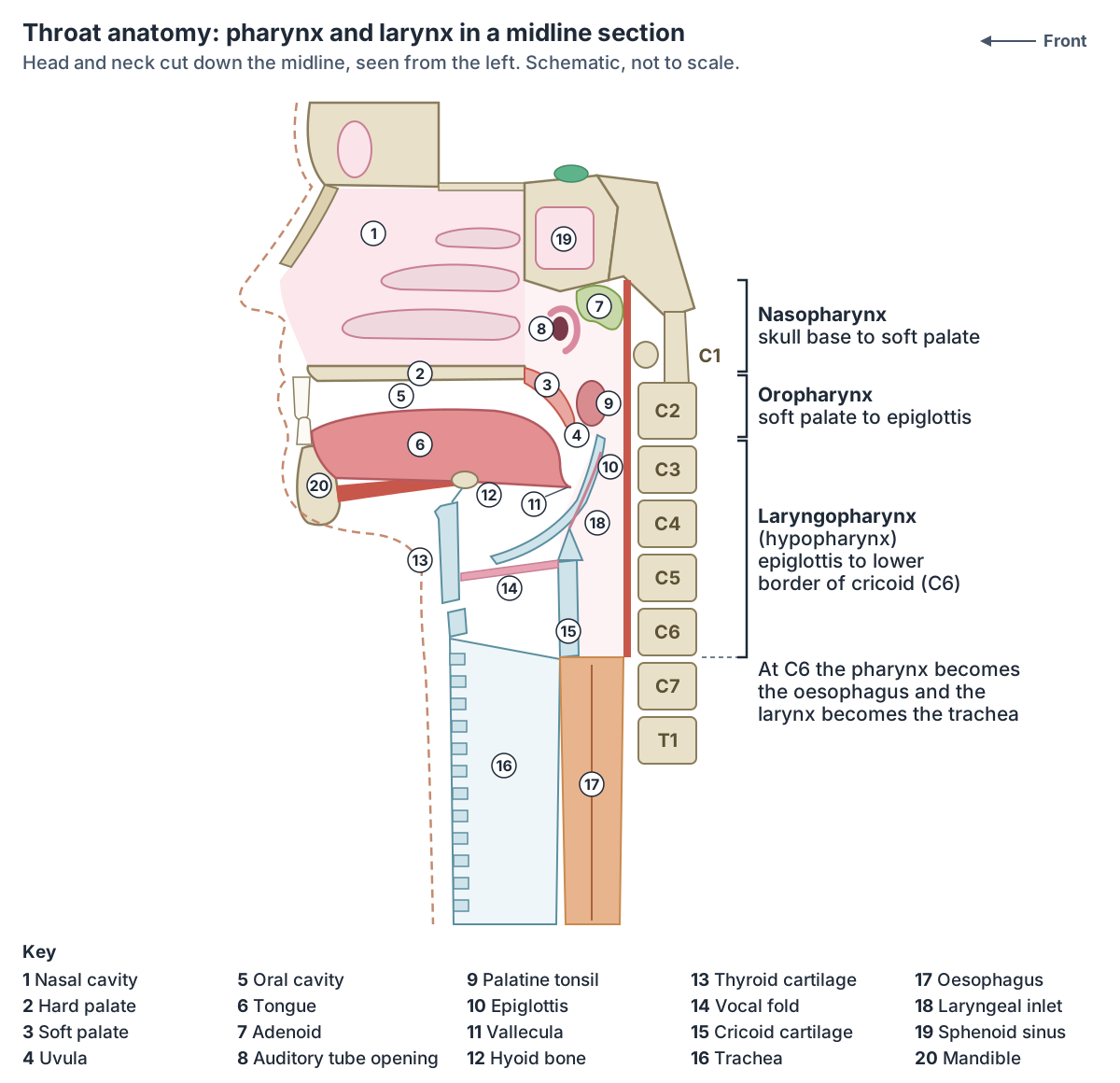
The three parts of the pharynx
| Part | From and to | Opens in front into | Main features | Lining |
|---|---|---|---|---|
| Nasopharynx | Base of the skull to the soft palate; back wall in front of C1 and C2 | Nasal cavity, through the choanae | Adenoid (pharyngeal tonsil), auditory tube openings, torus tubarius, pharyngeal recess | Ciliated pseudostratified columnar epithelium with goblet cells |
| Oropharynx | Soft palate to the tip of the epiglottis or the level of the hyoid | Mouth, through the oropharyngeal isthmus | Back third of the tongue with lingual tonsil, palatine tonsils between the arches, valleculae | Stratified squamous epithelium |
| Laryngopharynx (hypopharynx) | Tip of the epiglottis (hyoid level) to the lower border of the cricoid cartilage at C6 | Larynx, through the laryngeal inlet | Piriform fossae, postcricoid region, back wall; continues as the oesophagus | Mucosa over the inferior constrictor |
Boundaries from Radiopaedia and TeachMeAnatomy; linings from TeachMeAnatomy, OpenStax and Radiopaedia. Sources give the oropharynx's lower limit either as the tip of the epiglottis or as the level of the hyoid bone.
Nasopharynx anatomy
The nasopharynx is the top part of the throat, behind the nose. In front are the choanae and the back edge of the nasal septum (vomer). Its roof, the vault, is formed by the clivus and the floor of the sphenoid sinus, and its back wall lies in front of the first and second cervical vertebrae. Below, it reaches the junction of the hard and soft palates, and the upper surface of the soft palate forms its moving floor. (Radiopaedia: Nasopharynx.)
The opening of the auditory (Eustachian) tube sits in the middle of each side wall. Behind it the cartilage of the tube raises a ridge, the torus tubarius, and behind that is a pocket called the fossa of Rosenmüller (lateral pharyngeal recess). Lymphoid tissue around the tube opening forms the tubal tonsil, and the adenoid lies in the roof. (Radiopaedia: Nasopharynx.)
The nasopharynx conditions the air we breathe in and is lined by ciliated pseudostratified columnar epithelium with goblet cells. The adenoids (pharyngeal tonsil) sit in its upper back part. They enlarge between about 3 and 8 years of age and then shrink. (TeachMeAnatomy: The Pharynx.)
The auditory tube connects the middle ear to the nasopharynx. It equalises the air pressure on the two sides of the eardrum and drains secretions from the middle ear. Tensor veli palatini does most of the work of opening it, helped by levator veli palatini and salpingopharyngeus. (TeachMeAnatomy: The Eustachian Tube.)
During swallowing the soft palate and uvula swing upwards like a pendulum to close off the nasopharynx, so food cannot enter the nose. (OpenStax Anatomy and Physiology 2e, 22.1: Organs and Structures of the Respiratory System.)
The nasal cavity that opens into the nasopharynx through the choanae. Nose anatomy.
Anatomy of the oropharynx
The oropharynx is the part of the throat you see at the back of an open mouth. It lies between the soft palate and the upper border of the epiglottis. It contains the back third of the tongue, the lingual tonsil, the palatine tonsils in their tonsillar fossae between the palatoglossal and palatopharyngeal arches, and the superior constrictor muscle in its wall. (TeachMeAnatomy: The Pharynx.)
Radiopaedia lists its subsites as the base of the tongue with the lingual tonsils, the tonsils with their fossae and pillars, the underside of the soft palate and uvula, and the side and back walls. Its lower limit is the level of the hyoid bone or the tip of the epiglottis, and it includes the valleculae and the oropharyngeal isthmus between the palatoglossal arches. (Radiopaedia: Oropharynx.)
The valleculae are a pair of small pockets in front of the epiglottis and behind the base of the tongue, one on each side of the median glosso-epiglottic fold. They play a part in the swallowing of solid food. (Radiopaedia: Vallecula.)
The fauces is the opening between the mouth and the oropharynx. As the oropharynx carries food as well as air, its lining changes from the pseudostratified ciliated epithelium of the nasopharynx to tougher stratified squamous epithelium. (OpenStax Anatomy and Physiology 2e, 22.1: Organs and Structures of the Respiratory System.)
The oral cavity, palate and uvula in front of the oropharynx. Mouth anatomy.
Laryngopharynx (hypopharynx) anatomy
The laryngopharynx is the lowest part of the pharynx, behind the larynx. In front it opens into the larynx; below and behind it continues into the oesophagus. (OpenStax Anatomy and Physiology 2e, 22.1: Organs and Structures of the Respiratory System.)
It runs from the pharyngo-epiglottic folds at the level of the hyoid bone down to the lower edge of the cricoid cartilage at C6, where it becomes the cervical oesophagus. The inferior constrictor forms its back and side walls and the laryngeal cartilages its front wall. Radiologists divide it into three subsites: the piriform sinuses, the postcricoid region and the posterior wall. (Radiopaedia: Hypopharynx.)
The piriform fossa (piriform sinus or recess) is a pear-shaped pocket on each side of the laryngeal inlet. It is bounded medially by the aryepiglottic fold and the arytenoid and cricoid cartilages, and laterally by the thyroid cartilage and the thyrohyoid membrane. The internal laryngeal nerve runs just under the lining of its front wall. (Radiopaedia: Pyriform sinus.)
Foreign bodies such as fish bones can lodge in the piriform fossa and cause pain. (National University of Singapore, Department of Anatomy: HN32 The Pharynx.)
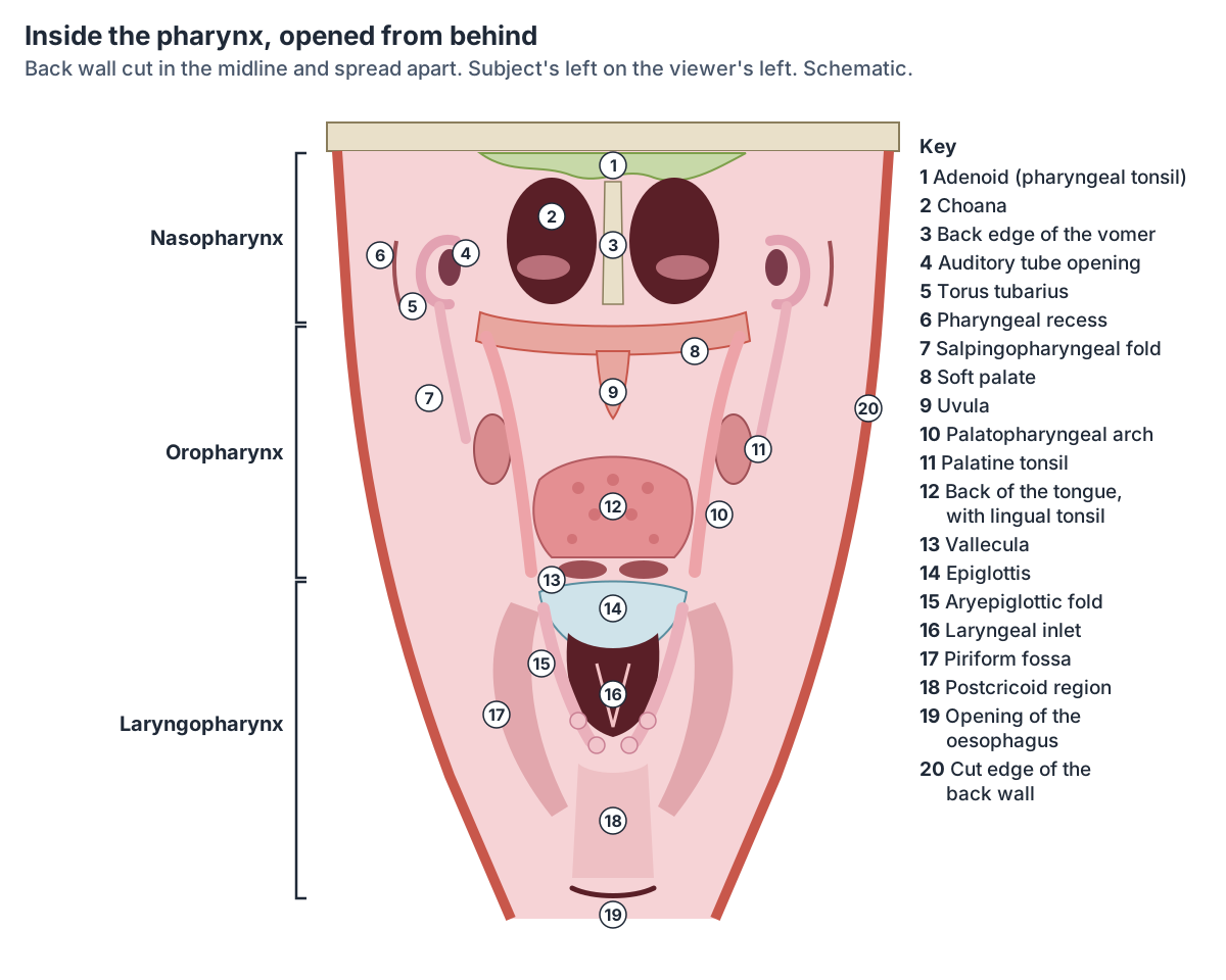
Tonsils and Waldeyer's ring
Waldeyer's ring is a ring of lymphoid tissue in the pharynx: one pharyngeal tonsil (adenoid) in the roof of the nasopharynx, two tubal tonsils around the auditory tube openings, two palatine tonsils in the oropharynx and one lingual tonsil on the back third of the tongue. The tonsils are mucosa-associated lymphoid tissue and are a first line of defence against microbes entering through the nose and mouth. (TeachMeAnatomy: The Tonsils and Adenoids (Waldeyer's Ring).)
Each palatine tonsil lies in the tonsillar bed on the side wall of the oropharynx, between the palatoglossal arch in front and the palatopharyngeal arch behind. Its outer surface has a fibrous capsule separated from the superior constrictor by loose connective tissue. Its main artery is the tonsillar branch of the facial artery, and it drains to the jugulodigastric and upper deep cervical lymph nodes. (TeachMeAnatomy: The Tonsils and Adenoids (Waldeyer's Ring).)
The palatine tonsils, adenoid and lingual tonsil in detail. Tonsils.
Muscles of the pharynx: constrictors and longitudinal muscles
The pharynx has six paired muscles. Three constrictors (superior, middle and inferior) form the outer circular layer; three longitudinal muscles (palatopharyngeus, salpingopharyngeus and stylopharyngeus) form the inner layer. The constrictors are lined on their inner side by the pharyngobasilar fascia and contract in sequence, like a wave of peristalsis, during swallowing. (Kenhub: Muscles of the pharynx.)
The constrictors contract from top to bottom to push food down. The inferior constrictor has two parts: thyropharyngeus, which arises from the thyroid cartilage, and cricopharyngeus, which arises from the cricoid cartilage. The longitudinal muscles shorten and widen the pharynx and raise the larynx. (TeachMeAnatomy: The Pharynx.)
Between and around the constrictors are four gaps in the pharyngeal wall where muscles, nerves and vessels enter. Stylopharyngeus and the glossopharyngeal nerve pass between the superior and middle constrictors, and the internal laryngeal nerve passes between the middle and inferior constrictors. (Kenhub: Muscles of the pharynx.)
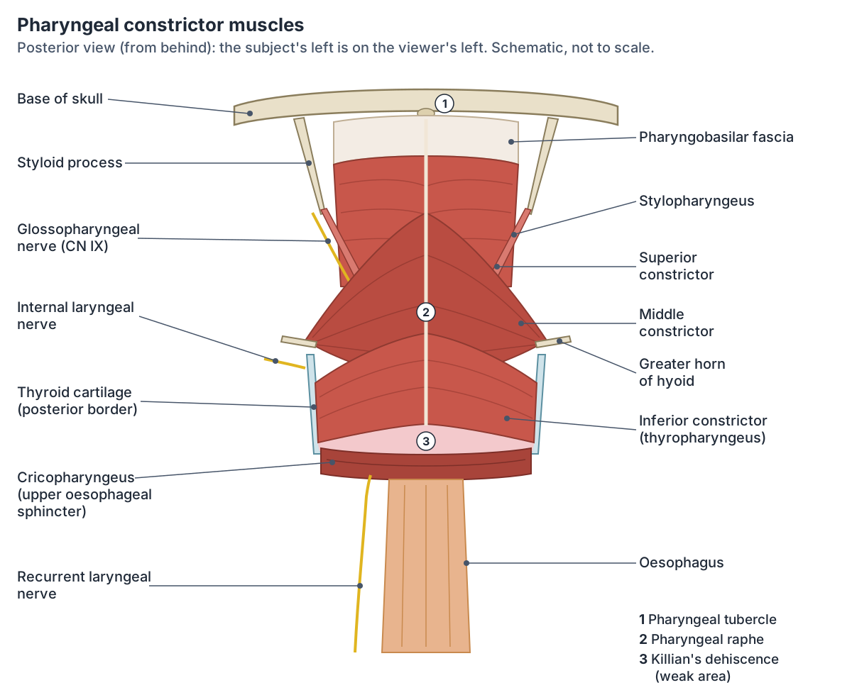
Muscles of the pharynx
| Muscle | Origin | Insertion | Nerve | Action |
|---|---|---|---|---|
| Superior constrictor | Pterygoid hamulus, pterygomandibular raphe, back of the mylohyoid line of the mandible | Pharyngeal tubercle of the occipital bone and pharyngeal raphe | Pharyngeal plexus (vagus, CN X) | Constricts the upper pharynx |
| Middle constrictor | Stylohyoid ligament and greater and lesser horns of the hyoid | Pharyngeal raphe | Pharyngeal plexus (vagus, CN X) | Constricts the middle pharynx |
| Inferior constrictor: thyropharyngeus | Oblique line of the thyroid cartilage | Pharyngeal raphe | Pharyngeal plexus (vagus, CN X) | Constricts the lower pharynx |
| Inferior constrictor: cricopharyngeus | Side of the cricoid cartilage | Blends with the circular muscle of the oesophagus | Pharyngeal plexus, plus external and/or recurrent laryngeal branches of the vagus | Upper oesophageal sphincter |
| Palatopharyngeus | Hard palate and palatine aponeurosis | Back border of the thyroid cartilage | Pharyngeal plexus (vagus, CN X) | Raises the pharynx; helps close the nasopharynx in swallowing |
| Salpingopharyngeus | Cartilage of the auditory tube | Blends with palatopharyngeus | Pharyngeal plexus (vagus, CN X) | Raises the pharynx; opens the auditory tube in swallowing |
| Stylopharyngeus | Medial base of the styloid process | Constrictors, lateral glosso-epiglottic fold, back border of the thyroid cartilage | Glossopharyngeal nerve (CN IX) | Raises the pharynx and larynx and widens the pharynx |
From Kenhub, cross-checked with TeachMeAnatomy. Stylopharyngeus is the only pharyngeal muscle supplied by the glossopharyngeal nerve.
Gaps in the pharyngeal wall and what passes through them
| Gap | Structures passing through |
|---|---|
| Between the superior constrictor and the skull base | Levator veli palatini, auditory (Eustachian) tube, ascending palatine artery |
| Between the superior and middle constrictors | Stylopharyngeus, glossopharyngeal nerve (CN IX) |
| Between the middle and inferior constrictors | Internal laryngeal nerve, superior laryngeal artery and vein |
| Below the inferior constrictor | Recurrent laryngeal nerve, inferior laryngeal artery |
From Kenhub. The gap above the superior constrictor is closed by the pharyngobasilar fascia.
The larynx: the voice box at the front of the throat
The larynx sits in the front of the neck in the midline, in front of the oesophagus, at the level of the third to sixth cervical vertebrae. It is continuous with the oropharynx above and the trachea below. Its skeleton is nine cartilages: the unpaired cricoid, thyroid and epiglottis and the paired arytenoid, corniculate and cuneiform cartilages. It conducts air, protects the airway and produces the voice. (Kenhub: Larynx.)
The epiglottis is a leaf-shaped cartilage that projects up into the pharynx above the laryngeal inlet. When you swallow, the larynx moves up and forward and the epiglottis swings down over the inlet, closing the airway and keeping food out. (Kenhub: Larynx.)
The larynx, or voice box, contains the vocal cords and forms the upper opening into the windpipe. (Children's Hospital of Philadelphia: Throat Anatomy and Physiology.)
The laryngeal cartilages, vocal folds and intrinsic muscles in detail. Larynx anatomy.
Where the trachea and oesophagus begin
The trachea begins at the lower margin of the cricoid cartilage, at C6. It is usually 10 to 13 cm long and is held open by 15 to 20 D-shaped rings of cartilage. The oesophagus lies directly behind it. (Radiopaedia: Trachea.)
The cervical oesophagus starts at the lower border of the cricoid cartilage (C5 to C6) at the upper oesophageal sphincter, which is formed by cricopharyngeus. This is the narrowest point of the oesophagus. The oesophagus lies behind the trachea, with the recurrent laryngeal nerves in the groove between them, and is about 22 to 24 cm long. (Radiopaedia: Oesophagus.)
The windpipe from the cricoid cartilage to the carina. Trachea.
What happens in the throat when you swallow?
Swallowing (deglutition) has three phases. In the voluntary phase, the tongue pushes the bolus up and back against the palate into the oropharynx. In the pharyngeal phase, receptors in the oropharynx signal the swallowing centre in the medulla oblongata; the soft palate and uvula close off the nasopharynx, the larynx is pulled up and the epiglottis folds over the glottis, breathing stops briefly, the constrictors move the bolus down and the upper oesophageal sphincter relaxes. In the oesophageal phase, peristalsis carries the bolus to the stomach. (OpenStax Anatomy and Physiology 2e, 23.3: The Mouth, Pharynx, and Esophagus.)
OpenStax gives a swallow of solid or semisolid food about 4 to 8 seconds, and very soft food or liquids about 1 second. (OpenStax Anatomy and Physiology 2e, 23.3: The Mouth, Pharynx, and Esophagus.)
Kenhub notes that swallowing coordinates 22 muscle groups in the mouth, pharynx and oesophagus. The vagus nerve (CN X) is the main nerve involved, helped by the trigeminal, facial, glossopharyngeal, accessory and hypoglossal nerves. If the epiglottis does not seal the airway, food or liquid can enter it, causing choking, voice changes and shortness of breath. (Kenhub: Stages of swallowing.)
The food pipe from the throat to the stomach, with its sphincters. Oesophagus (esophagus).
Nerves and blood supply of the throat
The vagus nerve (CN X) supplies almost all the pharyngeal muscles; stylopharyngeus, supplied by the glossopharyngeal nerve (CN IX), is the exception. The pharyngeal plexus on the middle constrictor carries pharyngeal branches of CN IX and CN X, branches of the external laryngeal nerve and sympathetic fibres from the superior cervical ganglion. (TeachMeAnatomy: The Pharynx.)
Sensation from most of the pharynx is carried by the glossopharyngeal nerve. The exceptions are the upper nasopharynx, supplied by a pharyngeal branch of the maxillary nerve (V2), the valleculae, supplied by the internal laryngeal nerve, and the rest of the laryngopharynx, which Radiopaedia assigns to the recurrent laryngeal nerve. Lymph from the pharynx drains mostly to the retropharyngeal nodes. (Radiopaedia: Pharynx.)
The internal branch of the superior laryngeal nerve, a branch of the vagus, gives sensation to the laryngopharynx and the upper larynx. (TeachMeAnatomy: The Vagus Nerve (CN X).)
The gag reflex tests these nerves: the glossopharyngeal nerve carries the sensory (afferent) limb from the oropharynx and the vagus nerve the motor (efferent) limb. An absent gag reflex suggests damage to CN IX. (TeachMeAnatomy: The Glossopharyngeal Nerve (CN IX).)
The arteries of the pharynx all come from the external carotid artery: the ascending pharyngeal artery and branches of the facial (including the tonsillar artery), lingual and maxillary arteries. The veins form a pharyngeal venous plexus that drains to the internal jugular vein. (Kenhub: Pharynx.)
The glossopharyngeal and vagus nerves that serve the throat. Cranial nerves.
Sore throat, adenoids and other clinical notes
A sore throat (pharyngitis) can come from tonsillitis, viral or bacterial infections such as flu or strep throat, allergies or mouth breathing. Cancers can arise in each part of the pharynx: nasopharyngeal, oropharyngeal and hypopharyngeal cancer. (Cleveland Clinic: Pharynx.)
Tonsillitis is usually viral; bacteria cause about a third of cases. A peritonsillar abscess (quinsy) is a collection of pus beside the tonsil, a complication of bacterial tonsillitis, and always needs drainage. (TeachMeAnatomy: The Tonsils and Adenoids (Waldeyer's Ring).)
Enlarged adenoids can block the openings of the auditory tubes and cause chronic otitis media with effusion (glue ear). (TeachMeAnatomy: The Pharynx.)
The fossa of Rosenmüller, the recess behind the torus tubarius, is the most common place for nasopharyngeal carcinoma to start. (Radiopaedia: Fossa of Rosenmüller.)
A pharyngeal pouch (Zenker diverticulum) is an outpouching of the back wall of the hypopharynx just above the upper oesophageal sphincter. It forms through Killian's dehiscence, a weak area between the thyropharyngeus and cricopharyngeus parts of the inferior constrictor. More than half of patients are 60 to 80 years old, and trapped food causes difficulty swallowing, regurgitation and bad breath. (Radiopaedia: Zenker diverticulum.)
In obstructive sleep apnoea the oropharynx collapses during sleep. Smoking, which swells the lining, and obesity, which adds fat around the airway, both make this more likely. (Kenhub: Muscles of the pharynx.)
Where the tonsils and pharynx drain, including the jugulodigastric node. Lymph nodes of the head and neck.
Throat anatomy in the 3D model
- Cricoid cartilage
- The cricoid cartilage is a complete signet-ring-shaped cartilage forming the lower part of the larynx.
- Epiglottis
- The epiglottis is a leaf-shaped elastic cartilage that covers the laryngeal inlet during swallowing.
- Hyoid bone
- A U-shaped bone in the neck.
- Inferior pharyngeal constrictor
- The lowest of the three constrictors of the pharynx.
- Levator veli palatini
- A muscle of the soft palate.
- Middle pharyngeal constrictor
- The middle of the three pharyngeal constrictors.
- Palatine tonsil
- The palatine tonsils are the two masses of lymphoid tissue at the back of the mouth, one on each side of the oropharynx. They are what most people mean by the tonsils.
- Palatopharyngeus
- A muscle of the soft palate and pharynx that forms the posterior pillar of the fauces.
- Pharyngeal raphe
- The pharyngeal raphe is a midline fibrous seam on the posterior wall of the pharynx.
- Salpingopharyngeus
- A thin muscle of the pharynx running from the pharyngotympanic tube.
- Soft palate
- The soft palate is the muscular back part of the roof of the mouth. Its free back edge carries the uvula.
- Stylohyoid ligament
- A fibrous band from the styloid process to the hyoid.
- Stylopharyngeus
- A slender pharyngeal muscle arising from the styloid process.
- Superior pharyngeal constrictor
- The uppermost of the three constrictors forming the pharyngeal wall.
- Tensor veli palatini
- A palatal muscle that tenses the soft palate.
- Thyroid cartilage
- The thyroid cartilage is the largest cartilage of the larynx, forming the laryngeal prominence.
- Uvular muscle
- A small paired muscle forming the core of the uvula.
Frequently asked questions
What are the parts of the throat?
The throat is made of the pharynx and the larynx. The pharynx has three parts: the nasopharynx behind the nose, the oropharynx behind the mouth and the laryngopharynx (hypopharynx) behind the larynx. The larynx, or voice box, lies in front of the laryngopharynx and leads down into the trachea. At C6 the pharynx continues as the oesophagus.
What is the difference between the pharynx and the larynx?
The pharynx is a shared passage for air and food that runs from the base of the skull to C6, behind the nose, mouth and larynx. The larynx is the cartilage-framed voice box in front of the lower pharynx, between the oropharynx and the trachea. It carries only air, contains the vocal folds and closes with the epiglottis during swallowing.
Where is the nasopharynx?
Behind the nose, above the soft palate. It opens in front into the nasal cavity through the two choanae, its roof lies under the sphenoid and the clivus, and its back wall is in front of the first two cervical vertebrae. It contains the adenoid and the openings of the auditory (Eustachian) tubes, and it carries only air.
Is the throat the same as the oesophagus?
No. The oesophagus (esophagus) begins where the throat ends, at the lower border of the cricoid cartilage at about C6, where cricopharyngeus forms the upper oesophageal sphincter. The pharynx above it carries both air and food; the oesophagus carries only food and liquid down to the stomach, behind the trachea.
What stops food from going down the wrong way?
During the pharyngeal phase of swallowing the soft palate and uvula rise to close off the nose, the larynx is pulled up and forward, the epiglottis tips down over the laryngeal inlet, and breathing stops for a moment. If this seal fails, food or liquid can enter the airway and cause choking.
Where are the tonsils in the throat?
The palatine tonsils, the ones usually called the tonsils, sit on each side of the oropharynx between the palatoglossal and palatopharyngeal arches. The adenoid (pharyngeal tonsil) is in the roof of the nasopharynx, small tubal tonsils surround the auditory tube openings, and the lingual tonsil covers the back third of the tongue. Together they form Waldeyer's ring.
How long is the pharynx?
Kenhub gives a length of 12 to 14 cm, from the base of the skull to the lower border of the cricoid cartilage at C6, while Cleveland Clinic gives about 4 inches (about 10 cm). The difference comes from how the ends are measured. Below the pharynx the oesophagus continues for another 22 to 24 cm to the stomach.
References
- Betts JG, et al. Anatomy and Physiology 2e, §23.3 The Mouth, Pharynx, and Esophagus. OpenStax, Rice University, 2022.
- Betts JG, et al. Anatomy and Physiology 2e, §22.1 Organs and Structures of the Respiratory System. OpenStax, Rice University, 2022.
- TeachMeAnatomy. The Pharynx: Subdivisions, Blood Supply.
- TeachMeAnatomy. The Tonsils and Adenoids (Waldeyer's Ring).
- TeachMeAnatomy. The Eustachian Tube.
- TeachMeAnatomy. The Glossopharyngeal Nerve (CN IX).
- TeachMeAnatomy. The Vagus Nerve (CN X).
- Kenhub. Pharynx (throat) anatomy: Muscles, arteries and nerves.
- Kenhub. Muscles of the pharynx: Anatomy, origins and insertions.
- Kenhub. Stages of swallowing (deglutition).
- Kenhub. Larynx.
- Radiopaedia. Pharynx (reference article).
- Radiopaedia. Nasopharynx (reference article).
- Radiopaedia. Oropharynx (reference article).
- Radiopaedia. Hypopharynx (reference article).
- Radiopaedia. Pyriform sinus (reference article).
- Radiopaedia. Vallecula (reference article).
- Radiopaedia. Fossa of Rosenmüller (reference article).
- Radiopaedia. Zenker diverticulum (reference article).
- Radiopaedia. Trachea (reference article).
- Radiopaedia. Oesophagus (reference article).
- Cleveland Clinic. Pharynx: Function and Anatomy.
- Children's Hospital of Philadelphia. Throat Anatomy and Physiology.
- National University of Singapore, Department of Anatomy. HN32: The Pharynx (anatomy museum e-guide).
- Mitsuhashi N. et al. (2009). BodyParts3D: 3D structure database for anatomical concepts. Nucleic Acids Research 37, D782–D785. Data © The Database Center for Life Science, CC BY 4.0.
- Browne K., Schlehlein H. (2026). 3D Reference Organ Set for Male, v1.10 (Visible Human Male). Human Reference Atlas 3D Reference Object Library, HuBMAP / NIH. CC BY 4.0.
Related anatomy
- LarynxLarynx anatomy with 3D model and diagrams: thyroid, cricoid, arytenoid and epiglottis cartilages, vocal folds…
- TonsilsTonsil anatomy with labelled diagrams and a 3D model: palatine tonsils, adenoid, lingual and tubal tonsils, Waldeyer's…
- Esophagus (oesophagus)Esophagus (oesophagus) anatomy: its course and levels, four constrictions, upper and lower sphincters, muscle layers…
- TracheaTrachea anatomy explained: where the windpipe runs, C-shaped rings, the carina, right vs left main bronchi and the…
- Neck anatomyNeck anatomy with labelled diagrams and a 3D model: landmarks, triangles, fascia, airway, thyroid, carotid sheath…
- Mouth anatomyMouth anatomy with a 3D model and labelled diagrams: lips, cheeks, hard and soft palate, uvula, tongue, teeth, gums…
Explore the whole body in the interactive 3D human anatomy model, or browse the A–Z list of human structures.