

What is male anatomy?
Whether an embryo develops male anatomy depends on a single gene on the Y chromosome, SRY. The early gonads appear about a month after conception and can become either testes or ovaries; SRY switches on the genes that build a testis, and without a working SRY gene the embryo develops as female. Soon after the testis forms, its Leydig cells start making testosterone. (OpenStax Anatomy and Physiology 2e, 27.3: Development of the Male and Female Reproductive Systems.)
Every embryo first has two duct systems. In males, testosterone makes the Wolffian duct grow into the epididymis, ductus deferens and seminal glands, while secretions from the testis make the Müllerian duct, which forms the uterus and uterine tubes in females, break down. The same tissue that becomes the glans of the penis when exposed to testosterone becomes the clitoris without it. (OpenStax Anatomy and Physiology 2e, 27.3: Development of the Male and Female Reproductive Systems.)
The 3D model above is the adult male BodyParts3D body, with the male reproductive and urinary organs and the thyroid cartilage highlighted. Lower the Skin slider to see inside, switch body systems on or off, or click any part to open its own page.
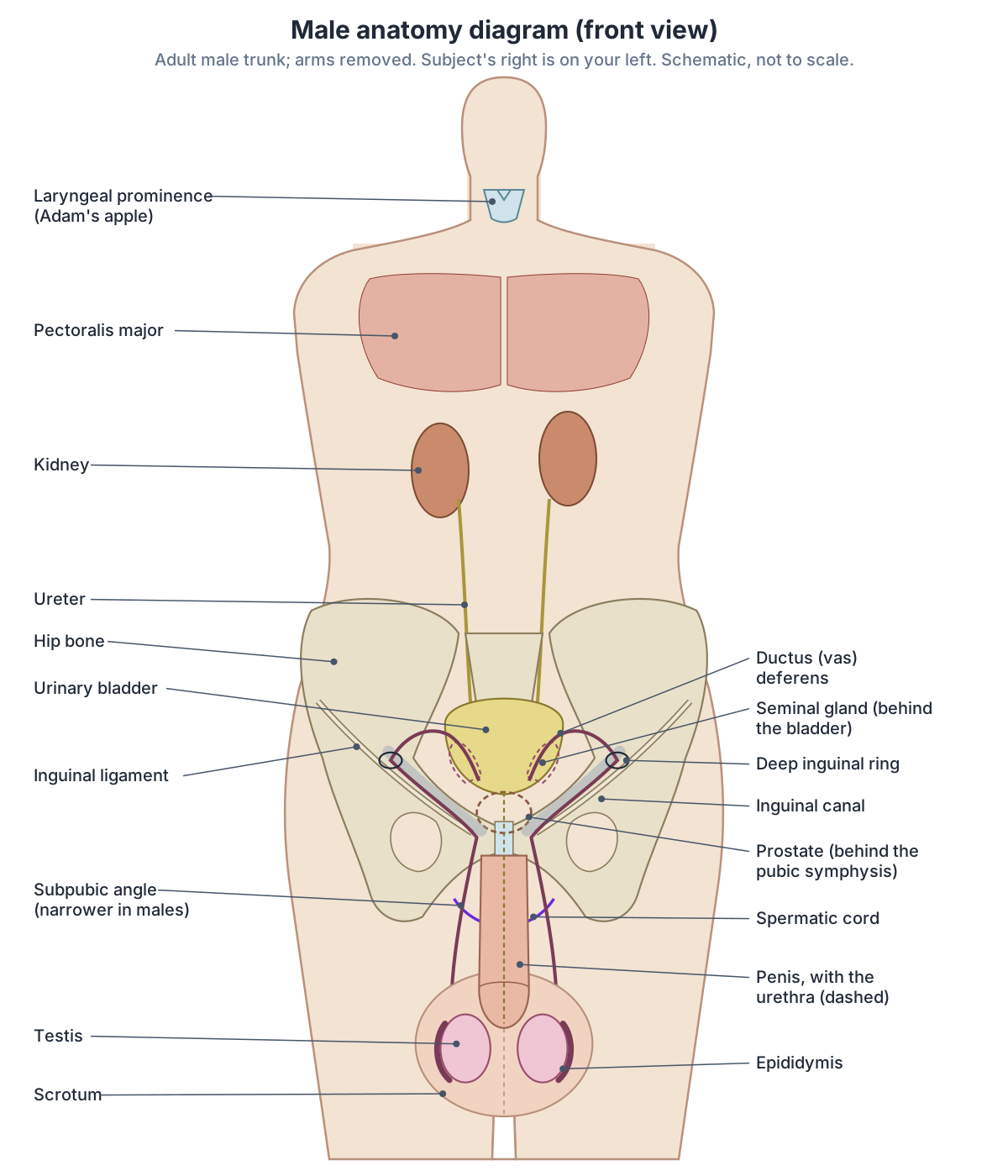
Male organs and structures by body system
| System | Male structure | Key fact |
|---|---|---|
| Reproductive | Testes, epididymides, ductus deferentes, seminal glands, prostate, bulbourethral glands, penis, scrotum | Testes make 100 to 300 million sperm a day, 2 to 4 °C below core temperature |
| Endocrine | Leydig cells of the testes | About 6 to 7 mg of testosterone a day, controlled by LH from the pituitary |
| Urinary | Urethra (prostatic, membranous, spongy); internal urethral sphincter | Urethra 15 to 20 cm long, shared by urine and semen |
| Skeletal | Android pelvis | Thicker, heavier bones; heart-shaped inlet; subpubic angle under 70 degrees |
| Respiratory | Larynx with laryngeal prominence | Adam's apple and larger vocal folds give a deeper voice |
| Muscular | Greater muscle mass | Strength difference largest in the upper body, from about age 15 |
| Pelvic floor | Levator ani with puboprostaticus | Two openings (urethra, anal canal) |
| Abdominal wall | Inguinal canal with the spermatic cord | Lifetime groin hernia risk 27 to 43% in men |
Summary of the sections below, from OpenStax, TeachMeAnatomy, Kenhub, Nuzzo 2023 and HerniaSurge 2018.
Male reproductive organs
Cleveland Clinic divides the male reproductive organs into external organs (the penis, scrotum and testicles) and internal organs (the vas deferens, ejaculatory ducts, urethra, seminal vesicles, prostate and bulbourethral glands). Together they make, store and carry sperm and semen, release sperm, and produce male sex hormones. (Cleveland Clinic: Male Reproductive System.)
The testes are about 4 to 5 cm long and sit in the scrotum, where they are kept 2 to 4 °C below core body temperature for sperm production. They make about 100 to 300 million sperm a day. Sperm take on average 12 days to pass through the coiled epididymis, where they mature, and leave through the ductus deferens, which runs in the spermatic cord. (OpenStax Anatomy and Physiology 2e, 27.1: Anatomy and Physiology of the Testicular Reproductive System.)
Sperm make up only about 5% of semen. OpenStax gives the seminal glands (seminal vesicles) about 60% of semen volume, the walnut-sized prostate an alkaline fluid that helps semen clot and then liquefy, and the bulbourethral glands a thick pre-ejaculate that lubricates the urethra. OpenStax gives the average erect penis as about 15 cm. (OpenStax Anatomy and Physiology 2e, 27.1: Anatomy and Physiology of the Testicular Reproductive System.)
Cleveland Clinic gives slightly different figures: seminal vesicles make up to 80% of the fluid, and the average penis is about 8.9 cm (3.5 inches) when flaccid and a little over 13 cm (5 inches) when erect. (Cleveland Clinic: Male Reproductive System.)
Testes, ducts, glands and penis in detail, with the path of sperm and a penis cross-section. Male reproductive system.
Testosterone and male puberty
Testosterone production is controlled by the brain. Pulses of gonadotropin-releasing hormone (GnRH) from the hypothalamus make the pituitary release luteinising hormone (LH), which stimulates Leydig cells to make testosterone, and follicle-stimulating hormone (FSH), which acts on Sertoli cells to support sperm production. Leydig cells make about 6 to 7 mg of testosterone a day, and its concentration in the testes is about 100 times that in the blood. Sertoli cells also release inhibin, which reduces FSH release, and testosterone and inhibin together feed back to damp down GnRH, LH and FSH. (OpenStax Anatomy and Physiology 2e, 27.1: Anatomy and Physiology of the Testicular Reproductive System.)
In boys, LH becomes detectable at about 8 or 9 years, mainly during sleep. The first physical sign of puberty is usually growth of the testes, followed by growth and darkening of the scrotum, growth of the penis, hair in the armpits, pubic area, chest and face, and growth of the larynx with thickening of the vocal folds, which lowers the voice. The growth spurt comes at about 11 to 13 years, first fertile ejaculations at about 15, and development can continue into the early 20s. (OpenStax Anatomy and Physiology 2e, 27.3: Development of the Male and Female Reproductive Systems.)
Cleveland Clinic lists the roles of testosterone in the adult as maintaining muscle mass and strength, fat distribution, bone mass and sex drive, and notes that sperm production may continue into a man's 80s or longer. (Cleveland Clinic: Male Reproductive System.)
Male urethra and bladder
The male urethra is about 15 to 20 cm long, against about 4 cm in females, and carries both urine and semen. It has a prostatic part, the widest, which receives the ejaculatory and prostatic ducts; a membranous part, the narrowest, surrounded by the external urethral sphincter; and a spongy (penile) part through the bulb and corpus spongiosum, which widens in the glans. Some authors add a short part in the bladder neck. Because of two bends, catheterising the male urethra is more complex, while the short female urethra makes women more prone to urinary tract infections. (TeachMeAnatomy: The Urethra.)
At the bladder neck, males have an internal urethral sphincter of circular smooth muscle under autonomic control, thought to stop semen flowing back into the bladder during ejaculation; females have no distinct internal sphincter. The voluntary external sphincter is more complex in males, linked with fibres of levator ani. In males, enlargement of the prostate (benign prostatic hyperplasia) is the most common cause of a blocked bladder outflow. (TeachMeAnatomy: The Bladder.)
Kenhub places the male external urethral sphincter (rhabdosphincter) just below the prostate. During orgasm, sympathetic nerves from the T11 to L2 segments of the spinal cord contract the internal sphincter, which stops semen travelling back into the bladder. (Kenhub: Urethral sphincters.)
In males the bladder lies behind the pubic symphysis with the rectovesical pouch and rectum behind it and the prostate below; in females the vesicouterine pouch, uterus, cervix and vagina lie behind it. Radiopaedia gives the normal functional volume of the adult bladder as 300 to 400 mL, whereas TeachMeAnatomy gives a storage capacity of up to 400 to 600 mL. (Radiopaedia: Urinary bladder.)
The rectovesical pouch is the lowest part of the peritoneal cavity in males. The male peritoneal cavity is completely closed, whereas in females the uterine tubes open into it. (TeachMeAnatomy: The Peritoneal Cavity.)
The bladder in 3D, with its position, capacity and sphincters. Urinary bladder.
Male pelvis vs female pelvis
The male pelvis is built for a heavier body and stronger muscles rather than for childbirth. Its bones are thicker and heavier, the pelvic inlet is heart-shaped, the subpubic angle under the pubic symphysis is less than 70 degrees, the pelvic outlet is smaller, and the greater sciatic notch is narrower and deeper. The female pelvis has lighter bones, a round or oval inlet, a subpubic angle greater than 80 degrees and a larger, rounder outlet. (OpenStax Anatomy and Physiology 2e, 8.3: The Pelvic Girdle and Pelvis.)
TeachMeAnatomy calls the typical male pelvis android and the typical female pelvis gynaecoid, and describes the female pelvis as wider and broader but lighter, adapted for childbirth. (TeachMeAnatomy: The Pelvic Girdle.)
Ilium, ischium and pubis in 3D, with a male vs female pelvis diagram. Pelvis bones.
Adam's apple and the male larynx
The thyroid cartilage is the largest cartilage of the larynx (voice box). Its front forms the laryngeal prominence, or Adam's apple, which tends to be more prominent in males. Male vocal folds tend to be larger than female ones, which gives a deeper voice. (OpenStax Anatomy and Physiology 2e, 22.1: Organs and Structures of the Respiratory System.)
Everyone has a thyroid cartilage, and it grows at puberty, typically more in males, but the size of the Adam's apple varies from person to person. Cleveland Clinic notes that it may help protect the voice box, that whether a larger Adam's apple deepens the voice is not proven, and that having a large or small one has no effect on health. (Cleveland Clinic: Adam's Apple.)
The cartilages of the voice box and the vocal folds, labelled. Larynx.
Muscle mass and body build
A 2023 review of sex differences found that males become notably stronger than females at about 15 years of age and that the difference is larger in upper-body muscles than in lower-body muscles. The extra strength is explained by greater muscle mass and larger type II (fast) muscle fibres, not by the nervous system activating the muscles more fully. (Nuzzo JL. Narrative review of sex differences in muscle strength, endurance, activation, size, fiber type, and strength training participation rates, preferences, motivations, injuries, and neuromuscular adaptations. J Strength Cond Res. 2023.)
Testosterone underlies these changes: circulating testosterone supports muscle development and bone growth as well as the secondary sex characteristics. (OpenStax Anatomy and Physiology 2e, 27.1: Anatomy and Physiology of the Testicular Reproductive System.)
OpenStax lists increased muscular development among the male secondary sex characteristics of puberty, alongside a larger larynx with a deeper voice and more facial, axillary, pubic and body hair. (OpenStax Anatomy and Physiology 2e, 27.3: Development of the Male and Female Reproductive Systems.)
The major muscles of the body in 3D. Muscular system.
Male chest and breast tissue
Gynaecomastia is a benign, usually reversible excess of breast glandular tissue in males, centred under the nipple and often one-sided or uneven. Without symptoms it is found in 60 to 90% of newborn boys, 50 to 60% of adolescents and up to 70% of men aged 50 to 69. It is not in itself a risk factor for male breast cancer, and breast enlargement caused by fat alone is called pseudogynaecomastia. (Radiopaedia: Gynaecomastia.)
The ribs, chest muscles and organs of the thorax. Chest anatomy.
The male pelvic floor and perineum
Males have the same pelvic floor muscles as females, but the pelvic floor has two openings in males (the urethra and anal canal) instead of three. The medial fibres of pubococcygeus that surround the prostate are called puboprostaticus. (Kenhub: Muscles of the pelvic floor.)
In males the pelvic floor holds the prostate in place and helps with erection and ejaculation as well as bladder and bowel control; prostate surgery can weaken previously strong muscles. (Cleveland Clinic: Pelvic Floor Muscles.)
The male external urethral sphincter is omega-shaped and lies between the two slings of levator ani, joined to them by fascia rich in elastic fibres, unlike the semicircular female sphincter. (Hinata N, Murakami G. The urethral rhabdosphincter, levator ani muscle, and perineal membrane: a review. Biomed Res Int. 2014.)
On the surface, the male perineum is the area between the scrotum and the anus, about 2 inches (about 5 cm) long or more. (Cleveland Clinic: Perineum.)
Levator ani, the perineal muscles and the male and female perineum, with labelled diagrams. Pelvic floor and perineum.
The male groin and inguinal canal
The testes form on the back wall of the abdomen and descend through the inguinal canal into the scrotum, guided by the gubernaculum, so in males the canal carries the spermatic cord. A pouch of peritoneum that descends with the testis, the processus vaginalis, normally closes; if it stays open it can cause an indirect inguinal hernia or a hydrocele. (TeachMeAnatomy: The Inguinal Canal.)
Both inguinal rings are larger in males, to let the testis through. (Kenhub: Inguinal canal.)
The lifetime risk of a groin hernia is 27 to 43% in men and 3 to 6% in women, and inguinal hernia repair is 8 to 10 times more common in males. Hernias peak at about 5 years of age, mainly indirect, and again at 70 to 80 years, mainly direct. (HerniaSurge Group. International guidelines for groin hernia management. Hernia. 2018.)
The inguinal ligament, inguinal canal, femoral triangle and hernia sites. Groin anatomy.
Male vs female anatomy: the main differences
Male and female reproductive organs develop from shared starting tissue. The early gonad becomes a testis or an ovary, and the same tissue forms the glans of the penis or the clitoris, depending on whether SRY and testosterone are present. (OpenStax Anatomy and Physiology 2e, 27.3: Development of the Male and Female Reproductive Systems.)
Some smaller structures are also matched. The paraurethral (Skene) glands beside the female urethra are considered homologous to the male prostate. (TeachMeAnatomy: The Urethra.)
The table below brings together the differences covered on this page, with the source for each in the caption.
The female body: reproductive organs, pelvis, breast and pelvic floor. Female anatomy.
Male vs female anatomy: the main differences
| Feature | Male | Female |
|---|---|---|
| Sex-determining gene | SRY on the Y chromosome present | No SRY; female development by default |
| Gonads | Two testes in the scrotum, outside the body cavity | Two ovaries in the pelvis |
| Duct system | Wolffian duct: epididymis, ductus deferens, seminal glands | Müllerian duct: uterine tubes, uterus, part of the vagina |
| Urethra | 15 to 20 cm; carries urine and semen | About 4 cm; carries urine only |
| Bladder neck | Internal urethral sphincter of smooth muscle | No distinct internal sphincter |
| Pelvis | Heart-shaped inlet; subpubic angle under 70 degrees; heavier bones | Round or oval inlet; subpubic angle over 80 degrees; lighter bones |
| Larynx | Larger, more prominent Adam's apple; larger vocal folds | Smaller laryngeal prominence |
| Pelvic floor | Two openings; perineum about 2 inches or more | Three openings; perineum about 1.5 inches; larger perineal body |
| Peritoneal cavity | Completely closed; lowest point is the rectovesical pouch | Open to the uterine tubes |
| Groin | Inguinal canal carries the spermatic cord; inguinal hernias far more common | Inguinal canal carries the round ligament; femoral hernias more common than in males |
Sources: OpenStax 8.3, 22.1, 27.1 and 27.3; TeachMeAnatomy (urethra, bladder, peritoneal cavity, inguinal canal, femoral canal); Kenhub (pelvic floor); Cleveland Clinic (perineum); Radiopaedia (perineal body); HerniaSurge 2018.
Common conditions linked to male anatomy
Cleveland Clinic lists the common conditions of the male reproductive system as testicular, penile and prostate cancer, sexually transmitted infections, premature ejaculation, male infertility, erectile dysfunction and priapism. (Cleveland Clinic: Male Reproductive System.)
Because the urethra runs through the prostate, an enlarged prostate can narrow it and restrict the flow of urine. (OpenStax Anatomy and Physiology 2e, 27.1: Anatomy and Physiology of the Testicular Reproductive System.)
In males, prostate enlargement (benign prostatic hyperplasia) is the most common obstructive cause of urinary retention, and the bladder's detrusor muscle thickens, with prominent ridges called trabeculae, as it works against the blockage. (TeachMeAnatomy: The Bladder.)
Groin hernias are another condition shaped by male anatomy: indirect inguinal hernias follow the path the testis took through the inguinal canal. This overview is educational and not a guide to diagnosis. (TeachMeAnatomy: The Inguinal Canal.)
Male anatomy in the 3D model
- Corpus cavernosum of penis
- The corpora cavernosa are the paired columns of erectile tissue forming the dorsal part of the penis.
- Corpus spongiosum of penis
- The corpus spongiosum is the single column of erectile tissue surrounding the penile urethra.
- Deferent duct
- The deferent duct (vas deferens) is a thick-walled muscular tube carrying sperm from the epididymis toward the urethra.
- Epididymis
- The epididymis is a coiled tube on the posterior testis where sperm mature and are stored.
- Glans penis
- The glans penis is the cone-shaped distal end of the penis.
- Kidney
- The kidney is a bean-shaped organ that filters blood to form urine and regulate fluid, electrolytes and blood pressure.
- Prostate
- The prostate is a walnut-sized gland surrounding the first part of the male urethra.
- Seminal vesicle
- The seminal vesicles are paired glands that produce much of the fluid of semen.
- Testicular artery
- A paired branch of the abdominal aorta to the testis.
- Testicular vein
- A vein draining the testis.
- Testis
- The testis is the male gonad that produces sperm and testosterone.
- Thyroid cartilage
- The thyroid cartilage is the largest cartilage of the larynx, forming the laryngeal prominence.
- Ureter
- The ureter is a muscular tube carrying urine from the renal pelvis to the urinary bladder.
- Urethra
- The urethra is the tube carrying urine out of the bladder; in males it also carries semen.
- Urinary bladder
- The urinary bladder is a hollow muscular organ that stores urine.
Frequently asked questions
What organs do males have that females do not?
The organs that differ are the reproductive organs. Males have two testes in the scrotum, the epididymides, the ductus (vas) deferentes, the ejaculatory ducts, the seminal glands, the prostate, the bulbourethral glands and the penis. Females have ovaries, uterine tubes, a uterus and a vagina instead. All other organs, including the heart, lungs, liver, kidneys and bladder, are present in both sexes.
What are the external and internal male reproductive organs?
The external male organs are the penis, the scrotum and the testicles (testes) inside it. The internal organs are the epididymis, vas deferens, ejaculatory ducts, urethra, seminal vesicles (seminal glands), prostate and bulbourethral (Cowper) glands. Together they make and store sperm, add the fluid of semen, carry semen out of the body and produce testosterone.
Why do men have an Adam's apple?
The Adam's apple is the laryngeal prominence, the front of the thyroid cartilage of the larynx. Everyone has this cartilage, but at puberty the larynx grows and the vocal folds thicken, usually more in males, so the prominence sticks out more and the voice deepens. Its size varies from person to person and has no effect on health.
How long is the male urethra?
The male urethra is about 15 to 20 cm long, compared with about 4 cm in females. It runs from the bladder neck through the prostate (prostatic urethra), through the external urethral sphincter (membranous urethra, the narrowest part) and along the penis (spongy urethra) to the external urethral orifice. It carries both urine and semen.
How is the male pelvis different from the female pelvis?
The male pelvis has thicker, heavier bones, a heart-shaped pelvic inlet, a smaller outlet, a narrower and deeper greater sciatic notch and a subpubic angle of less than 70 degrees. The female pelvis is lighter, with a round or oval inlet, a larger outlet and a subpubic angle greater than 80 degrees, adapted for childbirth.
What does testosterone do in the male body?
Testosterone, made by the Leydig cells of the testes, starts sperm production at puberty and drives the male secondary sex characteristics: growth of the penis and scrotum, facial, chest and pubic hair, growth of the larynx and a deeper voice, and greater muscle and bone development. In adults it helps maintain muscle mass, bone mass, fat distribution and sex drive.
Why are inguinal hernias more common in men?
In males the testes descend through the inguinal canal before birth, so the canal and its rings are larger and carry the spermatic cord. If the peritoneal pouch that follows the testis (the processus vaginalis) stays open, an indirect hernia can follow it. The lifetime risk of a groin hernia is 27 to 43% in men against 3 to 6% in women.
References
- OpenStax. Anatomy and Physiology 2e, §27.1 Anatomy and Physiology of the Testicular Reproductive System. Rice University, 2022.
- OpenStax. Anatomy and Physiology 2e, §27.3 Development of the Male and Female Reproductive Systems. Rice University, 2022.
- OpenStax. Anatomy and Physiology 2e, §8.3 The Pelvic Girdle and Pelvis. Rice University, 2022.
- OpenStax. Anatomy and Physiology 2e, §22.1 Organs and Structures of the Respiratory System. Rice University, 2022.
- Cleveland Clinic. Male Reproductive System.
- Cleveland Clinic. Adam's Apple.
- Cleveland Clinic. Pelvic Floor Muscles.
- Cleveland Clinic. Perineum.
- TeachMeAnatomy. The Urethra.
- TeachMeAnatomy. The Bladder.
- TeachMeAnatomy. The Peritoneal Cavity.
- TeachMeAnatomy. The Pelvic Girdle.
- TeachMeAnatomy. The Inguinal Canal.
- Kenhub. Urethral sphincters.
- Kenhub. Muscles of the pelvic floor.
- Kenhub. Inguinal canal.
- Radiopaedia. Urinary bladder.
- Radiopaedia. Gynaecomastia.
- Radiopaedia. Perineal body.
- TeachMeAnatomy. The Femoral Canal.
- HerniaSurge Group. International guidelines for groin hernia management. Hernia. 2018;22(1):1-165.
- Hinata N, Murakami G. The urethral rhabdosphincter, levator ani muscle, and perineal membrane: a review. Biomed Res Int. 2014;2014:906921.
- Nuzzo JL. Narrative review of sex differences in muscle strength, endurance, activation, size, fiber type, and strength training participation rates, preferences, motivations, injuries, and neuromuscular adaptations. J Strength Cond Res. 2023;37(2):494-536.
- Mitsuhashi N. et al. (2009). BodyParts3D: 3D structure database for anatomical concepts. Nucleic Acids Research 37, D782–D785. Data © The Database Center for Life Science, CC BY 4.0.
Related anatomy
- Male reproductive systemMale reproductive anatomy with labelled diagrams: scrotum, testes, epididymis, ductus deferens, seminal gland…
- PelvisPelvis and hip bone anatomy in 3D: ilium, ischium and pubis, ASIS and PSIS, ligaments, female pelvic anatomy and a…
- Pelvic floor and perineumPerineum and pelvic floor anatomy with labelled diagrams and a 3D model: levator ani, coccygeus, perineal body, both…
- GroinGroin anatomy with labelled diagrams and a 3D model: inguinal ligament and canal, femoral triangle, groin muscles…
- LarynxLarynx anatomy with 3D model and diagrams: thyroid, cricoid, arytenoid and epiglottis cartilages, vocal folds…
- Urinary bladderBladder anatomy with a labelled diagram and 3D model: apex, body, fundus, neck and trigone, detrusor and sphincters…
Explore the whole body in the interactive 3D human anatomy model, or browse the A–Z list of human structures.