

How big does the uterus get in pregnancy?
Outside pregnancy the uterus measures about 7.5 cm long, 5 cm wide at its upper part and 2.5 cm thick, and weighs about 30 to 40 g. In pregnancy it enlarges enormously and by the eighth month reaches the epigastric region below the ribs. It grows both by enlargement of existing smooth muscle fibres and by formation of new ones. (Radiopaedia: Uterus.)
A nursing textbook gives a heavier starting weight, about 60 g, and a weight of about 1,000 to 1,200 g at 40 weeks. As the fetus, placenta and amniotic fluid grow, the muscle cells of the myometrium stretch and thin, and most of the blood reaching the uterus goes to the placenta. (OpenStax Maternal-Newborn Nursing, 10.1: Physiologic Changes Due to Pregnancy.)
Before pregnancy the uterus is about the size of a fist, and it grows most during the second and third trimesters. A full-term pregnancy lasts about 270 days, or 38.5 weeks, from conception. (OpenStax Anatomy and Physiology 2e, 28.4: Changes During Pregnancy, Labor, and Birth.)
Cleveland Clinic compares the growth to a lemon becoming a watermelon. It counts pregnancy as about 40 weeks (280 days) from the last period, split into three trimesters of about 13 weeks: the first until week 13, the second from week 13 to 28 and the third from week 29 to 40. (Cleveland Clinic: Pregnancy: Gestation, Trimesters & What To Expect.)
The uterus grows out of the pelvis and into the abdomen: its top reaches the pubic symphysis by 12 weeks and the xiphoid process at about 36 weeks. After the 14th week the ovaries and uterine tubes are lifted with it and become abdominal organs. (OpenStax Maternal-Newborn Nursing, 10.1: Physiologic Changes Due to Pregnancy.)
In the 3D model the Female body shows the uterus, its fundus, body, isthmus and cervix, the uterine tubes, ovaries and their ligaments at their size before pregnancy, so you can see the starting point that the diagrams on this page build on. The hip bones and sacrum are highlighted too, so you can see the bony pelvis that the uterus rises out of after 12 weeks. Click any highlighted part to open its page.
The uterus, uterine tubes, ovaries and ligaments before pregnancy, in 3D. Female reproductive system.
Pregnancy body changes at a glance
| Structure or measure | Change by late pregnancy | Sources |
|---|---|---|
| Uterus weight | From about 30 to 40 g (Radiopaedia) or 60 g (OpenStax nursing text) to 1,000 to 1,200 g at 40 weeks | Radiopaedia; OpenStax |
| Top of the uterus | From inside the pelvis to just below the xiphoid process at about 36 weeks | Cleveland Clinic; TeachMeObGyn; OpenStax |
| Blood volume | Up 30% (1 to 2 L) to 40 to 50% (about 1,200 to 1,600 mL) | OpenStax; Soma-Pillay 2016 |
| Cardiac output | Up 30 to 50%; highest at about 20 to 28 weeks | OpenStax; Soma-Pillay 2016 |
| Heart rate | Up 10 to 20 beats per minute | Soma-Pillay 2016; OpenStax |
| Diaphragm | Raised about 4 to 5 cm | OpenStax; LoMauro 2015 |
| Minute ventilation | Up 40 to 50% | Soma-Pillay 2016; LoMauro 2015; OpenStax |
| Oxygen use | Up about 20% | Soma-Pillay 2016; OpenStax; LoMauro 2015 |
| Kidneys | 1 to 1.5 cm longer; GFR up 50 to 85% | Soma-Pillay 2016 |
| Placenta at term | About 22 cm across, 2 to 2.5 cm thick, 470 to 500 g | Radiopaedia; Cleveland Clinic |
| Umbilical cord at term | 50 to 60 cm long; two arteries and one vein | Spurway 2012; Cleveland Clinic; OpenStax |
| Amniotic fluid | Peaks at about 34 weeks, a little under 1 L | Cleveland Clinic |
Typical values; individual women vary. Where sources disagree, the range covers both.
Fundal height by week: where is the top of the uterus?
Fundal height is the distance from the pubic bone to the top of the uterus (the fundus), measured in centimetres with a tape at antenatal visits from about 20 weeks. From about 20 to 36 weeks the number of centimetres roughly matches the weeks of pregnancy, give or take 2 cm, so 30 to 34 cm is expected at 32 weeks. (Cleveland Clinic: Fundal Height.)
Cleveland Clinic gives three landmarks: at about 12 weeks the fundus is nearly at the pubic bone, at about 20 weeks it is at the belly button (umbilicus), and at about 36 weeks it reaches its highest point at the breastbone. After 36 weeks the measurement can fall as the baby drops into the pelvis before labour. (Cleveland Clinic: Fundal Height.)
TeachMeObGyn agrees: the uterus should be palpable above the pubic bone after 12 weeks, near the umbilicus at 20 weeks and near the xiphisternum at 36 weeks, with fundal height within 2 cm of the gestational age; tall or short women may differ slightly. (TeachMeObGyn: Obstetric Examination.)
A South African maternal care manual puts the umbilicus a little later. It places the fundus just above the symphysis at about 12 weeks, halfway between the symphysis and the umbilicus at about 16 weeks, one finger-width below the umbilicus at about 20 weeks, level with it at about 22 weeks and one finger-width above it at about 24 weeks, and it plots the symphysis-fundus measurement on a growth curve from 18 weeks. (Bettercare Maternal Care, 1B: Examination of the abdomen in pregnancy.)
Before 20 weeks the fundus is located by feeling for it, using the symphysis pubis and the umbilicus as landmarks; from 20 weeks it is measured in centimetres, and between 20 and 36 weeks the measurement should be close to the number of weeks. (OpenStax Maternal-Newborn Nursing, 11.3: Care in the Second Trimester of Pregnancy.)
A uterus can measure larger than expected because of a wrong due date, fibroids, twins or extra amniotic fluid, and smaller because of a petite build, little amniotic fluid, a baby that has already dropped or slow fetal growth. An ultrasound scan gives a more accurate size. (Cleveland Clinic: Fundal Height.)
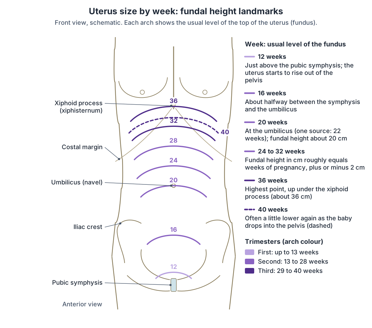
Fundal height landmarks by week
| Weeks | Usual level of the top of the uterus | Sources |
|---|---|---|
| 12 | Just above the pubic symphysis; first palpable in the abdomen | Cleveland Clinic; TeachMeObGyn; OpenStax; Bettercare |
| 16 | About halfway between the symphysis and the umbilicus | Bettercare |
| 20 | At the umbilicus (Bettercare: one finger-width below, level at 22 weeks) | Cleveland Clinic; TeachMeObGyn; Bettercare |
| 20 to 36 | Fundal height in cm about equal to weeks, plus or minus 2 cm | Cleveland Clinic; TeachMeObGyn; OpenStax |
| 36 | Highest point, near the xiphisternum (lower breastbone) | Cleveland Clinic; TeachMeObGyn; OpenStax |
| 37 to 40 | May fall slightly as the baby drops into the pelvis | Cleveland Clinic |
Where do the organs go during pregnancy?
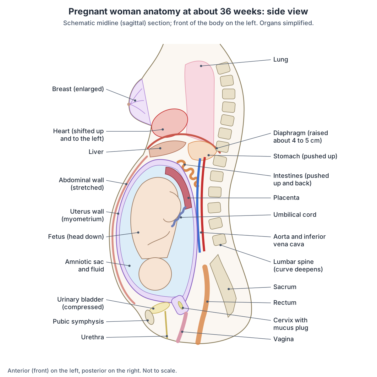
As the uterus fills the abdomen it pushes the intestines higher. In the third trimester it pushes the stomach up, and the heart shifts upwards and to the left, so a clinician may need to move the stethoscope to hear it. (OpenStax Maternal-Newborn Nursing, 10.1: Physiologic Changes Due to Pregnancy.)
A physiology review describes the stomach as increasingly displaced upwards, which changes its axis and raises the pressure inside it. (Soma-Pillay P, Nelson-Piercy C, Tolppanen H, Mebazaa A. Physiological changes in pregnancy. Cardiovasc J Afr. 2016.)
The same uterus pushes the intestines upwards against the diaphragm, which rises by as much as 4 cm while the rib cage widens by up to 6 cm. Pressure of the uterus on the bladder causes frequent urination throughout pregnancy. (OpenStax Maternal-Newborn Nursing, 10.3: Common Discomforts of Pregnancy.)
Sources weigh the causes of heartburn differently. OpenStax attributes late-pregnancy heartburn to pressure of the uterus on the stomach. (OpenStax Anatomy and Physiology 2e, 28.4: Changes During Pregnancy, Labor, and Birth.)
The nursing text puts progesterone first: it relaxes the cardiac sphincter at the top of the stomach and slows stomach emptying, and the heartburn becomes more severe in the final weeks because of the enlarged uterus. (OpenStax Maternal-Newborn Nursing, 10.3: Common Discomforts of Pregnancy.)
From about 28 weeks the enlarging uterus presses on the inferior vena cava when a woman lies flat on her back, which can make her feel dizzy, weak or sick; lying on one side avoids this. (OpenStax Maternal-Newborn Nursing, 10.3: Common Discomforts of Pregnancy.)
The physiology review puts a number on it: turning from lying on the side to lying on the back can reduce cardiac output by 25%. (Soma-Pillay P, Nelson-Piercy C, Tolppanen H, Mebazaa A. Physiological changes in pregnancy. Cardiovasc J Afr. 2016.)
The uterus also presses on the pelvic veins and slows the return of blood from the legs and pelvis, which can lead to varicose veins and haemorrhoids (US hemorrhoids). (OpenStax Anatomy and Physiology 2e, 28.4: Changes During Pregnancy, Labor, and Birth.)
Where the abdominal organs sit outside pregnancy, with regions and quadrants. Abdomen anatomy.
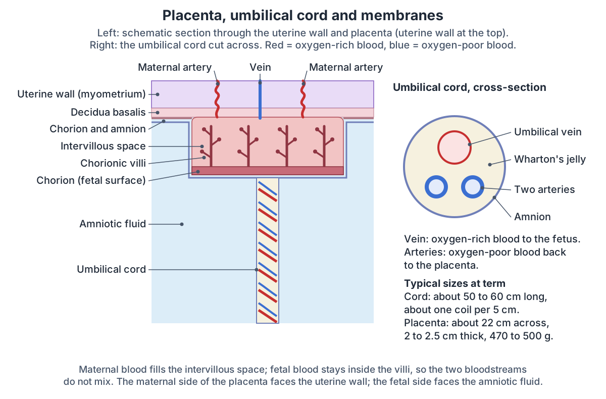
Placenta, umbilical cord and amniotic sac
The placenta is a temporary organ that forms in the uterus and connects to the fetus through the umbilical cord. It can attach to the back wall (posterior), the front wall (anterior), the top (fundal) or a side wall (lateral) of the uterus. A mature placenta is almost 22 cm long, about 2.5 cm thick at the centre and weighs a little over 500 g; its fetal side, where the cord attaches, is shiny and grey, and its maternal side is bumpy and deep red. (Cleveland Clinic: Placenta.)
Radiopaedia gives similar figures: a disc about 22 cm across and 2 to 2.5 cm thick, weighing about 470 g at term, with the cord usually inserting at its centre. Its maternal part, the decidua basalis, comes from the lining of the uterus, and its fetal part, the chorion frondosum, sends chorionic villi into the intervillous spaces. Its thickness in millimetres roughly matches the week of pregnancy. (Radiopaedia: Placenta.)
The embryo usually implants in the fundus or the back wall of the uterus. Between weeks 4 and 12 the placenta gradually takes over nourishing it, and OpenStax dates the completion of the placenta to weeks 14 to 16. Maternal and fetal blood never mix directly: oxygen, nutrients and wastes cross the thin walls of the villi. (OpenStax Anatomy and Physiology 2e, 28.2: Embryonic Development.)
A nursing text dates the placenta as usually complete by week 12. Its maternal side is divided into lobes called cotyledons, and it makes progesterone, oestrogen (US estrogen), human chorionic gonadotropin (hCG), human placental lactogen and relaxin. (OpenStax Maternal-Newborn Nursing, 10.4: Fetal Growth and Development.)
The umbilical cord normally holds two umbilical arteries, which carry oxygen-poor blood and wastes from the fetus to the placenta, and one umbilical vein, which carries oxygen-rich blood and nutrients to the fetus. The vessels are cushioned by a jelly-like connective tissue, Wharton's jelly, and the whole cord is wrapped in amnion. (OpenStax Anatomy and Physiology 2e, 28.2: Embryonic Development.)
At term the cord is on average 50 to 60 cm long, with a normal range of 30 to 100 cm, and under 30 cm counts as short. It usually has about one coil per 5 cm of length. (Spurway J, Logan P, Pak S. The development, structure and blood flow within the umbilical cord. Australas J Ultrasound Med. 2012.)
Cleveland Clinic gives the same 50 to 60 cm length and a thickness of 1 to 2 cm, and notes that the cord usually attaches to the centre of the placenta at one end and, at the other, to the spot on the fetal belly that becomes the navel. (Cleveland Clinic: Umbilical Cord.)
The amniotic sac, or bag of waters, is made of two membranes, the outer chorion and the inner amnion, and forms about 12 days after conception. It holds the most fluid at around 34 weeks, a little less than 1 litre, and the amount then falls towards the due date. The fluid cushions the fetus and helps keep its temperature steady. (Cleveland Clinic: Amniotic Sac.)
OpenStax explains where the fluid comes from: at first it is mostly filtered maternal plasma, and from about week 8 the fetal kidneys add urine to it. (OpenStax Anatomy and Physiology 2e, 28.2: Embryonic Development.)
How does the cervix change in pregnancy?
The cervix is the lower 3 to 4 cm of the uterus. Early in pregnancy it and the vagina soften (Goodell sign) and take on a bluish colour (Chadwick sign), and glands in the cervical canal make a thick mucus plug that helps keep bacteria out of the uterus. (OpenStax Maternal-Newborn Nursing, 10.1: Physiologic Changes Due to Pregnancy.)
The mucus plug loosens and comes away 1 to 2 days before true labour, with a little blood (the bloody show). Contractions then stretch the cervix so that it thins (effaces) and opens (dilates) until it is fully dilated at 10 cm, and relaxin helps it to dilate. (OpenStax Anatomy and Physiology 2e, 28.4: Changes During Pregnancy, Labor, and Birth.)
In the first stage of labour, the early phase lasts until the cervix is 6 cm dilated and the active phase from 6 to 10 cm. When it is 10 cm dilated and 100% effaced, the second stage begins. (OpenStax Maternal-Newborn Nursing, 15.2: Stages of Labor.)
After a vaginal birth the cervix is soft and slightly open and closes to less than 1 cm within a week. Its outer opening changes permanently from a round pinhole to a transverse slit after the first vaginal birth. (OpenStax Maternal-Newborn Nursing, 20.1: Physiologic Changes During the Postpartum Period.)
The vagina and the fornices that surround the cervix, in 3D. Vagina anatomy.
Breast changes in pregnancy
Oestrogen makes the milk ducts of the breast grow and branch, and prolactin from the anterior pituitary enlarges the mammary glands ready for milk production. (OpenStax Anatomy and Physiology 2e, 28.4: Changes During Pregnancy, Labor, and Birth.)
The breasts become larger and tender and their veins more visible. The nipples and areolae grow larger and darker, the Montgomery glands of the areola release a lubricant, and the milk-producing cells start making colostrum between 12 and 16 weeks, although oestrogen and progesterone hold back milk secretion until after the birth. (OpenStax Maternal-Newborn Nursing, 10.1: Physiologic Changes Due to Pregnancy.)
Cleveland Clinic lists tender, swollen breasts in the first trimester, darker areolae in the second, and colostrum, the first form of breast milk, sometimes leaking in the third. (Cleveland Clinic: Pregnancy: Gestation, Trimesters & What To Expect.)
After the birth, progesterone falls and prolactin rises, starting milk production. Colostrum changes to transitional milk around days 2 to 3, and the darker areola fades over the first 6 weeks. (OpenStax Maternal-Newborn Nursing, 20.1: Physiologic Changes During the Postpartum Period.)
Lobes, lactiferous ducts, areola and suspensory ligaments of the breast, in 3D. Breast anatomy.
Pelvis and ligaments: relaxin and the round ligament
Relaxin, made first by the corpus luteum and then by the placenta, makes the pubic symphysis and the pelvic ligaments more elastic. Late in pregnancy the pelvis becomes more flexible and the baby moves down into it, which is called lightening. (OpenStax Anatomy and Physiology 2e, 28.4: Changes During Pregnancy, Labor, and Birth.)
Relaxin softens ligaments in all joints. The ligaments of the pelvic joints stretch to widen the pelvic inlet for birth, the lower back curves more (lordosis) to balance the forward shift in the centre of gravity, and the pubic symphysis can separate. (OpenStax Maternal-Newborn Nursing, 10.1: Physiologic Changes Due to Pregnancy.)
Each round ligament runs from the uterine horn, where the uterine tube joins the uterus, through the inguinal canal to the labium majus; it is a remnant of the embryonic gubernaculum. As the uterus expands it pulls harder on these ligaments, which can be a source of pain in pregnancy. (TeachMeAnatomy: Ligaments of the Uterus.)
Round ligament pain is felt in the right or left lower abdomen and is most common between 14 and 27 weeks, when the uterus grows out of the pelvis and stretches the ligaments. Low back discomfort comes from the weight of the uterus increasing the curve of the spine, while progesterone and relaxin make the ligaments more elastic. (OpenStax Maternal-Newborn Nursing, 10.3: Common Discomforts of Pregnancy.)
The bones of the pelvis and how the female pelvis differs from the male pelvis. Pelvis anatomy.
Abdominal wall, skin and pelvic floor
The rectus abdominis muscles are held together in the midline by the linea alba. During pregnancy the uterus stretches this band until it thins and the two halves of the muscle pull apart, a condition called diastasis recti. It usually develops in the third trimester, affects about 6 in 10 women after childbirth and is defined clinically as a gap wider than 2 cm; one study found it in 45% of women six months after giving birth. (Cleveland Clinic: Diastasis Recti.)
Stretching of the skin can tear the connective tissue beneath it and leave stretch marks (striae) that fade to silvery white after the birth. Melanocyte-stimulating hormone and oestrogen darken the areolae, draw a dark line down the abdomen (the linea nigra) and can cause patches of facial pigment (chloasma). (OpenStax Anatomy and Physiology 2e, 28.4: Changes During Pregnancy, Labor, and Birth.)
The nursing text places the linea nigra from the pubic hairline through the umbilicus to the xiphoid process and lists stretch marks on the breasts, abdomen and buttocks. (OpenStax Maternal-Newborn Nursing, 10.1: Physiologic Changes Due to Pregnancy.)
Below, the pelvic floor muscles support the bladder, bowel, uterus and vagina, carry the weight of the growing fetus and take part in vaginal delivery. They can weaken during pregnancy and from childbirth. (Cleveland Clinic: Pelvic Floor Muscles.)
Levator ani, coccygeus and the perineum that support the pelvic organs. Pelvic floor.
Heart and blood volume in pregnancy
OpenStax says blood volume rises by about 30% by the time of birth, an extra 1 to 2 litres, and that pulse and blood pressure rise moderately. (OpenStax Anatomy and Physiology 2e, 28.4: Changes During Pregnancy, Labor, and Birth.)
The nursing text gives larger figures: total blood volume rises by about 1,500 mL, or 40 to 50%, by 30 to 32 weeks, cardiac output by 30 to 50% by 28 to 30 weeks, heart rate by 15 to 20 beats per minute and stroke volume by 25 to 30%. It also says blood pressure is lowest at around 28 weeks and back to its pre-pregnancy level by 36 weeks. (OpenStax Maternal-Newborn Nursing, 10.1: Physiologic Changes Due to Pregnancy.)
A physiology review agrees with the larger figures. Maternal blood volume rises by about 45%, 1,200 to 1,600 mL, with most of the rise in plasma, which outpaces the red cells and dilutes the haemoglobin. Cardiac output is already 20% higher by 8 weeks, rises by about 40% overall and peaks at 20 to 28 weeks; heart rate rises by 10 to 20 beats per minute, resistance in the blood vessels falls by 25 to 30%, and blood pressure falls in the first and second trimesters before returning to normal in the third. (Soma-Pillay P, Nelson-Piercy C, Tolppanen H, Mebazaa A. Physiological changes in pregnancy. Cardiovasc J Afr. 2016.)
So the sources differ on blood pressure: the general textbook describes a moderate rise, while the nursing text and the physiology review describe a fall in mid-pregnancy and a return to normal late on. (Soma-Pillay P, Nelson-Piercy C, Tolppanen H, Mebazaa A. Physiological changes in pregnancy. Cardiovasc J Afr. 2016.)
Chambers, valves and vessels of the heart that pump the extra blood volume. Heart anatomy.
Lungs and breathing in pregnancy
The diaphragm moves about 5 cm upwards as the uterus grows, and the angle of the lower ribs at the front (the subcostal angle) widens from 68.5 degrees early in pregnancy to 103.5 degrees at term. The amount of air breathed per minute (minute ventilation) rises by up to 48% from the first trimester, through deeper breaths rather than faster ones, while functional residual capacity falls by 9.5 to 25% late in pregnancy and oxygen use rises by up to 21%. (LoMauro A, Aliverti A. Respiratory physiology of pregnancy. Breathe. 2015.)
About 70% of healthy pregnant women report breathlessness during daily activities, starting in the first trimester. The review attributes the breathing changes of pregnancy mainly to hormones such as progesterone, which rises from 25 ng/mL at 6 weeks to 150 ng/mL at 37 weeks. (LoMauro A, Aliverti A. Respiratory physiology of pregnancy. Breathe. 2015.)
The nursing text gives a slightly smaller rise of the diaphragm, about 4 cm, with a 6 cm wider rib cage, a 35 to 50% larger tidal volume, a metabolic rate up to 15% higher and 20% more oxygen use. (OpenStax Maternal-Newborn Nursing, 10.1: Physiologic Changes Due to Pregnancy.)
The physiology review notes that although the raised diaphragm lowers functional residual capacity, the diaphragm still moves as far with each breath, so vital capacity is unchanged; minute ventilation rises by 40 to 50%. (Soma-Pillay P, Nelson-Piercy C, Tolppanen H, Mebazaa A. Physiological changes in pregnancy. Cardiovasc J Afr. 2016.)
OpenStax puts the rise in minute ventilation at 50% in the second half of pregnancy and says the raised diaphragm can cause breathlessness until the baby drops (lightening). Swelling of the lining of the airways can also cause a stuffy nose and nosebleeds. (OpenStax Anatomy and Physiology 2e, 28.4: Changes During Pregnancy, Labor, and Birth.)
Lobes, fissures and airways of the lungs, in 3D. Lung anatomy.
Kidneys, ureters and bladder in pregnancy
Blood flow to the kidneys rises and the kidneys grow 1 to 1.5 cm longer. More than 80% of women develop a harmless widening of the renal pelvis and ureters (physiological hydronephrosis), and renal plasma flow and glomerular filtration rate (GFR) rise by 40 to 65% and 50 to 85%. (Soma-Pillay P, Nelson-Piercy C, Tolppanen H, Mebazaa A. Physiological changes in pregnancy. Cardiovasc J Afr. 2016.)
The nursing text gives a 50% rise in kidney blood flow, which widens the renal pelvis and ureters. The bladder's muscle tone falls and its capacity rises, and slower flow of urine raises the risk of infection. (OpenStax Maternal-Newborn Nursing, 10.1: Physiologic Changes Due to Pregnancy.)
The uterus pressing down on the bladder causes frequent urination, and the kidneys produce more urine because they handle wastes from both mother and fetus. (OpenStax Anatomy and Physiology 2e, 28.4: Changes During Pregnancy, Labor, and Birth.)
Bladder anatomy, capacity and its position behind the pubic bone. Urinary bladder.
Where does pregnancy weight go?
OpenStax puts total weight gain at 10 to 16.3 kg (22 to 36 lb), made up of the fetus (3.2 to 3.6 kg), placenta and membranes, amniotic fluid, extra breast tissue, extra blood, the enlarged uterus and stored fat; the fat store is typically only about 2.3 kg and serves as a reserve for breastfeeding. (OpenStax Anatomy and Physiology 2e, 28.4: Changes During Pregnancy, Labor, and Birth.)
Cleveland Clinic gives a recommended gain of 25 to 35 lb (about 11 to 16 kg) for a pregnancy and notes that by about 17 weeks most women have gained at least 10 lb. (Cleveland Clinic: Pregnancy: Gestation, Trimesters & What To Expect.)
Where the weight of pregnancy goes
| Component | Typical gain |
|---|---|
| Fetus | 3.2 to 3.6 kg |
| Placenta and fetal membranes | 0.9 to 1.8 kg |
| Amniotic fluid | 0.9 to 1.4 kg |
| Breast tissue | 0.9 to 1.4 kg |
| Blood | 1.4 kg |
| Uterus | 0.9 to 2.3 kg |
| Fat | 0.9 to 4.1 kg |
| Total | 10 to 16.3 kg (22 to 36 lb) |
From OpenStax Anatomy and Physiology 2e, 28.4.
The 20-week anatomy scan: what it looks at
In England the 20-week screening scan, also called the mid-pregnancy or anomaly scan, usually happens between 18 and 21 weeks, and sometimes up to 23 weeks. It looks at the baby's bones, heart, brain, spinal cord, face, kidneys and abdomen, checks for 11 specific conditions and looks at the placenta, and usually takes about 30 minutes. The NHS notes that it cannot find every condition. (NHS: 20-week screening scan.)
A US nursing text places the second-trimester scan at 18 to 22 weeks. It images the skull and brain, face and neck, chest, abdomen, spine, limbs and external genitalia, measures the placenta and the amount of amniotic fluid, and parents can say whether they want to know the sex. (OpenStax Maternal-Newborn Nursing, 13.2: Prenatal Testing During the Second Trimester.)
Another chapter of the same text gives 16 to 20 weeks and notes that waiting until 20 weeks means fewer scans need repeating, because some anatomy is harder to see at 16 weeks. (OpenStax Maternal-Newborn Nursing, 11.3: Care in the Second Trimester of Pregnancy.)
After birth: how the body returns
The placenta and membranes are delivered in the third stage of labour, and a placenta still inside after about 30 minutes is called retained. The postpartum period lasts about 6 weeks. (OpenStax Anatomy and Physiology 2e, 28.4: Changes During Pregnancy, Labor, and Birth.)
In the first 24 to 48 hours the uterus shrinks to about the level of the umbilicus, then descends about 1 cm a day, is back inside the pelvis by day 10 and returns to about its pre-pregnancy size in about 6 weeks (involution). (OpenStax Maternal-Newborn Nursing, 20.1: Physiologic Changes During the Postpartum Period.)
Radiopaedia notes that after giving birth the uterus nearly regains its usual size, weighing about 42 g, but its cavity stays larger than before pregnancy. (Radiopaedia: Uterus.)
Pregnancy anatomy in the 3D model
- Breast
- The breast contains the mammary gland, a modified sweat gland that produces milk, embedded in fat and fibrous tissue.
- Broad ligament of uterus
- The broad ligament is a double fold of peritoneum that hangs from the sides of the uterus to the side walls and floor of the pelvis.
- Female urethra
- The female urethra is a short tube, about 4 cm long, that carries urine from the bladder to the outside.
- Hip bone
- The large irregular bone of the pelvis formed by the fusion of the ilium, ischium and pubis.
- Ovary
- The ovary is the female gonad, which produces oocytes and the hormones oestrogen and progesterone.
- Round ligament of uterus
- The round ligament is a fibromuscular cord from the uterus to the labium majus.
- Sacrum
- A triangular bone of five fused sacral vertebrae.
- Urinary bladder
- The urinary bladder is a hollow muscular organ that stores urine.
- Uterine tube
- The uterine tube is a muscular tube, about 10–12 cm long, that carries the oocyte from the ovary to the uterus.
- Uterosacral ligament
- The uterosacral ligament is a band of fibrous and smooth muscle tissue from the cervix to the sacrum.
- Uterus
- The uterus is a hollow, pear-shaped muscular organ in which a fetus develops.
- Vagina
- The vagina is a fibromuscular canal from the cervix to the vestibule of the vulva.
Frequently asked questions
Where do your organs go when you are pregnant?
The growing uterus pushes the intestines up and back, and in the third trimester it pushes the stomach upwards. The diaphragm rises by about 4 to 5 cm, the heart shifts up and to the left, and the uterus presses on the bladder, which causes frequent urination. Lying flat late in pregnancy lets the uterus press on the inferior vena cava.
How big does the uterus get during pregnancy?
Before pregnancy the uterus is about 7.5 cm long and weighs roughly 30 to 60 g, depending on the source. By 40 weeks it weighs about 1,000 to 1,200 g and reaches from the pelvis to just below the breastbone. Cleveland Clinic compares the change to a lemon growing into a watermelon. It shrinks back over about 6 weeks after the birth.
At how many weeks is the uterus at the belly button?
Most sources, including Cleveland Clinic and TeachMeObGyn, place the top of the uterus at the umbilicus at about 20 weeks. A South African maternal care manual puts it one finger-width below the umbilicus at 20 weeks and level with it at about 22 weeks. Before that it rises out of the pelvis at about 12 weeks and reaches halfway at about 16 weeks.
What is fundal height and how is it measured?
Fundal height is the distance from the pubic bone to the top of the uterus, measured with a tape in centimetres from about 20 weeks. Between about 20 and 36 weeks the number of centimetres roughly equals the weeks of pregnancy, within 2 cm. After 36 weeks it can fall slightly as the baby drops into the pelvis. An ultrasound gives a more accurate size.
What causes round ligament pain?
The round ligaments run from the top corners of the uterus through the inguinal canals to the labia majora. As the uterus grows out of the pelvis it stretches them, which can cause pain in the right or left lower abdomen. It is most common between 14 and 27 weeks. Relaxin also softens ligaments throughout the body in pregnancy.
How much does blood volume increase in pregnancy?
Sources give between about 30% (1 to 2 litres, OpenStax) and 40 to 50% (about 1,200 to 1,600 mL, a physiology review and a nursing textbook). Most of the rise is in plasma, which grows faster than the red cells and dilutes the haemoglobin. Cardiac output rises by about 30 to 50% and peaks in mid-pregnancy.
Why do pregnant women get short of breath?
About 70% of healthy pregnant women feel breathless during daily activities. Rising progesterone makes them breathe more deeply, so minute ventilation rises by 40 to 50%, and later the uterus pushes the diaphragm up by about 4 to 5 cm. The rib cage also widens, and vital capacity stays about the same. Breathing often eases when the baby drops.
What does the 20-week anatomy scan look at?
The mid-pregnancy scan, usually at 18 to 21 weeks in England and 18 to 22 weeks in US guidance, looks at the baby's brain, face, spine, heart, abdomen, kidneys, limbs and bones, and at the placenta and amniotic fluid. In England it screens for 11 specific conditions. It takes about 30 minutes and cannot detect every condition.
References
- OpenStax. Anatomy and Physiology 2e, §28.4 Changes During Pregnancy, Labor, and Birth. Rice University, 2022.
- OpenStax. Anatomy and Physiology 2e, §28.2 Embryonic Development. Rice University, 2022.
- OpenStax. Maternal-Newborn Nursing, §10.1 Physiologic Changes Due to Pregnancy. Rice University, 2024.
- OpenStax. Maternal-Newborn Nursing, §10.3 Common Discomforts of Pregnancy. Rice University, 2024.
- OpenStax. Maternal-Newborn Nursing, §10.4 Fetal Growth and Development. Rice University, 2024.
- OpenStax. Maternal-Newborn Nursing, §11.3 Care in the Second Trimester of Pregnancy. Rice University, 2024.
- OpenStax. Maternal-Newborn Nursing, §13.2 Prenatal Testing During the Second Trimester. Rice University, 2024.
- OpenStax. Maternal-Newborn Nursing, §15.2 Stages of Labor. Rice University, 2024.
- OpenStax. Maternal-Newborn Nursing, §20.1 Physiologic Changes During the Postpartum Period. Rice University, 2024.
- Cleveland Clinic. Fundal Height: Measurement, What It Means & Accuracy. Updated 2025.
- Cleveland Clinic. Placenta: Purpose, Anatomy, Function & Complications. Updated 2025.
- Cleveland Clinic. Umbilical Cord. Updated 2024.
- Cleveland Clinic. Amniotic Sac: Composition, Location & Function. Updated 2024.
- Cleveland Clinic. Pregnancy: Gestation, Trimesters & What To Expect. Updated 2024.
- Cleveland Clinic. Diastasis Recti. Updated 2025.
- Cleveland Clinic. Pelvic Floor Muscles: Anatomy, Function and Conditions.
- Mistry M. Obstetric Examination. TeachMeObGyn, updated 2024.
- Bettercare. Maternal Care, skills workshop 1B: Examination of the abdomen in pregnancy.
- Uterus. Radiopaedia reference article.
- Placenta. Radiopaedia reference article.
- Whitehead C. Ligaments of the Uterus. TeachMeAnatomy, updated 2025.
- Soma-Pillay P, Nelson-Piercy C, Tolppanen H, Mebazaa A. Physiological changes in pregnancy. Cardiovasc J Afr. 2016;27(2):89-94.
- LoMauro A, Aliverti A. Respiratory physiology of pregnancy: Physiology masterclass. Breathe (Sheff). 2015;11(4):297-301.
- Spurway J, Logan P, Pak S. The development, structure and blood flow within the umbilical cord with particular reference to the venous system. Australas J Ultrasound Med. 2012;15(3):97-102.
- NHS. 20-week screening scan. Reviewed 1 October 2024.
- Mitsuhashi N. et al. (2009). BodyParts3D: 3D structure database for anatomical concepts. Nucleic Acids Research 37, D782–D785. Data © The Database Center for Life Science, CC BY 4.0.
- Browne K., Schlehlein H. 3D Reference Organ Set for United, Female (Visible Human Female). Human Reference Atlas 3D Reference Object Library, HuBMAP / NIH. Data from the NLM Visible Human Project. CC BY 4.0.
Related anatomy
- Female reproductive systemLabelled female reproductive system diagrams: uterus anatomy and layers, uterine tube parts, ovaries, ligaments…
- Female anatomyFemale anatomy explained with a labelled body diagram and 3D model: reproductive organs, vulva, breasts, pelvis…
- PelvisPelvis and hip bone anatomy in 3D: ilium, ischium and pubis, ASIS and PSIS, ligaments, female pelvic anatomy and a…
- Pelvic floor and perineumPerineum and pelvic floor anatomy with labelled diagrams and a 3D model: levator ani, coccygeus, perineal body, both…
- Breast anatomyBreast anatomy with labelled diagrams and a 3D model: lobes, milk ducts, Cooper's ligaments, nipple, areola, blood…
- HeartHeart anatomy with labelled diagrams and a 3D model: location, pericardium, wall layers, four chambers, valves, great…
Explore the whole body in the interactive 3D human anatomy model, or browse the A–Z list of human structures.