

What is the deltoid muscle?
OpenStax describes the deltoid as the thick muscle that creates the rounded lines of the shoulder and the major abductor of the arm. It is a scapular muscle, one that lifts the arm at the shoulder. (OpenStax Anatomy and Physiology 2e, 11.5: Muscles of the Pectoral Girdle and Upper Limbs.)
The deltoid is shaped like an inverted triangle and is divided into anterior, middle and posterior parts. It belongs to the intrinsic (scapulohumeral) muscles of the shoulder, a group of six that also contains teres major and the four rotator cuff muscles, so the deltoid sits over the rotator cuff but is not part of it. (TeachMeAnatomy: The Intrinsic Muscles of the Shoulder.)
It is a superficial muscle of the shoulder that covers the coracoacromial ligament and the subacromial bursa. (Kenhub: Deltoid muscle.)
At the front of the shoulder it also covers the two origin tendons of the biceps brachii. (Kenhub: Biceps brachii muscle.)
The bones, joints, ligaments and all the muscles that move the shoulder. Shoulder anatomy.
The three parts: anterior, middle and posterior deltoid
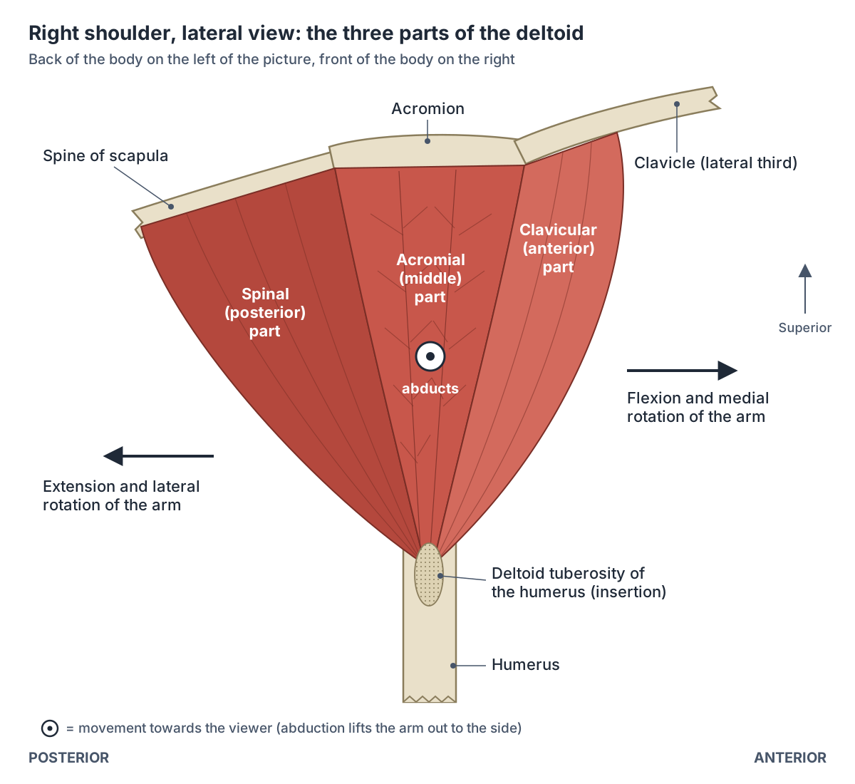
The deltoid has three origins in a continuous U around the shoulder. The clavicular (anterior) part arises from the superior surface and anterior border of the lateral third of the clavicle. The acromial (middle) part arises from the lateral margin and superior surface of the acromion. The spinal (posterior) part arises from the crest of the spine of the scapula. (Kenhub: Deltoid muscle.)
Sources differ on how much of the scapular spine the posterior part uses: Kenhub gives the lateral third of the spine, while Radiopaedia gives the lateral two-thirds. The University of Washington atlas simply lists the spine of the scapula. (Radiopaedia: Deltoid muscle.)
The parts are built differently. The acromial part is multipennate: its fibres attach at angles to several internal tendons. The clavicular and spinal parts are unipennate and run straight to the insertion tendon. (Kenhub: Deltoid muscle.)
The classic three parts may be too simple. A study of 60 cadaver shoulders divided the deltoid into seven segments (three anterior, one middle and three posterior) based on its internal tendons, and a PET imaging study in volunteers suggested these segments act as functional units. (Sakoma Y et al. Anatomical and functional segments of the deltoid muscle. J Anat. 2011.)
Radiopaedia similarly notes that electromyography suggests at least seven control regions in the deltoid that could act independently. (Radiopaedia: Deltoid muscle.)
Where does the deltoid insert?
All three parts converge on one insertion, the deltoid tuberosity, a roughened area about halfway down the lateral side of the shaft of the humerus. (Kenhub: Deltoid muscle.)
The shape of this insertion varies. In eight cadaver shoulders, the insertion averaged 39.45 mm in length (range about 25 to 48 mm). Three shoulders had a single broad insertion, two had three discrete insertions, one had a V-shaped pattern and two showed a new 'step-off' pattern. (Vohra A et al. Anatomic variations of the deltoid muscle insertion: a cadaveric study. JSES Int. 2024.)
The deltoid tuberosity, surgical neck and other landmarks of the upper arm bone. Humerus.
What does each part of the deltoid do?
Each part has its own action. The anterior part flexes and medially rotates the arm, the middle part abducts it, and the posterior part extends and laterally rotates it. (University of Washington Musculoskeletal Atlas: Deltoid.)
Orthobullets lists the same actions for each part. (Orthobullets: Deltoid.)
Radiopaedia adds abduction to the actions of the clavicular part, so the front fibres help the middle fibres when the arm is raised, and gives flexion and internal rotation for the front part and extension and external rotation for the back part. (Radiopaedia: Deltoid muscle.)
The deltoid does not start abduction on its own. TeachMeAnatomy describes supraspinatus as performing the first 0 to 15 degrees, after which the middle deltoid takes over as the main abductor and assists from 15 to 90 degrees. (TeachMeAnatomy: The Intrinsic Muscles of the Shoulder.)
Kenhub likewise says the acromial part can only abduct the arm once it is already abducted beyond 15 degrees, which is why the deltoid and axillary nerve are tested with the arm already slightly raised. (Kenhub: Deltoid muscle.)
Lifting the arm high above the head also needs the shoulder blade to turn upwards. That upward rotation of the scapula is produced by trapezius together with serratus anterior. (Kenhub: Trapezius muscle.)
Supraspinatus and the three other cuff muscles that start abduction and hold the humeral head in place. Rotator cuff.
Deltoid: origin, insertion, nerve, actions and blood supply of each part
| Part | Origin | Insertion | Nerve (roots) | Main actions | Blood supply |
|---|---|---|---|---|---|
| Clavicular (anterior) | Superior surface and anterior border of the lateral third of the clavicle | Deltoid tuberosity of the humerus | Axillary nerve, anterior branch (C5, C6) | Flexion and medial (internal) rotation of the arm; assists abduction | Shared supply of the whole muscle (see text) |
| Acromial (middle) | Lateral margin and superior surface of the acromion | Deltoid tuberosity of the humerus | Axillary nerve (C5, C6) | Abduction of the arm beyond about 15 degrees (main abductor) | Shared supply of the whole muscle (see text) |
| Spinal (posterior) | Crest of the spine of the scapula (lateral third per Kenhub, lateral two-thirds per Radiopaedia) | Deltoid tuberosity of the humerus | Axillary nerve, posterior branch (C5, C6) | Extension and lateral (external) rotation of the arm | Shared supply of the whole muscle (see text) |
Attachments and actions from Kenhub, the University of Washington atlas, Orthobullets and Radiopaedia; branches of the axillary nerve from TeachMeAnatomy. The arteries (thoracoacromial, anterior and posterior circumflex humeral, subscapular and deep brachial branches) are listed by Kenhub and Radiopaedia for the muscle as a whole, not part by part.
Nerve supply: the axillary nerve
The deltoid is supplied by the axillary nerve, from roots C5 and C6. The axillary nerve is a direct continuation of the posterior cord of the brachial plexus. (TeachMeAnatomy: The Axillary Nerve.)
The nerve leaves the axilla at the lower border of subscapularis through the quadrangular space, together with the posterior circumflex humeral artery and vein, and then winds around the surgical neck of the humerus. Its anterior branch supplies the front of the deltoid; its posterior branch supplies the back of the deltoid and teres minor. (TeachMeAnatomy: The Axillary Nerve.)
The axillary nerve also carries feeling from the skin over the lower deltoid through the upper lateral cutaneous nerve of the arm. This patch is called the regimental badge area, and numbness there is a quick sign of nerve damage. (TeachMeAnatomy: The Axillary Nerve.)
Because it winds around the surgical neck, the nerve is injured most often by a fracture of the surgical neck of the humerus or by shoulder dislocation, and both deltoid and teres minor are then affected. (TeachMeAnatomy: The Axillary Nerve.)
Kenhub lists the effects of axillary nerve injury as atrophy of the deltoid with weakness and loss of muscle tone, so abduction of the arm becomes weak. (Kenhub: Deltoid muscle.)
The quadrangular space that the nerve passes through is bounded by subscapularis (teres minor when seen from behind) above, teres major below, the long head of triceps medially and the surgical neck of the humerus laterally. Compression of the nerve and artery here causes quadrilateral (quadrangular) space syndrome, an uncommon condition that mostly affects athletes. (Kenhub: Anatomical spaces of the pectoral region.)
The brachial plexus, the posterior cord and the axillary and radial nerves. Nerves of the arm.
Blood supply of the deltoid
Kenhub lists several arteries: the acromial and deltoid branches of the thoracoacromial artery, the subscapular artery, the anterior and posterior circumflex humeral arteries and the deltoid branch of the deep brachial artery. All except the deep brachial artery are branches of the axillary artery. (Kenhub: Deltoid muscle.)
Radiopaedia gives the same fuller list: the acromial and deltoid branches of the thoracoacromial artery plus branches of the anterior and posterior humeral circumflex, subscapular and profunda brachii arteries. Short reference tables, such as the University of Washington atlas and Orthobullets, name only the deltoid branch of the thoracoacromial artery, so the difference is one of detail rather than a disagreement. (Radiopaedia: Deltoid muscle.)
The axillary artery, its thoracoacromial and circumflex humeral branches, and the cephalic vein. Arteries and veins of the arm.
Deltopectoral groove and what lies under the deltoid
The front border of the deltoid meets pectoralis major at the deltopectoral groove. The cephalic vein runs up the arm lateral to the biceps, in this groove, then pierces the clavipectoral fascia to drain into the first part of the axillary vein. (Radiopaedia: Cephalic vein.)
Deep to the deltoid lies the subacromial (subdeltoid) bursa, between the acromion and the tendon of supraspinatus. Inflammation of this bursa is called subacromial or subdeltoid bursitis. (Kenhub: Deltoid muscle.)
Pectoralis major, which meets the deltoid at the deltopectoral groove. Chest muscles.
Deltoid injection site anatomy
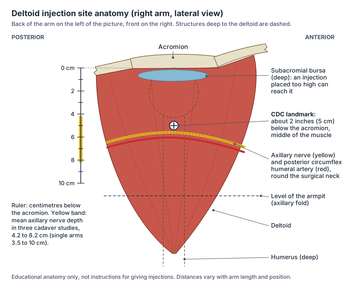
The deltoid is the recommended site for most adult vaccines. CDC guidance locates it by landmarks: find the acromion, the bony point at the end of the shoulder; the site is about 2 inches (5 cm) below it and above the armpit (axillary fold), in the middle and thickest part of the muscle, with the needle at 90 degrees. (CDC: Vaccine Administration, Intramuscular (IM) Injection, Adults 19 years of age and older (2025).)
Two structures under the muscle explain the landmarks. Kenhub notes that the course of the axillary nerve, which winds around the surgical neck of the humerus deep to the deltoid, matters for intramuscular injections, and the subacromial (subdeltoid) bursa lies deep to the upper part of the muscle, between the acromion and the supraspinatus tendon. (Kenhub: Deltoid muscle.)
Cadaver measurements of the nerve vary with the method. In a pilot study of 12 arms the axillary nerve lay a mean of 8.19 cm below the mid-acromion (range 7.5 to 10 cm) with the arm by the side, and about 1.1 cm higher with the arm abducted to 60 degrees. The authors advised injecting at or below the midpoint of the deltoid, never in its upper half. (Charmode S et al. The recommended deltoid intramuscular injection sites in the adult population: a cadaveric pilot study. Cureus. 2024.)
A follow-up study of 24 shoulders by the same group measured a mean of 6.81 cm from the mid-acromion to the nerve and found the nerve and artery consistently 6 to 7.5 cm below that point. (Charmode SH et al. Anthropometric cadaveric analysis of the quadrangular space and deltoid region. Maedica (Bucur). 2025.)
Other studies measure from different points of the acromion and find shorter distances. In 60 cadaver shoulders the nerve lay a mean of 5.22 cm from the acromion anteriorly (range 4.1 to 6.3 cm) and 4.17 cm posteriorly (range 3.5 to 4.9 cm), and the distance increased with upper arm length. (Patra A et al. Re-definition of position and calculation of safe area for axillary nerve in deltoid muscle. Anat Cell Biol. 2018.)
An ultrasound study of 30 volunteers proposed an alternative site where a vertical line from the mid-acromion crosses a line joining the front and back folds of the armpit, which the authors judged to lie away from the axillary nerve, the posterior circumflex humeral artery and the subacromial bursa. The nearest measured distance from this site to the artery was 3.2 cm in men and 2.8 cm in women. (Nakajima Y et al. Establishing a new appropriate intramuscular injection site in the deltoid muscle. Hum Vaccin Immunother. 2017.)
Injections placed too high can cause shoulder injury related to vaccine administration (SIRVA), defined as shoulder pain starting within 48 hours, lasting at least 7 days, with restricted movement and no previous symptoms. In one reported case the patient described the needle as placed too high and angled towards the subacromial space. (Moya D et al. Shoulder injury related to vaccine administration following misplaced SARS-CoV-2 vaccination. J Orthop Case Rep. 2022.)
How far below the acromion is the axillary nerve? Cadaver study results
| Study | Sample | Measured from | Distance to axillary nerve |
|---|---|---|---|
| Charmode et al. 2024 (Cureus) | 12 arms | Mid-acromion, arm by side | Mean 8.19 cm (range 7.5 to 10 cm); 7.09 cm at 60 degrees abduction |
| Charmode et al. 2025 (Maedica) | 24 shoulders | Mid-acromion | Mean 6.81 cm; nerve and artery 6 to 7.5 cm below |
| Patra et al. 2018 (Anat Cell Biol) | 60 shoulders | Acromion, anterior and posterior | Anterior mean 5.22 cm (4.1 to 6.3); posterior mean 4.17 cm (3.5 to 4.9) |
Distances differ with the reference point, arm position and arm length. CDC landmark guidance places the injection site about 2 inches (5 cm) below the acromion.
The deltoid in the 3D model
The model opens from the side and highlights the three parts of the deltoid separately: the clavicular part in front, the acromial part over the point of the shoulder and the spinal part behind. Click a part to open its page, then rotate the model to the front and back to see how the clavicular part borders pectoralis major and the spinal part covers the rotator cuff. The model, BodyParts3D from one adult male, has no nerves or bursae, so use the diagrams for the axillary nerve and the injection site.
When you turn the model to the front, the clavicular part forms the lateral wall of the deltopectoral groove, the channel where the cephalic vein runs between deltoid and pectoralis major. (Radiopaedia: Cephalic vein.)
Deltoid in the 3D model
- Acromial part of deltoid
- The middle fibres of the deltoid, forming the rounded contour of the shoulder.
- Clavicular part of deltoid
- The anterior fibres of the deltoid.
- Spinal part of deltoid
- The posterior fibers of deltoid arising from the scapular spine.
Frequently asked questions
What are the three parts of the deltoid?
The deltoid has a clavicular (anterior) part from the lateral third of the clavicle, an acromial (middle) part from the acromion and a spinal (posterior) part from the spine of the scapula. All three insert on the deltoid tuberosity of the humerus. The front part flexes and medially rotates the arm, the middle part abducts it, and the back part extends and laterally rotates it.
What nerve supplies the deltoid?
The axillary nerve, from spinal roots C5 and C6, supplies the deltoid. It continues from the posterior cord of the brachial plexus, passes through the quadrangular space with the posterior circumflex humeral artery and winds around the surgical neck of the humerus. It also supplies teres minor and the skin over the lower deltoid, the regimental badge area.
Does the deltoid start lifting the arm sideways?
No. Supraspinatus starts abduction over roughly the first 15 degrees, then the middle (acromial) deltoid takes over as the main abductor up to about 90 degrees. Raising the arm further needs the scapula to rotate upwards, which trapezius and serratus anterior do. This is why the deltoid is tested with the arm already slightly abducted.
Is the deltoid part of the rotator cuff?
No. The rotator cuff is four muscles: supraspinatus, infraspinatus, teres minor and subscapularis. The deltoid is a separate intrinsic shoulder muscle that lies over them like a cap. The cuff holds the head of the humerus in the socket while the deltoid provides much of the power for raising the arm.
Why are vaccines given in the deltoid?
CDC guidance names the deltoid as the recommended site for most adult intramuscular vaccines and locates it by landmarks: about 2 inches below the acromion and above the armpit, in the middle and thickest part of the muscle. Injecting much lower risks the axillary nerve, and injecting too high can reach the bursa under the acromion.
What happens if the axillary nerve is damaged?
The deltoid and teres minor weaken. Abduction of the arm becomes weak, the deltoid wastes and loses tone, and the skin over the lower deltoid (the regimental badge area) may go numb. The nerve is most often injured by a fracture of the surgical neck of the humerus or a shoulder dislocation, because it wraps around the bone there.
References
- OpenStax. Anatomy and Physiology 2e, §11.5 Muscles of the Pectoral Girdle and Upper Limbs. Rice University, 2022.
- Kenhub. Deltoid muscle: origin, insertion, innervation and function.
- Kenhub. Trapezius muscle.
- Kenhub. Anatomical spaces of the pectoral region.
- Kenhub. Biceps brachii muscle.
- TeachMeAnatomy. The Intrinsic Muscles of the Shoulder.
- TeachMeAnatomy. The Axillary Nerve.
- University of Washington Department of Radiology. Musculoskeletal Atlas: Deltoid.
- Radiopaedia. Deltoid muscle.
- Radiopaedia. Cephalic vein.
- Orthobullets. Deltoid.
- Sakoma Y, Sano H, Shinozaki N, et al. Anatomical and functional segments of the deltoid muscle. J Anat. 2011;218(2):185-190.
- Vohra A, Paul B, Saunders P, et al. Anatomic variations of the deltoid muscle insertion: a cadaveric study. JSES Int. 2024;8(3):546-550.
- Centers for Disease Control and Prevention. Vaccine Administration: Intramuscular (IM) Injection, Adults 19 years of age and older. 2025.
- Charmode S, Mehra S, Mishra AK. The recommended deltoid intramuscular injection sites in the adult population: a cadaveric pilot study with an orthopedic perspective. Cureus. 2024;16(8):e66391.
- Charmode SH, Mishra AK, Mehra S. Anthropometric cadaveric analysis of the quadrangular space and deltoid region: anatomical perspectives for clinical applications in the Indian population. Maedica (Bucur). 2025;20(2):316-324.
- Patra A, Singh M, Kaur H, Singla RK, Malhotra V. Re-definition of position and calculation of safe area for axillary nerve in deltoid muscle with its clinical relevance: a cadaveric study. Anat Cell Biol. 2018;51(2):93-97.
- Nakajima Y, Mukai K, Takaoka K, et al. Establishing a new appropriate intramuscular injection site in the deltoid muscle. Hum Vaccin Immunother. 2017;13(9):2123-2129.
- Moya D, Gómez D, Patiño P, et al. Shoulder injury related to vaccine administration following misplaced SARS-CoV-2 vaccination: a case report and review of literature. J Orthop Case Rep. 2022;12(3):100-103.
- Mitsuhashi N. et al. (2009). BodyParts3D: 3D structure database for anatomical concepts. Nucleic Acids Research 37, D782–D785. Data © The Database Center for Life Science, CC BY 4.0.
Related anatomy
- Shoulder jointShoulder anatomy with labelled diagrams and a 3D model: clavicle, scapula and humerus, the glenohumeral, AC and SC…
- Rotator cuffThe rotator cuff muscles explained: supraspinatus, infraspinatus, teres minor and subscapularis, with attachments…
- Arm musclesArm muscles labeled in 3D: biceps brachii, brachialis, coracobrachialis, triceps brachii, deltoid and anconeus, with…
- TrapeziusAnatomy of the trapezius with a 3D model and diagram: descending, transverse and ascending parts, attachments…
- Upper limb nervesBrachial plexus diagram with roots, trunks, cords and branches; axillary, musculocutaneous, median, ulnar and radial…
- HumerusHumerus bone labeled: head, anatomical and surgical neck, tubercles, deltoid tuberosity, radial groove, epicondyles…
Explore the whole body in the interactive 3D human anatomy model, or browse the A–Z list of human structures.