

Where is the trapezius muscle?
The trapezius is a large, broad, superficial muscle of the back of the neck and the upper back. Radiopaedia notes that it gains its name from its diamond shape. (Radiopaedia: Trapezius muscle.)
Together with latissimus dorsi, the trapezius is the most superficial of the superficial extrinsic back muscles. Its front edge forms the posterior border of the posterior triangle of the neck. (Kenhub: Trapezius muscle.)
OpenStax lists the trapezius among the muscles of the posterior thorax that position the pectoral girdle, with the actions of elevating the shoulders (shrugging), pulling the shoulder blades together and tilting the head backwards. (OpenStax Anatomy and Physiology 2e, 11.5: Muscles of the Pectoral Girdle and Upper Limbs.)
It is the only muscle of the upper limb that is not supplied by the brachial plexus; its motor nerve is the accessory nerve, a cranial nerve. (Kenhub: Trapezius muscle.)
The superficial, intermediate and deep back muscle layers, from trapezius and the lats to erector spinae. Back muscles.
The three parts: upper, middle and lower trapezius
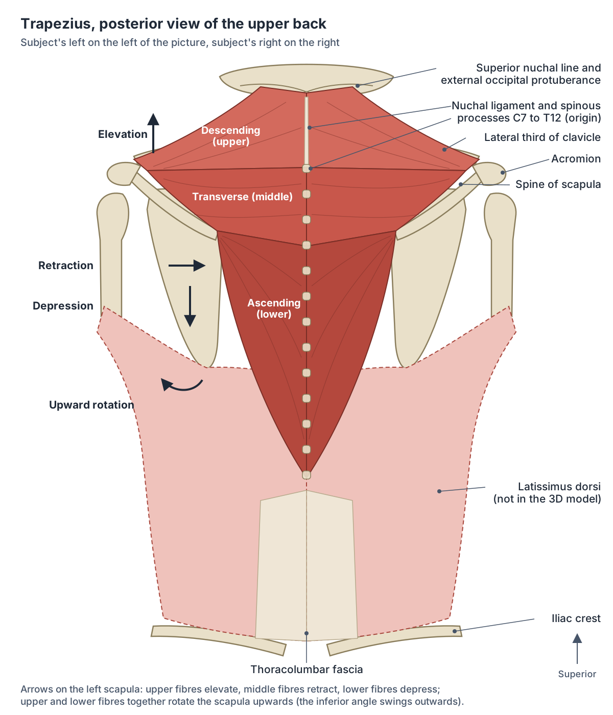
The trapezius has three functional parts, named for the direction of their fibres: the descending (upper) part, the transverse (middle) part and the ascending (lower) part. (Kenhub: Trapezius muscle.)
The descending part arises from the medial third of the superior nuchal line, the external occipital protuberance and the nuchal ligament, and inserts on the lateral third of the clavicle. Radiopaedia specifies the posterior aspect of the lateral third of the clavicle. (Kenhub: Trapezius muscle.)
The transverse part arises from the spinous processes and supraspinous ligaments of the upper thoracic vertebrae and inserts on the medial margin of the acromion and the superior crest of the spine of the scapula. (Kenhub: Trapezius muscle.)
The ascending part arises from the spinous processes and supraspinous ligaments of the lower thoracic vertebrae, about T4 to T12, and converges on the medial end of the spine of the scapula. (Kenhub: Trapezius muscle.)
Vertebral levels for each part differ between sources. Kenhub gives T1 to T4 (or C7 to T3) for the transverse part and T4 to T12 for the ascending part, while Radiopaedia describes the lower fibres as arising from T6 to T12. All sources agree that the muscle as a whole spans C7 to T12. (Radiopaedia: Trapezius muscle.)
Taken together, the origin runs from the medial third of the superior nuchal line and the external occipital protuberance, along the nuchal ligament and the spinous processes of C7 to T12; the insertion runs along the lateral third of the clavicle, the acromion and the spine of the scapula. (University of Washington Musculoskeletal Atlas: Trapezius.)
Trapezius: origin, insertion, nerve, action and blood supply of each part
| Part | Origin | Insertion | Nerve (roots) | Main actions | Blood supply (Kenhub) |
|---|---|---|---|---|---|
| Descending (upper) | Medial third of the superior nuchal line, external occipital protuberance, nuchal ligament | Lateral third of the clavicle (posterior border) | Spinal accessory nerve, CN XI (C1 to C5/C6); C3, C4 sensory | Elevates the scapula (shrug); upward rotation with the lower fibres; extends the head and neck | Occipital artery |
| Transverse (middle) | Spinous processes and supraspinous ligaments, T1 to T4 (or C7 to T3) | Medial margin of the acromion, superior crest of the spine of the scapula | Spinal accessory nerve, CN XI; C3, C4 sensory | Retracts the scapula; medial stabiliser | Superficial or transverse cervical artery |
| Ascending (lower) | Spinous processes and supraspinous ligaments, T4 to T12 (T6 to T12 per Radiopaedia) | Medial end of the spine of the scapula | Spinal accessory nerve, CN XI; C3, C4 sensory | Depresses the scapula; upward rotation with the upper fibres | Dorsal scapular artery |
Attachments and blood supply from Kenhub; levels compared with Radiopaedia; nerve from Radiopaedia and the University of Washington atlas; actions from TeachMeAnatomy, the University of Washington atlas and Kenhub. TeachMeAnatomy and the University of Washington atlas name the transverse cervical artery for the whole muscle.
What does the trapezius do?
The upper fibres elevate the scapula and rotate it during abduction of the arm, the middle fibres retract it (pull it towards the spine) and the lower fibres pull it downwards (depression). (TeachMeAnatomy: The Extrinsic Muscles of the Shoulder.)
The University of Washington atlas adds that the superior and inferior fibres act together to rotate the scapula upwards (superior rotation). (University of Washington Musculoskeletal Atlas: Trapezius.)
Radiopaedia notes that all fibres retract the scapula, and that with the shoulders fixed, both trapezius muscles contracting together extend the neck. (Radiopaedia: Trapezius muscle.)
Kenhub describes the overall role as stabilising the scapula in place, with the descending fibres also extending the head and neck and the transverse fibres helping serratus anterior turn the scapula upwards. (Kenhub: Trapezius muscle.)
Research on arm raising gives a more detailed picture. A 2009 review describes the upper trapezius as acting mainly to elevate and retract the clavicle, the middle trapezius as a medial stabiliser of the scapula, and the lower trapezius as the only part able to substantially rotate the scapula upwards. The middle and lower serratus anterior are the prime movers of this rotation. (Phadke V, Camargo PR, Ludewig PM. Scapular and rotator cuff muscle activity during arm elevation. Rev Bras Fisioter. 2009.)
How the scapula, clavicle and humerus move together when the arm is raised. Shoulder anatomy.
What nerve supplies the trapezius?
The motor supply of the trapezius is the spinal part of the accessory nerve (cranial nerve XI), which arises from the upper cervical spinal cord, C1 to C5 or C6. Branches of the cervical plexus from C3 and C4 carry pain and proprioceptive fibres from the muscle. (Radiopaedia: Trapezius muscle.)
Kenhub lists the C3 and C4 branches as motor and sensory, so sources differ on whether they add motor fibres; the University of Washington atlas, Radiopaedia and TeachMeAnatomy describe them as sensory (pain and proprioception). (Kenhub: Trapezius muscle.)
The spinal accessory nerve has an unusual course. Its fibres run up through the foramen magnum into the skull, leave again through the jugular foramen, supply sternocleidomastoid, and then cross the posterior triangle of the neck to reach the trapezius. It supplies only these two muscles. (TeachMeAnatomy: The Accessory Nerve (CN XI).)
In the posterior triangle the nerve lies close to the surface, covered only by skin and subcutaneous fascia, which is why it is easily injured there. (Gooding BWT et al. Scapular winging. Shoulder Elbow. 2014.)
All twelve cranial nerves, including the accessory nerve that supplies trapezius and sternocleidomastoid. Cranial nerves.
Blood supply of the trapezius
Kenhub gives a different main artery for each part: the occipital artery for the descending part, the superficial or transverse cervical artery for the transverse part, and the dorsal scapular artery for the ascending part. (Kenhub: Trapezius muscle.)
Shorter references, including TeachMeAnatomy and the University of Washington atlas, name the transverse cervical artery as the arterial supply of the muscle. (TeachMeAnatomy: Trapezius.)
What lies around the trapezius?
Radiopaedia describes the trapezius as lying superficial (posterior) to several deeper muscles of the neck and upper back, so it is the first muscle seen when the skin of the upper back is removed. (Radiopaedia: Trapezius muscle.)
Its anterior margin forms the posterior border of the posterior triangle of the neck, the space crossed by the accessory nerve. Its free inferior margin forms the medial boundary of the triangle of auscultation. (Kenhub: Trapezius muscle.)
Kenhub's latissimus dorsi page gives the borders of the triangle of auscultation as trapezius, the medial border of the scapula and latissimus dorsi. It is a spot where the lower lungs can be heard with a stethoscope. (Kenhub: Latissimus dorsi muscle.)
The deeper shoulder blade muscles, levator scapulae and the rhomboids, lie under the trapezius. Levator scapulae arises from the transverse processes of C1 to C4 and elevates the scapula; rhomboid minor (C7 to T1) and rhomboid major (T2 to T5) retract and rotate it. All three are supplied by the dorsal scapular nerve. (TeachMeAnatomy: The Superficial Back Muscles.)
The neck muscle under the upper trapezius that also elevates the scapula. Levator scapulae.
Trapezius weakness and accessory nerve injury
The most common cause of accessory nerve damage is iatrogenic, meaning it follows a medical procedure. Cervical lymph node biopsy and insertion of a line into the internal jugular vein are the typical examples. (TeachMeAnatomy: The Superficial Back Muscles.)
The accessory nerve is tested by asking the person to turn the head and shrug the shoulders. Signs of damage include a weak shrug, wasting of the muscle and an asymmetrical neckline. (TeachMeAnatomy: The Accessory Nerve (CN XI).)
A review of scapular winging describes the effects of trapezius palsy: the shoulder droops, the scapula wings laterally, and forward flexion and abduction of the arm are weak, limiting overhead activity. The winging may be slight at rest but increases with abduction and with resisted external rotation. In one series 14 of 22 patients were misdiagnosed at first. (Gooding BWT et al. Scapular winging. Shoulder Elbow. 2014.)
This lateral winging differs from the commoner medial winging caused by paralysis of serratus anterior after injury to the long thoracic nerve, which is made more prominent by pushing the arm forwards. (Gooding BWT et al. Scapular winging. Shoulder Elbow. 2014.)
Kenhub notes that when the trapezius is weak but sternocleidomastoid works normally, the accessory nerve has been damaged further along its course, after it has supplied sternocleidomastoid. The trapezius can also be used as a muscle flap in reconstructive surgery. (Kenhub: Trapezius muscle.)
The triangles of the neck, including the posterior triangle that the accessory nerve crosses. Neck anatomy.
Trapezius vs latissimus dorsi (traps vs lats)
Latissimus dorsi is the broad, triangular muscle of the lower back. OpenStax classes it with pectoralis major as one of the two axial muscles that move the humerus, whereas the trapezius moves the scapula and clavicle. (OpenStax Anatomy and Physiology 2e, 11.5: Muscles of the Pectoral Girdle and Upper Limbs.)
Latissimus dorsi arises from the spinous processes of T7 to T12, the thoracolumbar fascia, the iliac crest, the 9th to 12th ribs and, inconstantly, the inferior angle of the scapula. It inserts on the floor of the intertubercular sulcus of the humerus and is supplied by the thoracodorsal nerve (C6 to C8). It extends, adducts and internally rotates the arm and is used in climbing, rowing and swimming. (Kenhub: Latissimus dorsi muscle.)
TeachMeAnatomy gives the vertebral origin as T6 to T12 on one page and T7 to T12 on another, and the rib origin as the inferior three ribs, a small difference between sources. (TeachMeAnatomy: The Superficial Back Muscles.)
The lower trapezius overlaps the upper medial part of latissimus dorsi: Kenhub notes that latissimus dorsi covers almost all of the back muscles of the posterior trunk except the trapezius. Latissimus dorsi is not included in the BodyParts3D model used on this site, so the diagram shows it instead. (Kenhub: Latissimus dorsi muscle.)
The spine, ribs, shoulder blades and back muscle layers, including the lats. Back anatomy.
Trapezius vs latissimus dorsi
| Feature | Trapezius | Latissimus dorsi |
|---|---|---|
| Position | Back of the neck and upper back | Lower and middle back, up to the armpit |
| Origin | Skull (superior nuchal line, external occipital protuberance), nuchal ligament, spinous processes C7 to T12 | Spinous processes T7 to T12 (T6 per TeachMeAnatomy), thoracolumbar fascia, iliac crest, lower ribs, sometimes the inferior angle of the scapula |
| Insertion | Lateral third of the clavicle, acromion, spine of the scapula | Floor of the intertubercular sulcus of the humerus |
| Nerve | Spinal accessory nerve (CN XI); C3, C4 sensory | Thoracodorsal nerve (C6 to C8), from the brachial plexus |
| Main job | Moves and stabilises the scapula; extends the neck | Extends, adducts and internally rotates the arm |
| In the 3D model | Yes, as three parts | No |
Based on Kenhub (trapezius and latissimus dorsi), TeachMeAnatomy and OpenStax §11.5.
The trapezius in the 3D model
The model opens from behind and highlights the descending, transverse and ascending parts of the trapezius as three separate parts on each side. Click a part to open its own page. Follow the descending part up to the back of the skull and out to the clavicle, then the ascending part down to the lower thoracic spine. Rotate to the side to see the upper fibres forming the slope between neck and shoulder. Latissimus dorsi is missing from the model, so the lower back looks bare below the ascending fibres.
Use the attachments as landmarks while you rotate the view: the descending part ends on the lateral third of the clavicle, the transverse part on the acromion and the upper edge of the scapular spine, and the ascending part on the medial end of the spine. (Kenhub: Trapezius muscle.)
Trapezius in the 3D model
- Ascending part of trapezius
- The lower fibres of trapezius, running upward and laterally to the scapular spine.
- Descending part of trapezius
- The upper fibres of trapezius, running down from the skull and neck.
- Transverse part of trapezius
- The middle fibers of trapezius, running horizontally.
Frequently asked questions
What are the three parts of the trapezius?
The trapezius has a descending (upper) part from the back of the skull and the nuchal ligament to the clavicle, a transverse (middle) part from the upper thoracic spinous processes to the acromion and spine of the scapula, and an ascending (lower) part from the lower thoracic spinous processes to the medial end of the scapular spine. They elevate, retract and depress the scapula respectively.
What nerve supplies the trapezius?
The spinal accessory nerve, cranial nerve XI, supplies the trapezius with motor fibres. Its fibres come from the upper cervical spinal cord (about C1 to C5), pass up through the foramen magnum, leave the skull through the jugular foramen and cross the posterior triangle of the neck. Branches of C3 and C4 carry pain and position sense from the muscle.
What does the trapezius do?
The trapezius moves and steadies the shoulder blade. Its upper fibres lift the scapula, as in shrugging; its middle fibres pull the scapula towards the spine; its lower fibres pull it down. Upper and lower fibres together rotate the scapula upwards so the arm can be raised overhead, and both trapezius muscles acting together extend the neck.
Is the trapezius a neck muscle or a back muscle?
Both. The trapezius is a superficial back muscle whose upper part covers the back of the neck. It is one of the superficial extrinsic back muscles, and its front edge forms the back border of the posterior triangle of the neck. Its lower fibres reach down to the twelfth thoracic vertebra.
What happens if the accessory nerve is damaged?
The trapezius weakens. The shoulder droops, shrugging is weak, the muscle wastes and the scapula wings outwards (laterally), especially when the arm is abducted. Raising the arm overhead becomes difficult. The commonest cause is accidental injury during neck procedures such as lymph node biopsy, because the nerve lies just under the skin in the posterior triangle of the neck.
What is the difference between traps and lats?
The trapezius (traps) covers the neck and upper back, attaches to the skull, spine, clavicle and scapula, is supplied by the accessory nerve and mainly moves the scapula. Latissimus dorsi (lats) covers the lower back, runs from the lower thoracic spine, fascia, iliac crest and ribs to the humerus, is supplied by the thoracodorsal nerve and pulls the arm down and back.
References
- OpenStax. Anatomy and Physiology 2e, §11.5 Muscles of the Pectoral Girdle and Upper Limbs. Rice University, 2022.
- Kenhub. Trapezius muscle: origins, insertions, innervation and function.
- Kenhub. Latissimus dorsi muscle.
- TeachMeAnatomy. The Superficial Back Muscles.
- TeachMeAnatomy. The Extrinsic Muscles of the Shoulder.
- TeachMeAnatomy. Trapezius (encyclopaedia entry).
- TeachMeAnatomy. The Accessory Nerve (CN XI).
- University of Washington Department of Radiology. Musculoskeletal Atlas: Trapezius.
- Radiopaedia. Trapezius muscle.
- Gooding BWT, Geoghegan JM, Wallace WA, Manning PA. Scapular winging. Shoulder Elbow. 2014;6(1):4-11.
- Phadke V, Camargo PR, Ludewig PM. Scapular and rotator cuff muscle activity during arm elevation: a review of normal function and alterations with shoulder impingement. Rev Bras Fisioter. 2009;13(1):1-9.
- Mitsuhashi N. et al. (2009). BodyParts3D: 3D structure database for anatomical concepts. Nucleic Acids Research 37, D782–D785. Data © The Database Center for Life Science, CC BY 4.0.
Related anatomy
- Back musclesBack muscles anatomy with a labeled diagram and 3D model: the lats (latissimus dorsi), trapezius, rhomboids, erector…
- Back anatomyBack anatomy explained: spine regions, muscle layers, spinal cord and nerves, ribs, kidneys and the lower back, with…
- Shoulder jointShoulder anatomy with labelled diagrams and a 3D model: clavicle, scapula and humerus, the glenohumeral, AC and SC…
- DeltoidDeltoid anatomy with a 3D model and labelled diagrams: clavicular, acromial and spinal parts, attachments, axillary…
- Neck musclesNeck muscles explained: sternocleidomastoid, suprahyoid, infrahyoid, scalene, prevertebral and suboccipital groups…
- Levator scapulaeLevator scapulae muscle anatomy: origin on C1 to C4, insertion on the scapula, actions on the shoulder blade and neck…
Explore the whole body in the interactive 3D human anatomy model, or browse the A–Z list of human structures.