

What are the leg bones? Leg vs thigh in anatomy
In anatomy, the word leg (crus) is reserved for the part of the lower limb between the knee and the ankle, and the part between the hip and the knee is the thigh (femoral region). By that definition the leg has two bones, the tibia and fibula, and the thigh has one, the femur. (OpenStax Anatomy and Physiology 2e, 1.6: Anatomical Terminology.)
In everyday speech, as Kenhub points out, people use 'leg' for the entire lower extremity. So when most people say leg bones they mean four bones: the femur in the thigh, the patella at the front of the knee, and the tibia and fibula below it. The tibia and fibula are two long bones that run parallel to each other, the tibia on the medial (inner) side and the thinner fibula on the lateral (outer) side. (Kenhub: Lower leg and knee anatomy.)
For the muscles, nerves, vessels and regions of the whole lower limb, see the leg anatomy guide.
How many bones are in the leg?
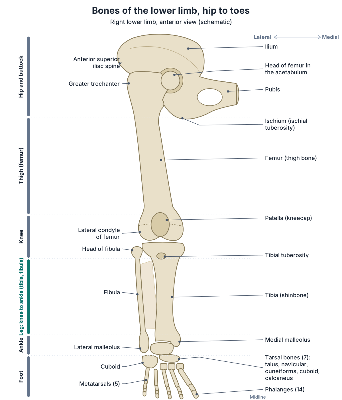
Each lower limb contains 30 bones: the femur, the patella, the tibia, the fibula, 7 tarsal bones, 5 metatarsal bones and 14 phalanges. The leg in the everyday sense, from hip to ankle, has four of them; the leg in the strict anatomical sense, from knee to ankle, has two, the tibia and fibula. (OpenStax Anatomy and Physiology 2e, 8.4: Bones of the Lower Limb.)
The hip bone is not one of the 30. OpenStax places it in the pelvic girdle: a single bone on each side, formed by the ilium, ischium and pubis, which fuse in the late teens. It is the attachment point for each lower limb, and it is joined to the sacrum of the vertebral column. (OpenStax Anatomy and Physiology 2e, 8.3: The Pelvic Girdle and Pelvis.)
Kenhub groups the same bones by region: the femur and patella with the thigh, the tibia and fibula in the leg, and 26 bones in the foot. The foot's 26 are 7 tarsals, 5 metatarsals and 14 phalanges, with two phalanges in the big toe and three in each of the other toes. (Kenhub: Lower extremity anatomy.)
For every bone of the body grouped by region, see the skeletal system guide.
Bones of one lower limb
| Region | Bones | Number |
|---|---|---|
| Thigh | Femur | 1 |
| Front of the knee | Patella (kneecap) | 1 |
| Leg (knee to ankle) | Tibia and fibula | 2 |
| Ankle and back of the foot | Tarsal bones: talus, calcaneus, navicular, cuboid and three cuneiforms | 7 |
| Front of the foot | Metatarsal bones | 5 |
| Toes | Phalanges: 2 in the big toe, 3 in each other toe | 14 |
| Total | 30 |
Sources: OpenStax Anatomy and Physiology 2e, 8.4 (the count of 30); Kenhub, Lower extremity anatomy (the groups). The hip bone belongs to the pelvic girdle and is not counted.
Leg bones diagram: labelling the femur, patella, tibia and fibula
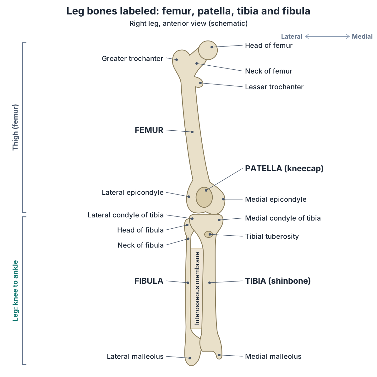
A labelled leg bones diagram is easiest to read from the top down. The femur's rounded head fits into the acetabulum of the hip bone, and the greater trochanter projects upwards on the lateral side above the base of the neck. At the knee the medial and lateral condyles of the femur rest on the condyles of the tibia, and the patella sits in front, against the groove called the patellar surface. (OpenStax Anatomy and Physiology 2e, 8.4: Bones of the Lower Limb.)
Below the knee, the tibia is the thick bone on the medial side and the fibula is the slender bone on the lateral side. The head of the fibula sits just below the lateral condyle of the tibia, and the fibula's lower end forms the lateral malleolus, the bump on the outer side of the ankle. The tibia's lower end forms the medial malleolus on the inner side. (OpenStax Anatomy and Physiology 2e, 8.4: Bones of the Lower Limb.)
The diagram on this page shows the right leg from the front (anterior view), so its lateral side, with the fibula and the greater trochanter, is on the viewer's left. A left leg seen from the front is its mirror image. To check which side an unlabelled picture shows, find the fibula: it is always on the lateral side of the leg. (OpenStax Anatomy and Physiology 2e, 8.4: Bones of the Lower Limb.)
Leg bones at a glance
| Bone | Location | Key landmarks | Articulations |
|---|---|---|---|
| Femur | Thigh, from hip to knee | Head and fovea, neck, greater and lesser trochanters, linea aspera, medial and lateral condyles and epicondyles, adductor tubercle, patellar surface | Hip bone (hip joint); tibia and patella (knee) |
| Patella | Front of the knee, inside the quadriceps tendon | Base, apex, medial and lateral facets | Femur only (patellofemoral) |
| Tibia | Medial side of the leg (the shin) | Medial and lateral condyles (tibial plateau), intercondylar eminence, tibial tuberosity, anterior border, soleal line, medial malleolus, fibular notch | Femur (knee); fibula (proximal and distal tibiofibular joints); talus (ankle) |
| Fibula | Lateral side of the leg | Head, neck, shaft, lateral malleolus | Tibia (proximal and distal tibiofibular joints); talus (ankle) |
Sources: OpenStax Anatomy and Physiology 2e, 8.4 and 9.6; TeachMeAnatomy; Kenhub.
Femur: the thigh bone
The femur is the single bone of the thigh and the longest and strongest bone in the body. Its upper end articulates with the acetabulum of the hip bone at the hip joint, and its lower end articulates with the tibia and the patella at the knee. (OpenStax Anatomy and Physiology 2e, 8.4: Bones of the Lower Limb.)
At the top of the femur are the head, a smooth ball covered with articular cartilage with a small pit (the fovea) where the ligament of the head of the femur (ligamentum teres) attaches, and the narrower neck below it. The greater trochanter, on the lateral side, is the attachment site for many of the gluteal muscles, and the lesser trochanter, on the medial side, receives iliopsoas. The intertrochanteric line joins the two trochanters in front and the intertrochanteric crest joins them behind. (TeachMeAnatomy: The Femur.)
Down the back of the shaft runs a rough ridge, the linea aspera (Latin for 'rough line'). Near the top it becomes the gluteal tuberosity, where gluteus maximus attaches. At the lower end, the medial and lateral condyles articulate with the tibia, and the epicondyles, bony bumps on the non-articular sides of the condyles, anchor the collateral ligaments of the knee. The adductor tubercle sits at the top of the medial epicondyle, and the intercondylar fossa is the deep notch between the condyles at the back. (TeachMeAnatomy: The Femur.)
For every landmark, muscle attachment, the blood supply of the head and femur fractures, see the femur guide.
Patella: the kneecap
The patella (kneecap) is the largest sesamoid bone in the body, a sesamoid bone being one that forms inside a tendon where the tendon crosses a joint. It lies within the tendon of quadriceps femoris in front of the knee, and it articulates with the patellar surface of the femur but not with the tibia. (OpenStax Anatomy and Physiology 2e, 8.4: Bones of the Lower Limb.)
The patella is triangular. Its base points upwards and receives the quadriceps tendon, and its apex points downwards and is joined to the tibial tuberosity by the patellar ligament (often called the patellar tendon). Its back surface carries a medial and a lateral facet, which glide on the medial and lateral condyles of the femur. (TeachMeAnatomy: The Patella.)
It does two jobs: it protects the front of the knee joint, and it increases the leverage that quadriceps can exert when it straightens the knee, acting like a pulley. (TeachMeAnatomy: The Patella.)
For the facets, development and dislocation of the kneecap, see the patella guide.
Tibia: the shinbone
The tibia (shinbone) is the medial bone of the leg and its main weight-bearing bone. It is the second-largest bone in the body after the femur, and it takes part in two major joints, the knee above and the ankle below. (Kenhub: Tibia.)
Its upper end widens into the medial and lateral condyles, whose flat upper surfaces form the tibial plateau. Between the condyles rises the intercondylar eminence, and the upper part of the front border is marked by the tibial tuberosity, where the patellar ligament attaches. (TeachMeAnatomy: The Tibia.)
The shaft is prism-shaped, with three borders and three surfaces. Its front (anterior) border can be felt just under the skin all the way down the front of the leg as the shin. On the back of the shaft, a ridge called the soleal line is where part of the soleus muscle of the calf arises. (TeachMeAnatomy: The Tibia.)
At its lower end the tibia forms the medial malleolus, the large bony bump on the inner side of the ankle, which articulates with the talus as part of the ankle joint. On its lateral side, a wide groove called the fibular notch receives the lower end of the fibula at the distal tibiofibular joint. (OpenStax Anatomy and Physiology 2e, 8.4: Bones of the Lower Limb.)
For the tibial plateau, muscle attachments, development and tibia fractures, see the tibia guide.
Fibula: the thin outer bone of the lower leg
The fibula lies on the lateral side of the leg, parallel to the tibia. It is not a main weight-bearing bone: its main jobs are to give attachment to muscles and to help stabilise the ankle. (TeachMeAnatomy: The Fibula.)
Its upper end is the head, which carries a facet on its medial surface for the lateral condyle of the tibia at the proximal tibiofibular joint. Below the head is the narrow neck. The common fibular (common peroneal) nerve winds around the back and outer side of the fibular neck, close to the bone. (TeachMeAnatomy: The Fibula.)
The long, slender shaft is joined to the tibia along its length by the interosseous membrane, a sheet of dense connective tissue. The lower end forms the lateral malleolus, the bump on the outer side of the ankle, whose deep surface articulates with the talus as part of the ankle joint. (OpenStax Anatomy and Physiology 2e, 8.4: Bones of the Lower Limb.)
For the head, shaft, lateral malleolus, muscle attachments and fibula fractures, see the fibula guide.
Tibia and fibula: how the two lower leg bones are joined
The tibia and fibula are joined in three places. At the top, the head of the fibula meets the lateral condyle of the tibia at the superior (proximal) tibiofibular joint, a plane synovial joint in which the bone surfaces glide over each other. (Kenhub: Tibiofibular joints.)
Along their shafts, the interosseous membrane connects the two bones; Kenhub calls it the middle tibiofibular joint. At the bottom, the fibula sits in the fibular notch of the tibia at the inferior (distal) tibiofibular joint, a fibrous joint of the type called a syndesmosis, in which the bones are bound by tough fibrous tissue and move very little. (Kenhub: Tibiofibular joints.)
Below that joint, the lower ends of the tibia and fibula form a socket, the ankle mortise, that grips the talus of the foot. The tibia and fibula together with the talus are the three bones of the ankle joint. (TeachMeAnatomy: The Ankle Joint.)
For the ankle mortise, its ligaments and sprains, see ankle anatomy.
Joints formed by the leg bones: hip, knee and ankle
The leg bones take part in three large joints. The hip joint is a ball-and-socket joint between the head of the femur and the acetabulum of the hip bone. The knee, which OpenStax calls the largest joint of the body, joins the femur, the tibia and the patella. The ankle (talocrural) joint is formed by the talus of the foot and the lower ends of the tibia and fibula. (OpenStax Anatomy and Physiology 2e, 9.6: Anatomy of Selected Synovial Joints.)
The fibula is not part of the knee joint, which TeachMeAnatomy describes as formed by articulations between the patella, femur and tibia: the head of the fibula articulates only with the tibia, just below the knee. Inside the knee, the femur meets the tibia at the tibiofemoral articulation and the patella at the patellofemoral articulation. (TeachMeAnatomy: The Knee Joint.)
For the ligaments, menisci and bursae that hold these bones together at the knee, see knee anatomy.
How to feel the leg bones on your own leg
Several parts of the leg bones lie just under the skin. The greater trochanter can be felt on the outer side of the upper thigh. The anterior border and medial surface of the tibia can be followed down the whole length of the shin, and the medial and lateral malleoli form the bony bumps on the inner and outer sides of the ankle. (OpenStax Anatomy and Physiology 2e, 8.4: Bones of the Lower Limb.)
The fibula is harder to find. OpenStax notes that only its upper and lower ends can be felt: the head on the outer side of the leg just below the knee, and the lateral malleolus at the ankle. The rest of it is largely surrounded by muscles, because its main role is muscle attachment. (OpenStax Anatomy and Physiology 2e, 8.4: Bones of the Lower Limb.)
Common leg bone fractures and injuries
Fractures of the femoral neck tend to happen in older people after low-energy falls. A fracture inside the hip joint capsule can damage the blood supply to the femoral head, which may then die (avascular necrosis). Fractures of the femoral shaft, by contrast, are typically high-energy injuries. (TeachMeAnatomy: The Femur.)
The tibia is the most commonly fractured long bone in the body, and in many tibial shaft fractures the fibula breaks as well. High-energy injuries such as road traffic collisions are a classic cause, but lower-energy injuries, for example a twisting fall while skiing, can also break the shaft. (AAOS OrthoInfo: Tibia (Shinbone) Shaft Fractures.)
High-energy injuries are more likely to cause severe soft-tissue damage. (Radiopaedia: Tibial shaft fracture.)
Lower-energy injuries are also common: in one population-based study most tibial shaft fractures happened while walking, during indoor activities or in sport. (Larsen P et al. Incidence and epidemiology of tibial shaft fractures. Injury. 2015.)
The lateral malleolus of the fibula is a common site of fracture, for example when the foot is forced into external rotation and the talus presses on it. A fracture of the fibular neck can injure the common fibular nerve, which winds around it. (TeachMeAnatomy: The Fibula.)
The patella most often dislocates laterally, out of its groove on the femur, and it can break from a direct blow or a sudden contraction of quadriceps. These notes explain the anatomy only; anyone with a leg injury should see a clinician. (TeachMeAnatomy: The Patella.)
For the joint that the head and neck of the femur form with the pelvis, see hip anatomy.
Foot bones in brief
Below the ankle, each foot has 26 bones: seven tarsal bones in the back half of the foot, five metatarsal bones in the front half and 14 phalanges in the toes. The tarsals include the talus and the heel bone (calcaneus). The talus is the foot's link with the leg bones: its upper surface articulates with the tibia and its sides with the medial and lateral malleoli. (OpenStax Anatomy and Physiology 2e, 8.4: Bones of the Lower Limb.)
For all 26 foot bones with a labelled diagram and their joints and arches, see the foot bones guide.
How to use the 3D model of the leg bones
The 3D model uses BodyParts3D, built from an adult male body. It highlights the femur, patella, tibia and fibula of both legs on a ghosted skeleton, with key labels on the left leg. The Female option uses the same leg bones. The model shows the outer shape of each bone only, with no internal structure such as the marrow cavity; the knee ligaments and menisci are highlighted on the knee anatomy page instead.
Use the model to find the landmarks you can feel on yourself. The greater trochanter is at the top of the femur on the outer side, the front border of the tibia runs down the shin, and the two malleoli form the bumps at the ankle: the medial one on the tibia and the lateral one on the fibula. (OpenStax Anatomy and Physiology 2e, 8.4: Bones of the Lower Limb.)
Leg bones in the 3D model
Frequently asked questions
How many bones are in the leg?
Each lower limb has 30 bones: the femur, patella, tibia, fibula, 7 tarsals, 5 metatarsals and 14 phalanges. In everyday speech the leg, from hip to ankle, has four bones: the femur, patella, tibia and fibula. In strict anatomical terms the leg is only the part between the knee and the ankle, and it has two bones, the tibia and fibula.
What is the lower leg bone called?
The lower leg has two bones. The tibia, or shinbone, is the thick bone on the inner (medial) side and carries the body's weight; you can feel its front border and medial surface down your shin. The fibula is the slender bone on the outer (lateral) side. Only its ends can be felt: the head just below the knee and the lateral malleolus at the ankle.
What is the strongest bone in the leg?
The femur, the thigh bone, is the longest and strongest bone in the body. It runs from the hip, where its head fits into the acetabulum of the hip bone, to the knee, where its condyles rest on the tibia. The tibia comes next: it is the second-largest bone in the body and the main weight-bearing bone of the lower leg.
Does the fibula bear weight?
Only a small share. Textbooks such as OpenStax describe the fibula as non-weight-bearing, and Kenhub says it is not the principal weight-bearing bone of the leg; the tibia does that job. Cadaver loading studies show the fibula still carries a minority of the load passing down the leg, roughly 7 to 17% in different studies, with the share changing with ankle position and load. The fibula anchors many leg muscles, forms the lateral malleolus that helps stabilise the ankle, and is bound to the tibia at both ends and along its length by the interosseous membrane.
Is the kneecap a bone?
Yes. The kneecap, or patella, is a bone, and it is the largest sesamoid bone in the body: it forms inside the tendon of quadriceps femoris in front of the knee. It articulates with the femur but not with the tibia. It protects the front of the knee and increases the leverage of quadriceps when the knee straightens.
Which side of the leg is the fibula on?
The fibula is on the lateral side, the outer side of the leg, away from the midline; the tibia is on the medial, inner side. So in a front view of a right leg the fibula appears on the viewer's left, and in a front view of a left leg it appears on the viewer's right. The lateral malleolus, the bump on the outside of the ankle, is the fibula's lower end.
References
- OpenStax. Anatomy and Physiology 2e, §8.4 Bones of the Lower Limb. Rice University, 2022.
- OpenStax. Anatomy and Physiology 2e, §8.3 The Pelvic Girdle and Pelvis. Rice University, 2022.
- OpenStax. Anatomy and Physiology 2e, §9.6 Anatomy of Selected Synovial Joints. Rice University, 2022.
- OpenStax. Anatomy and Physiology 2e, §1.6 Anatomical Terminology. Rice University, 2022.
- TeachMeAnatomy. The Femur.
- TeachMeAnatomy. The Tibia.
- TeachMeAnatomy. The Fibula.
- TeachMeAnatomy. The Patella.
- TeachMeAnatomy. The Knee Joint.
- TeachMeAnatomy. The Ankle Joint.
- Kenhub. Lower leg and knee anatomy.
- Kenhub. Lower extremity anatomy.
- Kenhub. Femur.
- Kenhub. Tibia.
- Kenhub. Fibula.
- Kenhub. Tibiofibular joints.
- Kenhub. Patella.
- American Academy of Orthopaedic Surgeons. Tibia (Shinbone) Shaft Fractures. OrthoInfo.
- Radiopaedia. Tibial shaft fracture.
- Larsen P, Elsoe R, Hansen SH, Graven-Nielsen T, Laessoe U, Rasmussen S. Incidence and epidemiology of tibial shaft fractures. Injury. 2015;46(4):746–750.
- Wang Q, Whittle M, Cunningham J, Kenwright J. Fibula and its ligaments in load transmission and ankle joint stability. Clin Orthop Relat Res. 1996;(330):261–270.
- Goh JC, Mech AM, Lee EH, Ang EJ, Bayon P, Pho RW. Biomechanical study on the load-bearing characteristics of the fibula and the effects of fibular resection. Clin Orthop Relat Res. 1992;(279):223–228.
- Mitsuhashi N. et al. (2009). BodyParts3D: 3D structure database for anatomical concepts. Nucleic Acids Research 37, D782–D785. Data © The Database Center for Life Science, CC BY 4.0.
Related anatomy
- Leg anatomyLeg anatomy explained: lower limb regions, its 30 bones, hip, knee and ankle joints, muscle groups, nerves and…
- FemurFemur bone labelled: head, neck, trochanters, linea aspera, condyles and epicondyles, with muscle attachments, blood…
- TibiaTibia anatomy explained: where the shinbone is, the proximal tibia, tibial plateau, shaft and medial malleolus, its…
- FibulaFibula anatomy: the head of fibula, shaft and lateral malleolus, the proximal and distal tibiofibular joints and…
- PatellaWhat is the patella? The kneecap explained: base, apex, facets, quadriceps tendon and patellar ligament, why it…
- Foot bonesFoot bone anatomy with a labeled diagram and 3D model: all 26 bones of the foot, the 7 tarsals, 5 metatarsals and 14…
Explore the whole body in the interactive 3D human anatomy model, or browse the A–Z list of human structures.