

What is the orbit (eye socket)?
The orbits are two bony cavities in the upper face, one on each side of the root of the nose. Each one holds and protects an eyeball together with the optic nerve, the extraocular muscles, the lacrimal apparatus, fascia, nerves, vessels and orbital fat. The word orbit can mean the bony socket alone or the socket with its contents. (Kenhub: Bones of the orbit.)
Each orbit is shaped like a pyramid lying on its side. The base faces forwards and opens onto the face as the orbital rim, and the apex points backwards towards the optic canal. The orbit has four walls (roof, floor, medial wall and lateral wall) and is closed at the front by the eyelids and the orbital septum. (TeachMeAnatomy: The Bony Orbit.)
The adult orbit holds about 30 mL, of which the eyeball itself takes up only about 6.5 mL. The rest is filled by muscles, nerves, vessels, the lacrimal gland and fat. The eye does not rest on the orbital floor: it is slung in a hammock of fascia called the suspensory ligament of Lockwood. (Radiopaedia: Orbit.)
The eyeball inside the orbit: its layers, lens, retina and optic nerve. Eye anatomy.
The 7 bones of the orbit
Seven bones form the walls of each orbit: the frontal, sphenoid, zygomatic, maxilla, palatine, ethmoid and lacrimal bones. OpenStax summarises them by wall: the frontal bone forms the roof, the zygomatic bone the lateral wall and lateral floor, the maxilla most of the floor with a small part from the palatine bone, the ethmoid and lacrimal bones much of the medial wall, and the sphenoid the back of the orbit. (OpenStax Anatomy and Physiology 2e, 7.2: The Skull.)
Kenhub gives a mnemonic for the seven bones: Many Friendly Zebras Enjoy Lazy Summer Picnic (maxilla, frontal, zygomatic, ethmoid, lacrimal, sphenoid, palatine). The bones also carry crests, ridges and tubercles for muscles and ligaments, and hollows (fossae) for the lacrimal gland and the lacrimal sac. (Kenhub: Bones of the orbit.)
Only a part of each bone faces the orbit: the orbital plate of the frontal bone, the orbital surface of the maxilla, the orbital process of the palatine and zygomatic bones, the lamina papyracea of the ethmoid, the lacrimal bone, and the greater and lesser wings and body of the sphenoid. (Radiopaedia: Orbit.)
All 22 bones of the skull and the sutures that join them. Skull bones.
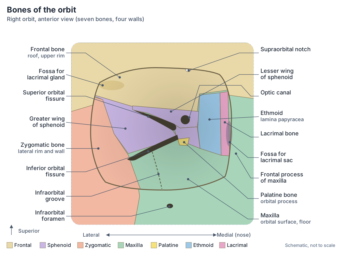
Walls of the orbit: roof, floor, medial and lateral wall
The roof (superior wall) is made of the frontal bone and the lesser wing of the sphenoid. It separates the orbit from the anterior cranial fossa inside the skull. Near its lateral edge is a hollow for the lacrimal gland, and near its medial edge a small trochlear fossa for the pulley of the superior oblique muscle. (EyeWiki (American Academy of Ophthalmology): Bony Anatomy of the Orbit.)
The lateral wall is made of the zygomatic bone in front and the greater wing of the sphenoid behind, and EyeWiki describes it as the strongest wall. The floor (inferior wall) is made of the maxilla, zygomatic and palatine bones and is also the roof of the maxillary sinus. (EyeWiki (American Academy of Ophthalmology): Bony Anatomy of the Orbit.)
The medial wall is made of four bones, from front to back the frontal process of the maxilla, the lacrimal bone, the ethmoid and the sphenoid. Most of it is the lamina papyracea of the ethmoid, a paper-thin plate that separates the orbit from the ethmoid air cells. Because of this, the medial wall is the thinnest wall of the orbit. (EyeWiki (American Academy of Ophthalmology): Bony Anatomy of the Orbit.)
TeachMeAnatomy points out that the medial wall and the floor are the two weakest walls, and that the orbit's neighbours matter clinically: the anterior cranial fossa above, the maxillary sinus below and the ethmoid sinus medially. Infection and fractures can pass across these thin partitions. (TeachMeAnatomy: The Bony Orbit.)
The maxillary and ethmoid sinuses that border the orbital floor and medial wall. Paranasal sinuses.
Walls of the orbit: bones and neighbours
| Wall | Bones | What lies on the other side | Notes |
|---|---|---|---|
| Roof (superior wall) | Orbital plate of the frontal bone; lesser wing of the sphenoid | Anterior cranial fossa (frontal lobe of the brain) | Fossa for the lacrimal gland laterally; trochlear fossa medially |
| Lateral wall | Zygomatic bone; greater wing of the sphenoid | Temporal fossa (temporalis muscle) | Strongest wall |
| Floor (inferior wall) | Maxilla (most); zygomatic bone; orbital process of the palatine bone | Maxillary sinus | Most common site of blowout fracture; carries the infraorbital groove |
| Medial wall | Frontal process of the maxilla, lacrimal bone, orbital plate of the ethmoid (lamina papyracea), sphenoid (front to back) | Ethmoid air cells and nasal cavity | Thinnest wall; fossa for the lacrimal sac at the front |
Compiled from TeachMeAnatomy, EyeWiki and Ophthalmology Review. Some texts list the lesser wing, others the body of the sphenoid, at the back of the medial wall.
The orbital rim (orbital margin)
The orbital rim is the thick, rounded edge around the opening of the orbit. Three bones form it: the frontal bone above, the zygomatic bone laterally and the maxilla below. The rim is much stronger than the thin walls behind it and protects the eyeball from blows. (EyeWiki (American Academy of Ophthalmology): Bony Anatomy of the Orbit.)
In the medial third of the upper rim is the supraorbital notch or foramen for the supraorbital nerve and vessels. Just inside the lateral rim, on the zygomatic bone, is a small bump called Whitnall's tubercle, which anchors the lateral canthal tendon of the eyelids and several other ligaments. (EyeWiki (American Academy of Ophthalmology): Bony Anatomy of the Orbit.)
At the front of the medial wall is the fossa for the lacrimal sac. On the lacrimal bone it is bounded behind by the vertical posterior lacrimal crest, where part of orbicularis oculi attaches. Below, the fossa leads into the nasolacrimal canal, which carries the nasolacrimal duct. (Radiopaedia: Lacrimal bone.)
The orbital apex and optic canal
The orbital apex is the narrow back end of the orbit where the four walls converge. Here the orbit communicates with the inside of the skull through two openings, the optic canal and the superior orbital fissure. (Radiopaedia: Orbit.)
The optic canal (optic foramen) is a cylindrical tunnel running obliquely through the lesser wing of the sphenoid, close to where the wing joins the body of the bone. It carries the optic nerve and the ophthalmic artery with its sympathetic fibres; inside the canal the artery lies below and lateral to the nerve. (Radiopaedia: Optic canal.)
The ophthalmic artery is a branch of the internal carotid artery. It arises just as the carotid leaves the cavernous sinus, enters the orbit through the optic canal and gives an ocular group of branches (including the central retinal artery) and an orbital group (lacrimal, supraorbital, ethmoidal, palpebral, supratrochlear and dorsal nasal arteries). (Radiopaedia: Ophthalmic artery.)
The openings of the cranial base, including the optic canal and superior orbital fissure. Skull foramina.
What passes through the superior orbital fissure?
The superior orbital fissure is a slit between the greater and lesser wings of the sphenoid, lateral to the optic canal, that connects the cavernous sinus with the orbital apex. It is bounded medially by the body of the sphenoid and laterally by the frontal bone. From top to bottom it carries the lacrimal and frontal nerves, the superior ophthalmic vein, the trochlear nerve, the superior division of the oculomotor nerve, the nasociliary nerve, the inferior division of the oculomotor nerve, the abducens nerve and a branch of the inferior ophthalmic vein. (Radiopaedia: Superior orbital fissure.)
The common tendinous ring (annulus of Zinn), the shared origin of the four rectus muscles, straddles the lower and medial part of the fissure and encloses the optic canal. The superior and inferior divisions of the oculomotor nerve, the nasociliary nerve and the abducens nerve pass inside the ring, together with the optic nerve and ophthalmic artery in the optic canal. (Radiopaedia: Tendinous ring (annulus of Zinn).)
The lacrimal and frontal branches of the ophthalmic nerve, the trochlear nerve and the superior ophthalmic vein pass through the fissure above the ring, so they enter the orbit outside the cone formed by the rectus muscles. (EyeWiki (American Academy of Ophthalmology): Bony Anatomy of the Orbit.)
Ophthalmology Review gives the same split: lacrimal, frontal and trochlear nerves and the superior ophthalmic vein above the annulus, and the oculomotor nerve, nasociliary nerve, sympathetic fibres and abducens nerve within it. Knowing which nerves run inside the ring helps explain which eye movements fail together when the apex is injured. (Ophthalmology Review: Orbital Anatomy (BCSC fundamentals).)
The four rectus muscles that arise from the common tendinous ring, the obliques and their nerves. Eye muscles.
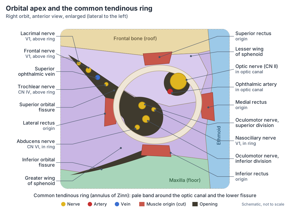
Inferior orbital fissure and other openings of the orbit
The inferior orbital fissure lies in the back of the orbital floor, below the superior fissure. It is bounded by the greater wing of the sphenoid above, the maxilla and the orbital process of the palatine bone below, and the zygomatic bone laterally. It carries the infraorbital and zygomatic nerves (from the maxillary nerve, V2), the infraorbital artery and vein, branches of the inferior ophthalmic vein to the pterygoid venous plexus, and small orbital branches of the pterygopalatine ganglion. (Radiopaedia: Inferior orbital fissure.)
The inferior orbital fissure links the orbit with the pterygopalatine and infratemporal fossae. Other small openings are the anterior and posterior ethmoidal foramina in the medial wall, which carry the anterior and posterior ethmoidal nerves and vessels, and the zygomatic foramina, which carry the zygomaticofacial and zygomaticotemporal nerves. (Ophthalmology Review: Orbital Anatomy (BCSC fundamentals).)
The infraorbital nerve, from V2, runs forwards in the floor of the orbit and through the infraorbital canal of the maxilla to the infraorbital foramen below the rim. On the face it supplies the lower eyelid, the front of the cheek, the side of the nose and the upper lip. The zygomaticofacial nerve supplies the prominence of the cheek and the zygomaticotemporal nerve the skin of the temple. (EyeWiki (American Academy of Ophthalmology): Bony Anatomy of the Orbit.)
The nasolacrimal canal runs down from the front of the medial wall to the nasal cavity. It carries the nasolacrimal duct, which drains tears into the inferior meatus of the nose, below the inferior nasal concha. (TeachMeAnatomy: The Nasal Cavity.)
Openings of the orbit and what passes through them
| Opening | Where it is | Main contents |
|---|---|---|
| Optic canal | Lesser wing of the sphenoid, at the apex | Optic nerve (CN II), ophthalmic artery, sympathetic fibres |
| Superior orbital fissure | Between the greater and lesser wings of the sphenoid | Above the tendinous ring: lacrimal, frontal and trochlear (CN IV) nerves, superior ophthalmic vein. Inside the ring: superior and inferior divisions of the oculomotor nerve (CN III), nasociliary nerve, abducens nerve (CN VI) |
| Inferior orbital fissure | Between the lateral wall and the floor | Infraorbital and zygomatic nerves (from V2), infraorbital artery and vein, branches of the inferior ophthalmic vein, orbital branches of the pterygopalatine ganglion |
| Supraorbital notch or foramen | Upper orbital rim, frontal bone | Supraorbital nerve (V1), artery and vein |
| Infraorbital groove, canal and foramen | Floor of the orbit to the face below the rim, maxilla | Infraorbital nerve (V2), artery and vein |
| Anterior and posterior ethmoidal foramina | Medial wall, between frontal and ethmoid bones | Anterior and posterior ethmoidal nerves and vessels |
| Zygomatic foramina | Zygomatic bone | Zygomaticofacial and zygomaticotemporal nerves |
| Nasolacrimal canal | Front of the medial wall and floor | Nasolacrimal duct, draining tears to the inferior meatus of the nose |
Sources: Radiopaedia, EyeWiki, Ophthalmology Review and TeachMeAnatomy. V1 = ophthalmic nerve, V2 = maxillary nerve.
What is inside the orbit?
Radiopaedia lists the contents of the orbit as the eyeball, the extraocular muscles, cranial nerves II, III, IV, V1, V2 and VI, autonomic nerves and the ciliary ganglion, the ophthalmic artery and its branches, the ophthalmic veins, orbital fat, the lacrimal gland and Tenon's capsule (the fascial sheath of the eyeball). (Radiopaedia: Orbit.)
Apart from the eyeball and optic nerve, the contents are divided by the cone of rectus muscles and their fascia into an intraconal space, inside the cone, and an extraconal space outside it. Radiologists use this split to describe where an orbital mass or abscess lies. (Radiopaedia: Orbit.)
Six extraocular muscles move the eyeball within the orbit. The lacrimal gland, which produces tears, sits just inside the orbit above and lateral to the eyeball. (OpenStax Anatomy and Physiology 2e, 14.1: Sensory Perception.)
The superior ophthalmic vein forms at the front of the orbit from the angular, supraorbital and supratrochlear veins. It runs back between the superior rectus and the optic nerve, leaves through the superior orbital fissure above the tendinous ring and drains directly into the cavernous sinus. Because its tributary, the angular vein, also drains into the facial vein, the superior ophthalmic vein connects the veins of the face with the cavernous sinus. (Radiopaedia: Superior ophthalmic vein.)
The optic, oculomotor, trochlear, trigeminal and abducens nerves that enter the orbit. Cranial nerves.
How big is the eye socket?
EyeWiki gives the average adult orbit a volume of about 30 mL, an entrance about 35 mm high and 40 mm wide, a medial wall about 45 mm long and a lateral wall about 35 mm long. (EyeWiki (American Academy of Ophthalmology): Bony Anatomy of the Orbit.)
Ophthalmology Review gives similar figures: a volume of 30 cm3, a height of 35 mm, a width of 45 mm and a depth of 40 to 45 mm. The small differences in width reflect where the rim is measured and normal variation between people. (Ophthalmology Review: Orbital Anatomy (BCSC fundamentals).)
Orbital fractures: rim and blowout fractures
An orbital rim fracture breaks the strong outer edge, usually at the sutures where the maxilla, zygomatic and frontal bones meet. A blowout fracture is different: it is a partial herniation of orbital contents through one of the walls, usually after blunt force, and the medial wall and floor are the weakest. Contents herniate into the ethmoid sinus through the medial wall and into the maxillary sinus through the floor. (TeachMeAnatomy: The Bony Orbit.)
Radiopaedia explains that a blow to the orbit suddenly raises the pressure inside it, so a wall breaks while the rim stays intact. The floor is the most common site, with fat and sometimes the inferior rectus muscle prolapsing into the maxillary sinus. Fractures of the medial wall (lamina papyracea) into the ethmoid air cells are the second most common, while pure roof and lateral wall fractures are uncommon. Clinical signs include a sunken eye (enophthalmos), double vision from trapped muscle, numbness of the cheek from injury to the infraorbital nerve, and air in the orbit. (Radiopaedia: Orbital blowout fracture.)
On CT, fat hanging into the roof of the maxillary sinus gives the teardrop sign. In children the floor can crack and spring back into place, trapping the inferior rectus in a trapdoor fracture; muscle entrapment is an indication for surgery. (Radiopaedia: Orbital blowout fracture.)
Orbital cellulitis and orbital apex syndrome
Preseptal (periorbital) cellulitis is infection of the eyelid tissues in front of the orbital septum. Orbital (postseptal) cellulitis lies behind the septum inside the orbit and is more serious. It usually spreads from sinusitis, most often from the ethmoid air cells through the thin lamina papyracea, and it typically affects children and young adults. (Radiopaedia: Orbital cellulitis.)
Complications of orbital cellulitis include thrombosis of the superior ophthalmic vein and of the cavernous sinus, loss of vision, meningitis and brain abscess. They follow directly from the anatomy: the ophthalmic veins drain backwards into the cavernous sinus. (Radiopaedia: Orbital cellulitis.)
Orbital apex syndrome is a group of signs caused by disease at the orbital apex, where the optic nerve, the oculomotor, trochlear and abducens nerves and the ophthalmic nerve are packed closely together. It causes eye movement palsies, drooping of the eyelid, numbness of the forehead and loss of vision. Involvement of the optic nerve is what separates it from superior orbital fissure syndrome and cavernous sinus syndrome. (Radiopaedia: Orbital apex syndrome.)
The orbit in the 3D model
The 3D viewer on this page highlights the seven bones of the orbit (frontal, sphenoid, zygomatic, maxilla, palatine, ethmoid and lacrimal) together with the optic nerve, the ophthalmic artery, the common tendinous ring, the lacrimal gland, the ciliary ganglion and the branches of the ophthalmic, oculomotor and trochlear nerves that run in the orbit. The model is BodyParts3D, built from an adult male. Turn the model to look into the socket from the front, then hide the skeletal system to see the nerves at the apex. The model has no abducens nerve, ophthalmic veins or orbital fat, and the fissures are not labelled, so use the diagrams above for the openings. (Mitsuhashi N et al. BodyParts3D: 3D structure database for anatomical concepts. Nucleic Acids Res 2009.)
Orbit in the 3D model
- Ciliary ganglion
- The ciliary ganglion is a small parasympathetic ganglion in the orbit that supplies the intrinsic muscles of the eye.
- Common tendinous ring
- A fibrous ring at the back of the orbit giving origin to the recti muscles of the eye.
- Ethmoid
- A delicate cube-shaped bone between the orbits.
- Frontal bone
- The bone of the forehead and the roof of the orbits.
- Frontal nerve
- The frontal nerve is the largest branch of the ophthalmic nerve and supplies the forehead and upper eyelid.
- Inferior branch of oculomotor nerve
- The inferior division of the oculomotor nerve supplies three extraocular muscles and carries parasympathetic fibres to the ciliary ganglion.
- Lacrimal bone
- A small, delicate bone of the medial orbit wall.
- Lacrimal gland
- The lacrimal gland is the main tear-secreting gland of the eye.
- Lacrimal nerve
- The lacrimal nerve is the smallest branch of the ophthalmic nerve and supplies the lacrimal gland and adjacent skin and conjunctiva.
- Maxilla
- The upper jaw bone, paired.
- Nasociliary nerve
- The nasociliary nerve is a branch of the ophthalmic nerve that supplies the eyeball, nasal cavity and parts of the face.
- Ophthalmic artery
- A branch of the internal carotid artery supplying the eye and orbit.
- Ophthalmic nerve
- The ophthalmic nerve is the first and smallest division of the trigeminal nerve and is purely sensory.
- Optic nerve
- The optic nerve (CN II) carries visual information from the retina to the brain.
- Palatine bone
- An L-shaped bone behind the maxilla.
- Sphenoid bone
- A butterfly-shaped bone at the base of the skull.
- Superior branch of oculomotor nerve
- The superior division of the oculomotor nerve supplies the superior rectus and the levator palpebrae superioris.
- Trochlear nerve
- The trochlear nerve (CN IV) is a motor nerve supplying the superior oblique muscle of the eye.
- Zygomatic bone
- The cheekbone.
Frequently asked questions
What are the 7 bones of the orbit?
The seven bones of the orbit are the frontal, sphenoid, zygomatic, maxilla, palatine, ethmoid and lacrimal bones. The frontal bone and lesser wing of the sphenoid form the roof, the zygomatic bone and greater wing of the sphenoid the lateral wall, the maxilla, zygomatic and palatine bones the floor, and the maxilla, lacrimal, ethmoid and sphenoid the medial wall.
Which wall of the orbit is the weakest?
The medial wall is the thinnest, because most of it is the paper-thin lamina papyracea of the ethmoid bone. The floor is also weak and is the most common site of a blowout fracture, in which orbital fat and sometimes the inferior rectus drop into the maxillary sinus. The lateral wall, made of the zygomatic bone and greater wing of the sphenoid, is the strongest.
What passes through the superior orbital fissure?
The lacrimal, frontal and trochlear nerves and the superior ophthalmic vein pass through the superior orbital fissure above the common tendinous ring. The superior and inferior divisions of the oculomotor nerve, the nasociliary nerve and the abducens nerve pass through it inside the ring. A branch of the inferior ophthalmic vein and sympathetic fibres also use the fissure.
What passes through the optic canal?
The optic canal, which runs through the lesser wing of the sphenoid at the apex of the orbit, carries the optic nerve (cranial nerve II) and the ophthalmic artery with its sympathetic fibres. Inside the canal the ophthalmic artery lies below and lateral to the optic nerve. Both are enclosed by the common tendinous ring as they enter the orbit.
Which bones form the floor of the orbit?
The floor of the orbit is formed mainly by the orbital surface of the maxilla, with the zygomatic bone laterally and a small orbital process of the palatine bone at the back. The floor is also the roof of the maxillary sinus and carries the infraorbital groove, which leads forwards into the infraorbital canal and foramen for the infraorbital nerve.
How big is the eye socket?
An adult orbit holds about 30 mL. Its entrance is about 35 mm high and 40 to 45 mm wide, and it is about 40 to 45 mm deep, with a medial wall about 45 mm long and a lateral wall about 35 mm long. The eyeball takes up only about 6.5 mL; the rest is muscles, nerves, vessels, fat and the lacrimal gland.
Is the orbit the same as the eye socket?
Yes. Orbit is the anatomical name for the eye socket, the bony cavity of the skull that holds the eyeball. The word is used both for the bony socket alone (the bony orbit) and for the socket together with its contents: the eyeball, extraocular muscles, nerves, vessels, fat and lacrimal gland.
References
- Betts JG, et al. Anatomy and Physiology 2e, §7.2 The Skull. OpenStax, Rice University, 2022.
- Betts JG, et al. Anatomy and Physiology 2e, §14.1 Sensory Perception. OpenStax, Rice University, 2022.
- TeachMeAnatomy. The Bony Orbit. Accessed 2026-10-23.
- TeachMeAnatomy. The Nasal Cavity. Accessed 2026-10-23.
- EyeWiki, American Academy of Ophthalmology. Bony Anatomy of the Orbit. Accessed 2026-10-23.
- Ophthalmology Review. Orbital Anatomy (BCSC fundamentals). Accessed 2026-10-23.
- Kenhub. Bones of the orbit. Accessed 2026-10-23.
- Radiopaedia. Orbit. Accessed 2026-10-23.
- Radiopaedia. Superior orbital fissure. Accessed 2026-10-23.
- Radiopaedia. Inferior orbital fissure. Accessed 2026-10-23.
- Radiopaedia. Optic canal. Accessed 2026-10-23.
- Radiopaedia. Tendinous ring (annulus of Zinn). Accessed 2026-10-23.
- Radiopaedia. Ophthalmic artery. Accessed 2026-10-23.
- Radiopaedia. Superior ophthalmic vein. Accessed 2026-10-23.
- Radiopaedia. Lacrimal bone. Accessed 2026-10-23.
- Radiopaedia. Orbital blowout fracture. Accessed 2026-10-23.
- Radiopaedia. Orbital cellulitis. Accessed 2026-10-23.
- Radiopaedia. Orbital apex syndrome. Accessed 2026-10-23.
- Mitsuhashi N, et al. BodyParts3D: 3D structure database for anatomical concepts. Nucleic Acids Res. 2009;37:D782-D785.
- Mitsuhashi N. et al. (2009). BodyParts3D: 3D structure database for anatomical concepts. Nucleic Acids Research 37, D782–D785. Data © The Database Center for Life Science, CC BY 4.0.
Related anatomy
- Eye anatomyEyeball anatomy with a labeled 3D eye model: sclera, cornea, choroid, iris, lens, retina and chambers, the 3 layers…
- Eye musclesEye muscles explained: the six extraocular muscles, levator palpebrae superioris and superior tarsal muscle, with the…
- Skull bonesSkull anatomy with labelled diagrams and a 3D model: the 8 cranial and 14 facial bones, sutures, fontanelles, the…
- Skull foraminaSkull foramina explained: the three cranial fossae, foramen magnum, jugular foramen and every other opening, with a…
- Paranasal sinusesParanasal sinuses anatomy: the maxillary, frontal, ethmoid and sphenoid sinuses, where each drains, the nasal conchae…
- Lacrimal boneLacrimal bone: A small, delicate bone of the medial orbit wall. See where it is in an interactive 3D human body, what…
Explore the whole body in the interactive 3D human anatomy model, or browse the A–Z list of human structures.