

Where is the thyroid gland?
The thyroid gland lies in the front of the neck, in front of the trachea and just below the larynx. It is butterfly-shaped: a narrow middle part, the isthmus, joins two wing-shaped lobes on either side. The small parathyroid glands are embedded in the back of the lobes. (OpenStax Anatomy and Physiology 2e, 17.4: The Thyroid Gland.)
In terms of the skeleton, the gland extends from C5 to T1. It lies in front of the thyroid and cricoid cartilages and the first five or six tracheal rings, with its isthmus attached to the second to fourth tracheal rings. (Radiopaedia: Thyroid gland.)
You can find your own thyroid by putting one finger on your Adam's apple and another on the top of your breastbone: the gland lies in the gap between them, under the skin and muscles. It bobs up and down when you swallow. (Nemours KidsHealth: Your Thyroid.)
Landmarks, triangles, fascia, vessels and nerves of the whole neck. Neck anatomy.
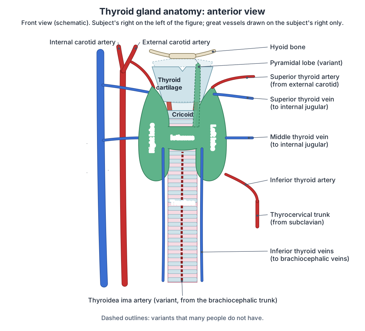
Parts of the thyroid: lobes, isthmus and pyramidal lobe
Each lobe is cone-shaped, with its apex pointing up and out and its base down and in, and measures about 5 cm long, 3 cm wide and 2 cm from front to back. The isthmus lies over the second or third tracheal cartilages. Kenhub gives the whole gland a weight of 15 to 25 g, making it the largest endocrine gland, and notes that it is slightly heavier in females after the age of 15. (Kenhub: Thyroid gland.)
Radiopaedia gives slightly different figures: lobes about 4 cm long in dissection and 4 to 6 cm long and under 2 cm deep on ultrasound, an isthmus under 0.5 cm thick, and an average weight of 25 g. On ultrasound, a normal gland measures 10 to 15 mL in women and 12 to 18 mL in men. Each lobe has a narrower superior pole and a wider inferior pole. (Radiopaedia: Thyroid gland.)
Many people have a third, small pyramidal lobe that projects upwards from the isthmus towards the hyoid bone. Radiopaedia puts it at about 20% of people (range 10 to 30%). It usually arises from the right or left side of the isthmus and only rarely (about 2%) from the midline. It is a persistent remnant of the thyroglossal duct, and a fibrous band, sometimes containing muscle (levator glandulae thyroideae), may tie it to the hyoid. (Radiopaedia: Pyramidal lobe of thyroid.)
Thyroid gland size and variants: what the sources say
| Feature | Kenhub | Radiopaedia | Other sources |
|---|---|---|---|
| Weight | 15 to 25 g; slightly heavier in females after age 15 | Average 25 g; may increase in menstruation and pregnancy | No more than about 20 g on examination (Loyola) |
| Length | Each lobe about 5 cm (3 cm wide, 2 cm deep) | Each lobe about 4 cm; 4 to 6 cm on ultrasound, under 2 cm deep | Gland about 2 inches long (Cleveland Clinic) |
| Isthmus | Over the 2nd or 3rd tracheal cartilages; about 1.25 cm | On the 2nd to 4th tracheal rings; under 0.5 cm thick | |
| Volume (ultrasound) | Women 10 to 15 mL, men 12 to 18 mL | ||
| Pyramidal lobe | May be absent; more often from the left side | About 20% (range 10 to 30%); midline origin about 2% | |
| Thyroidea ima artery | Inconsistent, from the brachiocephalic trunk | About 3% | About 10% (TeachMeAnatomy) |
Figures differ because they come from different methods (dissection, ultrasound) and populations. Empty cells: the source gives no figure.
Capsule, fascia and Berry's ligament: why the thyroid moves when you swallow
The thyroid has two coverings. A true fibrous capsule sends septa into the gland, and a false capsule, the thyroid sheath, comes from the pretracheal fascia. The space between them holds fat, nerves, vessels and the parathyroid glands. The false capsule thickens on each side into the suspensory ligament, and at the back Berry's ligament runs from the capsule to the cricoid cartilage and upper tracheal rings, enclosing part of the recurrent laryngeal nerve. (Radiopaedia: Thyroid gland.)
Kenhub describes the suspensory ligament of Berry as fixing the gland to the cricoid cartilage and the first two tracheal rings, while the lateral thyroid ligaments attach it to the cricoid. A fibromuscular band, the levator glandulae thyroideae, can anchor the isthmus or pyramidal lobe to the hyoid bone. (Kenhub: Thyroid gland.)
That is why doctors ask a patient to swallow water while they look at and feel the thyroid: in a person with a slender neck the gland rises together with the thyroid and cricoid cartilages. A normal gland is soft and weighs no more than about 20 g, and it is often not palpable in older people. (Loyola University Chicago, Physical Diagnosis: Method of thyroid examination.)
Relations: what lies around the thyroid gland
In front, the gland is covered by skin, platysma and the superficial and investing fascia, with sternothyroid lying directly on it and sternohyoid, the superior belly of omohyoid and the edge of sternocleidomastoid further out. Its medial surface lies against the larynx and trachea, cricothyroid and the inferior pharyngeal constrictors, and the external laryngeal nerve runs along it. The carotid sheath lies behind and to the side, and the thoracic duct passes behind and below the left lobe. (Kenhub: Thyroid gland.)
Radiopaedia describes the area behind and medial to each lobe as the tracheo-oesophageal groove, which contains lymph nodes, the recurrent laryngeal nerve and the parathyroid glands, together with the inferior thyroid artery. The carotid space lies behind and lateral to the gland. (Radiopaedia: Thyroid gland.)
TeachMeAnatomy places the gland in the visceral compartment of the neck with the trachea, oesophagus and pharynx, enclosed by the pretracheal fascia. Its lobes wrap around the cricoid cartilage and upper tracheal rings, and the carotid sheath, with the common carotid artery, internal jugular vein and vagus nerve, lies lateral to them. (TeachMeAnatomy: The Thyroid Gland.)
The windpipe that the thyroid wraps around, from the cricoid to the carina. Trachea.
Relations of the thyroid gland
| Side | Structures |
|---|---|
| In front | Skin, platysma, superficial and investing fascia; sternothyroid (directly on the gland), sternohyoid, superior belly of omohyoid, front edge of sternocleidomastoid |
| Medially (deep) | Larynx and trachea, cricothyroid and the inferior pharyngeal constrictor, pharynx and oesophagus, external laryngeal nerve |
| Behind and medially | Tracheo-oesophageal groove: recurrent laryngeal nerve, parathyroid glands, lymph nodes; inferior thyroid artery |
| Behind and laterally | Carotid sheath: common carotid artery, internal jugular vein, vagus nerve |
| Below and behind the left lobe | Thoracic duct |
From Kenhub, Radiopaedia and TeachMeAnatomy.
Blood supply: superior and inferior thyroid arteries and the thyroidea ima
The thyroid is supplied by two pairs of arteries. The superior thyroid artery is the first branch of the external carotid artery and runs near the external branch of the superior laryngeal nerve. The inferior thyroid artery comes from the thyrocervical trunk of the subclavian artery and runs near the recurrent laryngeal nerve. TeachMeAnatomy adds a thyroidea ima artery in around 10% of people, arising from the brachiocephalic trunk and supplying the front of the gland and the isthmus. (TeachMeAnatomy: The Thyroid Gland.)
Radiopaedia gives a lower figure for the thyroidea ima artery, about 3%, and notes that when present it may replace the inferior thyroid artery. It also notes that an inferior thyroid artery arising directly from the subclavian artery is called an accessory inferior thyroid artery. (Radiopaedia: Thyroid gland.)
Kenhub describes the superior thyroid artery dividing into anterior and posterior branches and the inferior thyroid artery into ascending and inferior branches. These vessels connect with each other inside and around the gland. (Kenhub: Thyroid gland.)
The external carotid branches, including the superior thyroid artery, and the thyrocervical trunk. Carotid arteries.
Thyroid veins and lymph drainage
Three pairs of veins drain a plexus around the gland. The superior thyroid vein leaves the upper pole and the middle thyroid vein the side of the gland, and both end in the internal jugular vein. The inferior thyroid veins arise from a venous plexus in front of the trachea: on the left they drain into the left brachiocephalic vein, and on the right into the right brachiocephalic vein or directly into the superior vena cava. (Kenhub: Thyroid gland.)
Lymph from the thyroid drains in several directions: first to nodes around the gland, then to the prelaryngeal, pretracheal and paratracheal nodes of the central neck (level VI). (Radiopaedia: Thyroid gland.)
Kenhub adds that the prelaryngeal (Delphian) nodes above the isthmus are often the first stop, that the deep cervical nodes receive lymph from the sides of the gland, and that some lymph vessels bypass the nodes and drain straight into the thoracic duct. (Kenhub: Thyroid gland.)
Neck node levels I to VI, including the central (level VI) nodes around the thyroid. Lymph nodes of the head and neck.
Blood vessels of the thyroid gland
| Vessel | Origin or drainage | Notes |
|---|---|---|
| Superior thyroid artery | First branch of the external carotid artery | Reaches the upper pole; runs with the external laryngeal nerve |
| Inferior thyroid artery | Thyrocervical trunk of the subclavian artery | Reaches the back of the lower gland; closely related to the recurrent laryngeal nerve; main supply of the parathyroids |
| Thyroidea ima artery (variant) | Brachiocephalic trunk | Supplies the isthmus and front of the gland; about 3% (Radiopaedia) to about 10% (TeachMeAnatomy) |
| Superior thyroid vein | Internal jugular vein | Drains the upper pole |
| Middle thyroid vein | Internal jugular vein | Drains the side of the gland |
| Inferior thyroid vein | Brachiocephalic vein (left); right one may enter the superior vena cava | Arises from a venous plexus in front of the trachea |
From TeachMeAnatomy, Kenhub and Radiopaedia.
Nerves: the recurrent laryngeal nerve and thyroid surgery
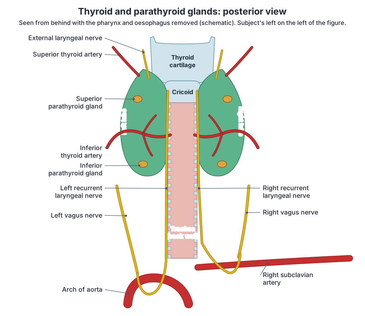
The recurrent laryngeal nerve is a branch of the vagus. On the right it leaves the vagus in front of the subclavian artery and hooks under it; on the left it leaves at the aortic arch and loops under it behind the ligamentum arteriosum. Each nerve then climbs vertically in the tracheo-oesophageal groove against the medial surface of the thyroid lobe and enters the larynx below the inferior pharyngeal constrictor. It supplies all the intrinsic muscles of the larynx except cricothyroid, and sensation below the vocal cords. (Radiopaedia: Recurrent laryngeal nerve.)
Two nerve and artery pairs matter in thyroid surgery. The recurrent laryngeal nerve is most often related to the posterior branch of the inferior thyroid artery, and the external laryngeal nerve, which supplies cricothyroid, lies close to the superior thyroid artery at the upper pole. The tubercle of Zuckerkandl, which marks where the ultimobranchial body fused with the gland in the embryo, also lies close to the recurrent laryngeal nerve. (Kenhub: Thyroid gland.)
In about 0.7% of people (reported range 0.6 to 0.8%) the right nerve does not loop under the subclavian artery but runs straight from the vagus to the larynx. This non-recurrent laryngeal nerve is linked to an aberrant right subclavian artery; on the left it is much rarer, about 0.04%. A review estimated the risk of nerve injury at 12.9% when the nerve is non-recurrent, compared with 1.8% for a normal nerve. (Inversini D et al. Non-recurrent Laryngeal Nerve During Intraoperative Neuromonitoring Thyroidectomy: A Case Report and Literature Review. J Investig Med High Impact Case Rep 2024.)
Damage to one recurrent laryngeal nerve causes a hoarse voice (dysphonia); damage to both causes loss of voice and stridor, an inspiratory wheeze. Paralysis of these nerves is usually caused by cancer of the larynx or thyroid or by surgical complications. (TeachMeAnatomy: The Vagus Nerve (CN X).)
The gland itself receives sympathetic fibres from the cervical ganglia of the sympathetic trunk. These nerves do not control how much hormone the gland makes; that is regulated by the pituitary gland. (TeachMeAnatomy: The Thyroid Gland.)
The laryngeal muscles that the recurrent and external laryngeal nerves supply. Larynx.
Parathyroid glands: number, position and blood supply
About 80% of people have four parathyroid glands, a superior and an inferior pair, but there can be up to twelve: about 15% have extra glands and fewer than 3% have fewer than four. Each gland is about 5 to 6 by 4 by 1 to 2 mm and weighs 40 to 60 mg. The superior glands lie behind the upper thyroid at the level of the first tracheal ring, above the inferior thyroid artery; the inferior glands lie below the lower pole and the inferior thyroid artery, lateral to the recurrent laryngeal nerve. (Radiopaedia: Parathyroid glands.)
The superior glands develop from the fourth pharyngeal pouch and the inferior glands from the third. Because the inferior glands migrate further down the neck before birth, they are more often found in unusual (ectopic) places, which happens in about 15% of people. Both pairs are usually supplied by the inferior thyroid artery. (Radiopaedia: Parathyroid glands.)
The parathyroids sit outside the thyroid but inside the pretracheal fascia, the superior pair about 1 cm above the point where the inferior thyroid artery enters the gland. They make parathyroid hormone, which raises blood calcium. Because they can be damaged or removed by accident during thyroid surgery, causing a sudden drop in calcium with cramps, tetany and tingling of the fingers, toes and mouth, calcium and hormone levels are checked after the operation. (TeachMeAnatomy: The Parathyroid Glands.)
Thyroid histology: follicles, colloid and C cells
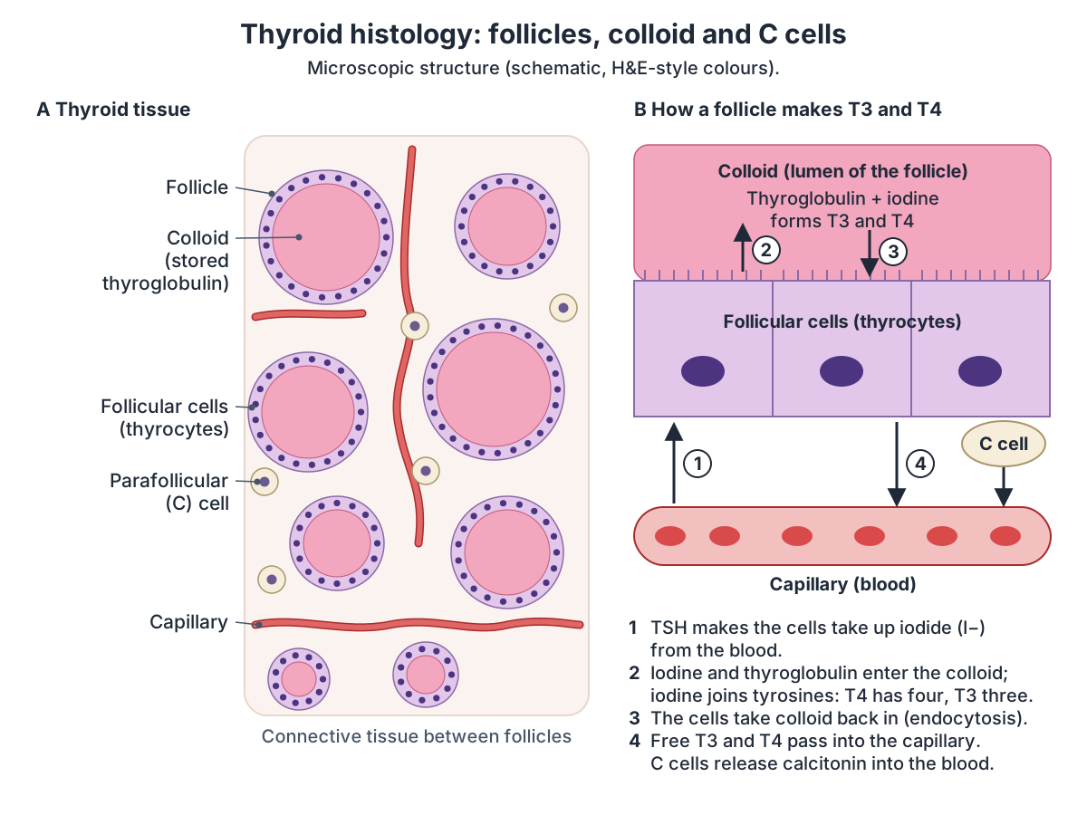
Most of the thyroid is made of follicles: hollow spheres lined by follicle cells and filled with a sticky fluid called colloid, where hormone is made. Larger parafollicular cells, or C cells, lie between the follicles and produce calcitonin. (OpenStax Anatomy and Physiology 2e, 17.4: The Thyroid Gland.)
Kenhub describes the follicle lining as simple cuboidal or low columnar epithelium that becomes flatter when the follicle is inactive. The colloid is stored thyroglobulin and stains bright pink with the usual H&E stain. C cells are slightly larger and paler than the follicle cells (thyrocytes), and a dense network of fenestrated capillaries runs through the thin connective tissue between the follicles. (Kenhub: Thyroid gland.)
What does the thyroid gland do? T4, T3 and calcitonin
Follicle cells take up iodide from the blood and attach iodine to tyrosines in thyroglobulin. Molecules with four iodines form T4 (thyroxine) and with three form T3 (triiodothyronine). T4 is made in larger amounts, but T3 is more potent, and many cells convert T4 into T3. About 99% of both hormones travel in the blood bound to carrier proteins. (OpenStax Anatomy and Physiology 2e, 17.4: The Thyroid Gland.)
Secretion is controlled by negative feedback. When T3 and T4 are low, the hypothalamus releases thyrotropin-releasing hormone (TRH), which makes the anterior pituitary release thyroid-stimulating hormone (TSH); TSH drives the thyroid, and rising T3 and T4 then reduce TRH and TSH. The hormones raise the basal metabolic rate and heat production and are essential for growth and for development of the nervous system. (OpenStax Anatomy and Physiology 2e, 17.4: The Thyroid Gland.)
Calcitonin from the C cells helps regulate blood calcium. The thyroid needs iodine from the diet to make its hormones, which affect metabolism, heart rate, breathing, digestion, body temperature, brain development, skin and bone maintenance and fertility. (Cleveland Clinic: Thyroid.)
The gland that releases TSH and controls the thyroid. Pituitary gland.
Development: the thyroglossal duct and ectopic thyroid
The thyroid starts as an outgrowth at the foramen caecum at the base of the tongue and descends to the front of the neck by about week 7, leaving a track called the thyroglossal duct, which normally disappears by about week 10. If part of the duct persists it can form a thyroglossal cyst: a midline lump in the front of the neck that rises when the tongue is stuck out. (TeachMeAnatomy: The Thyroid Gland.)
The calcitonin-producing C cells come from the ultimobranchial body, an outgrowth of the fourth pharyngeal pouch. Variants of development include a missing lobe, the pyramidal lobe, accessory thyroid tissue and ectopic thyroid, such as a lingual thyroid at the base of the tongue. (Radiopaedia: Thyroid gland.)
The tongue and the foramen caecum, where the thyroid begins. Tongue anatomy.
Goitre, thyroid nodules and other thyroid conditions
Without enough iodine the gland cannot make enough T3 and T4, so TSH rises and colloid builds up, enlarging the thyroid into a goitre. Hypothyroidism (too little hormone) slows metabolism and causes weight gain and cold hands and feet; hyperthyroidism speeds it up and causes weight loss, sweating, tremor and a fast heart rate. Graves disease is an autoimmune cause of hyperthyroidism in which antibodies overstimulate the follicle cells. (OpenStax Anatomy and Physiology 2e, 17.4: The Thyroid Gland.)
Cleveland Clinic estimates that about 20 million people in the US have some type of thyroid disorder and that women are five to eight times more likely to be diagnosed. A goitre affects about 5% of people in the US, and about 53,000 people there are diagnosed with thyroid cancer each year, papillary cancer making up to 80% of cases. (Cleveland Clinic: Thyroid.)
Lumps in the thyroid, called nodules, are common. They can be felt in 4 to 7% of the population, but ultrasound finds them in 19 to 67% of people. About 5% of palpable nodules are cancerous, so a nodule is usually assessed with a TSH blood test and, if hormone levels are normal, a fine-needle aspiration. (Welker MJ, Orlov D. Thyroid Nodules. Am Fam Physician 2003.)
The thyroid region in the 3D model
BodyParts3D, the adult male model used in the viewer, was built from MRI scans and does not include the thyroid or parathyroid glands. The viewer therefore highlights the gland's bed: the hyoid bone, epiglottis and thyroid and cricoid cartilages, the middle and inferior pharyngeal constrictors, the trachea, the common and internal carotid arteries and internal jugular veins of the carotid sheath, the inferior thyroid arteries and thyrocervical trunks, the vertebral arteries, and the subclavian and brachiocephalic vessels. The lobes would fill the space beside the trachea and cricoid, between the carotid arteries, so compare the view with the anterior diagram. (Mitsuhashi N et al. BodyParts3D: 3D structure database for anatomical concepts. Nucleic Acids Res 2009.)
Thyroid gland in the 3D model
- Brachiocephalic artery
- The brachiocephalic artery (trunk) is the first branch of the aortic arch and supplies the right head, neck and arm.
- Brachiocephalic vein
- The brachiocephalic veins are two large veins that unite to form the superior vena cava.
- Common carotid artery
- The common carotid artery is the main artery of the neck and splits into the external and internal carotid arteries.
- Cricoid cartilage
- The cricoid cartilage is a complete signet-ring-shaped cartilage forming the lower part of the larynx.
- Epiglottis
- The epiglottis is a leaf-shaped elastic cartilage that covers the laryngeal inlet during swallowing.
- Hyoid bone
- A U-shaped bone in the neck.
- Inferior pharyngeal constrictor
- The lowest of the three constrictors of the pharynx.
- Inferior thyroid artery
- The inferior thyroid artery supplies the thyroid and parathyroid glands.
- Internal carotid artery
- The internal carotid artery supplies the brain and the eye.
- Internal jugular vein
- The internal jugular vein is the main vein draining the head and neck.
- Middle pharyngeal constrictor
- The middle of the three pharyngeal constrictors.
- Subclavian artery
- The major artery to the upper limb, neck and part of the brain.
- Subclavian vein
- The large vein draining the upper limb into the brachiocephalic vein.
- Thyrocervical trunk
- A short branch of the subclavian artery to the neck.
- Thyroid cartilage
- The thyroid cartilage is the largest cartilage of the larynx, forming the laryngeal prominence.
- Trachea
- The trachea (windpipe) is a flexible air tube extending from the larynx to the bronchi.
- Vertebral artery
- A branch of the subclavian artery supplying the brain and spinal cord.
Frequently asked questions
Where is the thyroid gland located?
The thyroid gland is in the front of the neck, below the larynx (Adam's apple) and above the top of the breastbone. Its lobes lie on each side of the trachea and cricoid cartilage, and its isthmus crosses the front of the upper tracheal rings. In terms of the spine it spans the fifth cervical to the first thoracic vertebra (C5 to T1).
What are the parts of the thyroid gland?
The thyroid has a right lobe and a left lobe, each with an upper and a lower pole, joined in front of the trachea by a narrow isthmus. Many people, about one in five on ultrasound, also have a small pyramidal lobe rising from the isthmus towards the hyoid bone. Inside, the gland is divided into lobules of follicles filled with colloid.
Can you feel your thyroid gland?
A normal thyroid is soft and often hard to feel, especially in older people. In a slender neck it can be felt below the Adam's apple and seen or felt to rise when you swallow, because it is attached to the larynx and trachea. A lump, swelling or pain in the area should be checked by a doctor.
What is the difference between the thyroid and parathyroid glands?
The thyroid is one large gland of 15 to 25 g that makes T4, T3 and calcitonin. The parathyroid glands are usually four tiny glands of 40 to 60 mg each on the back of the thyroid lobes. They make parathyroid hormone, which raises blood calcium. They have separate origins in the embryo and only share the same location.
What arteries supply the thyroid gland?
The superior thyroid artery, the first branch of the external carotid artery, supplies the upper pole, and the inferior thyroid artery, from the thyrocervical trunk of the subclavian artery, supplies the lower and back parts. A small number of people also have a thyroidea ima artery from the brachiocephalic trunk; sources give about 3% to about 10%.
Which nerve is at risk in thyroid surgery?
The recurrent laryngeal nerve, which climbs behind the thyroid lobe in the groove between the trachea and oesophagus and crosses the inferior thyroid artery, is the main nerve at risk. Injury to one nerve causes hoarseness. The external laryngeal nerve near the superior thyroid artery is also at risk, and the parathyroid glands can be damaged too.
What does the thyroid gland do?
The thyroid makes thyroxine (T4) and triiodothyronine (T3), which raise the body's metabolic rate and heat production and are essential for growth and brain development. It needs iodine from the diet to make them. Its C cells also make calcitonin, which helps regulate blood calcium. The pituitary hormone TSH controls how much T4 and T3 it releases.
References
- Betts JG, et al. Anatomy and Physiology 2e, §17.4 The Thyroid Gland. OpenStax, Rice University, 2022.
- Kenhub. Thyroid gland. Accessed 2026-10-24.
- TeachMeAnatomy. The Thyroid Gland. Accessed 2026-10-24.
- TeachMeAnatomy. The Parathyroid Glands. Accessed 2026-10-24.
- TeachMeAnatomy. The Vagus Nerve (CN X). Accessed 2026-10-24.
- Radiopaedia. Thyroid gland. Accessed 2026-10-24.
- Radiopaedia. Pyramidal lobe of thyroid. Accessed 2026-10-24.
- Radiopaedia. Parathyroid glands. Accessed 2026-10-24.
- Radiopaedia. Recurrent laryngeal nerve. Accessed 2026-10-24.
- Cleveland Clinic. Thyroid: What It Is, Function & Problems. Accessed 2026-10-24.
- Nemours KidsHealth. Your Thyroid. Accessed 2026-10-24.
- Chandrasekhar A, Braithwaite S. Physical Diagnosis: Method of thyroid examination. Loyola University Chicago Stritch School of Medicine. Accessed 2026-10-24.
- Welker MJ, Orlov D. Thyroid Nodules. Am Fam Physician. 2003;67(3):559-567.
- Inversini D, et al. Non-recurrent Laryngeal Nerve During Intraoperative Neuromonitoring Thyroidectomy: A Case Report and Literature Review. J Investig Med High Impact Case Rep. 2024;12:23247096241273099.
- Mitsuhashi N, et al. BodyParts3D: 3D structure database for anatomical concepts. Nucleic Acids Res. 2009;37:D782-D785.
- Mitsuhashi N. et al. (2009). BodyParts3D: 3D structure database for anatomical concepts. Nucleic Acids Research 37, D782–D785. Data © The Database Center for Life Science, CC BY 4.0.
Related anatomy
- Neck anatomyNeck anatomy with labelled diagrams and a 3D model: landmarks, triangles, fascia, airway, thyroid, carotid sheath…
- LarynxLarynx anatomy with 3D model and diagrams: thyroid, cricoid, arytenoid and epiglottis cartilages, vocal folds…
- TracheaTrachea anatomy explained: where the windpipe runs, C-shaped rings, the carina, right vs left main bronchi and the…
- Pituitary glandPituitary gland (hypophyseal gland) anatomy: the hypophyseal fossa and sella turcica, relations, lobes, portal blood…
- Lymph nodes of the head and neckLymph node map of the head and neck: where neck lymph nodes are, submental and submandibular nodes, levels I to VI…
Explore the whole body in the interactive 3D human anatomy model, or browse the A–Z list of human structures.