

What is the mouth (oral cavity)?
The oral cavity, or mouth, is the entry point of the digestive tract. It opens at the front through the oral fissure, the gap between the lips, and at the back it leads into the oropharynx through the oropharyngeal isthmus. Its three jobs are digestion, communication (shaping the sound from the larynx into speech) and breathing, as a second airway alongside the nose. (TeachMeAnatomy: The Oral Cavity.)
Kenhub describes the mouth as the first part of the digestive system, lying on the front of the face under the nasal cavities, with a roof, a floor and lateral walls. Its bony framework comes from the paired maxillae, palatine and temporal bones and the unpaired mandible, sphenoid and hyoid bone. (Kenhub: Oral cavity.)
Radiologists and surgeons divide the mouth into subsites: the lips, the lining of the cheeks (buccal mucosa), the upper and lower alveolar ridges (gums), the retromolar area behind the last molars, the floor of the mouth, the front two-thirds of the tongue and the hard palate. The back third of the tongue belongs to the oropharynx, not the mouth. (Radiopaedia: Oral cavity.)
Parts of the mouth: vestibule and oral cavity proper
The upper and lower dental arches divide the mouth into two parts. The oral vestibule is the horseshoe-shaped space between the lips and cheeks on the outside and the teeth and gums on the inside. It communicates with the rest of the mouth through the space behind the third molar teeth. (TeachMeAnatomy: The Oral Cavity.)
The oral cavity proper is the main open space, running from the gums and teeth to the fauces. OpenStax defines the fauces as the opening where the oral cavity joins the oropharynx. (OpenStax Anatomy and Physiology 2e, 23.3: The Mouth, Pharynx, and Esophagus.)
Radiopaedia draws the back boundary of the oral cavity proper as a ring formed by the circumvallate papillae of the tongue, the front tonsillar pillars (palatoglossal arches) and the junction of the hard and soft palates. It also names the gingivobuccal sulcus, the groove where the gums meet the lining of the cheek. (Radiopaedia: Oral cavity.)
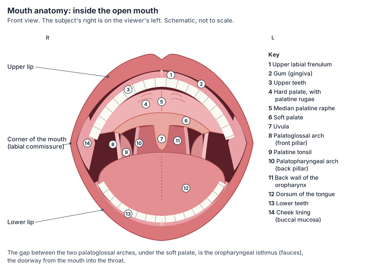
Parts of the mouth at a glance
| Part | Where it is | Lining or tissue | Sensory nerve |
|---|---|---|---|
| Lips | Front of the mouth, around the oral fissure | Skin outside, red vermilion, oral mucosa inside; orbicularis oris in the core | Upper lip: maxillary nerve (V2). Lower lip: mandibular nerve (V3) |
| Oral vestibule | Between lips and cheeks outside and teeth and gums inside | Non-keratinised oral mucosa | V2 above, V3 below |
| Cheeks | Side walls of the mouth | Skin outside, non-keratinised mucosa inside, buccinator between | Trigeminal nerve branches |
| Gums (gingivae) | Over the alveolar processes and around the necks of the teeth | Keratinised mucosa firmly bound to bone | Upper: V2. Lower: V3 |
| Hard palate | Front two-thirds of the roof | Bone covered by oral mucosa with palatine glands | Greater palatine and nasopalatine nerves (V2) |
| Soft palate and uvula | Back third of the roof | Muscle and palatine aponeurosis covered by mucosa | Lesser palatine nerves (V2) |
| Tongue (front two-thirds) | Floor of the oral cavity proper | Muscle covered by mucosa with papillae | General: lingual nerve (V3). Taste: facial nerve (VII) |
| Floor of the mouth | Under the tongue, above mylohyoid | Mucosa over the sublingual glands and ducts | Mandibular nerve (V3) |
From TeachMeAnatomy, OpenStax, Kenhub, Radiopaedia and Dentalcare (see references). The back third of the tongue belongs to the oropharynx and is supplied by the glossopharyngeal nerve (IX).
Lip anatomy
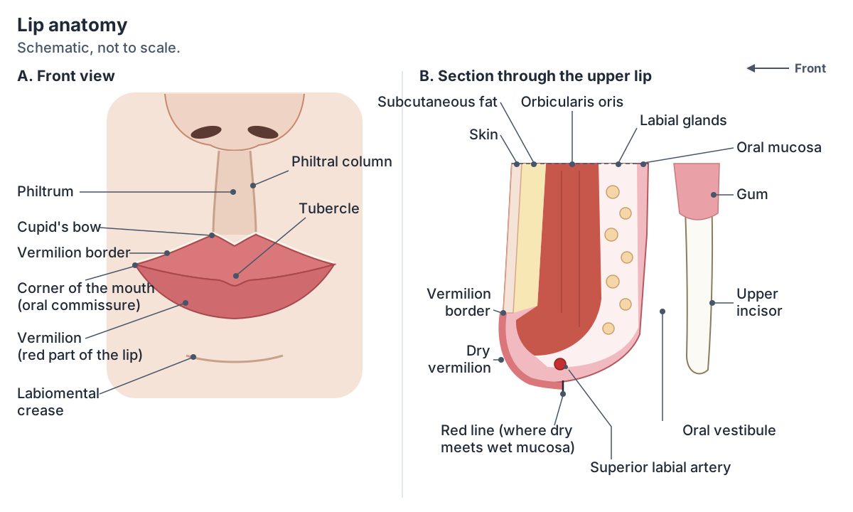
The lips (labia) are covered by skin on the outside and by mucous membrane on the inside. OpenStax explains their red colour by a rich blood supply under a thin layer of keratin. The orbicularis oris muscle in each lip controls what enters and leaves the mouth, and a midline fold of mucosa, the labial frenulum, ties the inside of each lip to the gum. (OpenStax Anatomy and Physiology 2e, 23.3: The Mouth, Pharynx, and Esophagus.)
The red part of the lip is the vermilion, a modified mucosa. A pale white line, the vermilion border, separates it from the facial skin, and a red line marks where the dry vermilion meets the wet lining of the mouth. Wu and colleagues describe the vermilion epithelium as non-keratinised stratified squamous epithelium. (Wu WT, et al. Lip Sonoanatomy and Relevance to Aesthetic Filler Injections: A Pictorial Review. J Cosmet Dermatol 2025.)
The upper lip carries the philtrum, a central groove bordered by two vertical philtral ridges (columns). The lower margin of the philtrum forms the downward arch of the Cupid's bow, and the central bulge of the upper lip is the tubercle. The lips meet at the corners of the mouth (oral commissures), and the labiomental crease separates the lower lip from the chin. (Wu WT, et al. Lip Sonoanatomy and Relevance to Aesthetic Filler Injections: A Pictorial Review. J Cosmet Dermatol 2025.)
In section, the lip has skin, a layer of fat, the orbicularis oris muscle, a submucosa and the oral lining. The superior and inferior labial arteries usually branch from the facial artery and most often run in the submucosa, deep to orbicularis oris, near the junction of the vermilion and the wet mucosa. (Wu WT, et al. Lip Sonoanatomy and Relevance to Aesthetic Filler Injections: A Pictorial Review. J Cosmet Dermatol 2025.)
Orbicularis oris arises from the maxilla and the neighbouring cheek muscles and inserts into the skin and mucous membrane of the lips; it closes the lips. Small muscles above and below the mouth, such as the zygomatic muscles, levator labii superioris, depressor anguli oris and mentalis, raise or lower the lips. All are muscles of facial expression supplied by the facial nerve (CN VII). (TeachMeAnatomy: Muscles of Facial Expression.)
Sensation from the upper lip travels in the maxillary division of the trigeminal nerve (V2) and from the lower lip in the mandibular division (V3). (TeachMeAnatomy: The Trigeminal Nerve (CN V).)
Lymph from the centre of the lower lip drains to the submental nodes under the chin. Lymph from the upper lip and the outer parts of the lower lip drains to the submandibular nodes under the jaw. (TeachMeAnatomy: Lymphatics of the Head and Neck.)
Orbicularis oris and the muscles that raise and lower the lips. Facial muscles.
Cheeks and the inside of the cheek
The cheeks form the side walls of the mouth. Their outer surface is skin and their inner surface is non-keratinised stratified squamous mucosa (the buccal mucosa). Between the two lies the buccinator muscle. (OpenStax Anatomy and Physiology 2e, 23.3: The Mouth, Pharynx, and Esophagus.)
Buccinator presses the cheek against the teeth during chewing so that food stays between the teeth instead of collecting in the vestibule. It is supplied by the buccal branches of the facial nerve (CN VII). The duct of the parotid gland opens into the vestibule on the inside of the cheek, opposite the upper second molar. (TeachMeAnatomy: The Oral Cavity.)
Roof of the mouth: hard palate, soft palate and uvula
The roof of the mouth is the palate. The bony hard palate makes up the front two-thirds and the muscular soft palate the back third. The palatine processes of the maxillae form the front three-quarters of the hard palate and the horizontal plates of the palatine bones the back quarter. The uvula is a cone-shaped projection from the middle of the soft palate's free back edge. (Kenhub: The palate.)
The lining of the hard palate has landmarks you can feel with your tongue. The incisive papilla is a fleshy bump just behind the upper front teeth, over the incisive foramen. The median palatine raphe, a ridge left where the two halves of the palate fused, runs back from it in the midline, and transverse palatine rugae (ridges) radiate out from the raphe, most prominent at the front. Hundreds of small mucous glands lie in the palatal lining. (Kenhub: Hard palate.)
Three groups of openings pierce the hard palate. The incisive foramen at the front transmits the nasopalatine nerve. The greater palatine foramina, at the back corners, transmit the greater palatine nerve and vessels, which supply most of the hard palate. The lesser palatine foramina behind them transmit the lesser palatine nerves and vessels. The nasal surface of the palate is covered by respiratory mucosa and the oral surface by oral mucosa. (Radiopaedia: Hard palate.)
The soft palate is a mobile fold attached to the back edge of the hard palate. Its oral surface contains palatine glands and some taste buds. It rises against the back wall of the pharynx to close off the nasopharynx when we swallow, it helps with speech and the gag reflex, and its arteries are the lesser palatine, ascending palatine and palatine branches of the ascending pharyngeal artery. (Radiopaedia: Soft palate.)
During swallowing the soft palate and uvula swing up to keep food and drink out of the nasal cavity. The uvula can also contribute to snoring. (OpenStax Anatomy and Physiology 2e, 23.3: The Mouth, Pharynx, and Esophagus.)
The L-shaped bone that forms the back of the hard palate. Palatine bone.
Muscles of the soft palate
| Muscle | From | To | Action | Nerve |
|---|---|---|---|---|
| Tensor veli palatini | Medial pterygoid plate of the sphenoid | Palatine aponeurosis | Tenses the soft palate | Nerve to medial pterygoid (mandibular nerve, V3) |
| Levator veli palatini | Petrous temporal bone and auditory tube | Palatine aponeurosis | Raises the soft palate | Pharyngeal branch of the vagus (X) |
| Palatoglossus | Palatine aponeurosis | Side of the tongue | Pulls the soft palate towards the tongue | Pharyngeal branch of the vagus (X) |
| Palatopharyngeus | Palatine aponeurosis and hard palate | Thyroid cartilage | Tenses the soft palate; draws the pharynx forward in swallowing | Pharyngeal branch of the vagus (X) |
| Musculus uvulae | Posterior nasal spine and palatine aponeurosis | Mucosa of the uvula | Shortens the uvula | Pharyngeal branch of the vagus (X) |
Attachments and actions from TeachMeAnatomy; the nerve supply agrees with Radiopaedia and Kenhub. All five muscles are paired.
The tongue in the mouth
The tongue fills much of the oral cavity. Its base is attached to the floor of the mouth and its apex is free. It has eight muscles: four intrinsic muscles (superior and inferior longitudinal, transverse and vertical) that change its shape, and four extrinsic muscles (genioglossus, hyoglossus, styloglossus and palatoglossus) that move it. (Kenhub: Oral cavity.)
The hypoglossal nerve (CN XII) supplies all the tongue muscles except palatoglossus, which is supplied by the vagus nerve (CN X). On the front two-thirds, general sensation travels in the lingual nerve (a branch of V3) and taste in the facial nerve (CN VII); the back third is supplied for both by the glossopharyngeal nerve (CN IX). Filiform papillae have a mechanical role; the fungiform, vallate and foliate papillae carry taste buds. (Kenhub: Oral cavity.)
The lingual frenulum is the fold that tethers the underside of the tongue to the floor of the mouth. In ankyloglossia (tongue tie) it is too short or malformed, and severe cases can affect speech. (OpenStax Anatomy and Physiology 2e, 23.3: The Mouth, Pharynx, and Esophagus.)
The dorsum, underside, papillae and muscles of the tongue in detail. Tongue anatomy.
Teeth and gums
Adults have 32 permanent teeth: 8 incisors, 4 canines, 8 premolars and 12 molars, the third molars being the wisdom teeth. Children have 20 deciduous (milk) teeth, which first appear at about 6 months and are replaced between about 6 and 12 years. Each tooth sits in a socket of the jaw, held by the periodontal ligament. (OpenStax Anatomy and Physiology 2e, 23.3: The Mouth, Pharynx, and Esophagus.)
The gums (gingivae) are the mucosa that covers the alveolar processes of the maxilla and mandible and ends at the neck of each tooth. The free (marginal) gingiva forms a collar about 1.5 mm wide around the tooth, and in a healthy mouth a probe enters the gingival sulcus between gum and tooth no more than about 3 mm. (Kenhub: Gingiva.)
The attached gingiva is keratinised and bound firmly to the bone. Its height varies from 3 to 12 mm in different parts of the mouth (Kenhub gives a typical width of 4 to 5 mm). A scalloped mucogingival junction separates it from the redder, looser, non-keratinised alveolar mucosa, which lets the lips and cheeks move. Between neighbouring teeth the gum forms the interdental papilla. (Dentalcare: Anatomy of the Periodontium.)
Types of teeth, the numbering chart and tooth surfaces. Teeth.
Floor of the mouth and under the tongue
The floor of the mouth is a muscular diaphragm formed by the two mylohyoid muscles, with the geniohyoid muscles above them. Both pull the larynx forward during swallowing. The tongue is attached to the floor by the lingual frenulum, and the salivary glands and their ducts lie here. (TeachMeAnatomy: The Oral Cavity.)
Under the tongue, the submandibular duct opens by one to three openings on the sublingual papilla (caruncle), a small bump on each side of the base of the lingual frenulum. (TeachMeAnatomy: The Submandibular Gland.)
The two sublingual glands meet in a horseshoe around the frenulum. Their upper edge raises the sublingual fold, which runs back from the sublingual papilla on each side, and 8 to 20 small ducts from each gland open along it. (TeachMeAnatomy: The Sublingual Gland.)
Salivary glands and where they open
There are three pairs of major salivary glands. The parotid is the largest and makes purely serous saliva, about 25 to 30% of the daily output. The submandibular gland makes mixed serous and mucous saliva, up to about 70%. The sublingual gland is the smallest and makes mainly mucous saliva, up to about 5%. About 800 to 1,000 minor salivary glands in the lips, cheeks, tongue and palate add 1% or less. (Kenhub: Salivary glands.)
An adult makes about 1 to 1.5 litres of saliva a day. It is more than 99% water (99.4% in OpenStax), and the rest includes ions, mucus, antibodies (immunoglobulin A), the antibacterial enzyme lysozyme and salivary amylase, which starts the digestion of starch. OpenStax gives the pH of saliva as 6.35 to 6.85. (OpenStax Anatomy and Physiology 2e, 23.3: The Mouth, Pharynx, and Esophagus.)
The parotid duct (Stensen's duct) leaves the front of the gland, crosses the surface of the masseter, pierces the buccinator and opens into the mouth beside the upper second molar. The facial nerve divides into its five terminal branches inside the parotid gland. (TeachMeAnatomy: The Parotid Gland.)
The submandibular gland is hook-shaped and wraps around the back edge of mylohyoid: its larger superficial part lies under the jaw and its deep part lies in the floor of the mouth. Its duct (Wharton's duct) is about 5 cm long and runs forward to the sublingual papilla. (TeachMeAnatomy: The Submandibular Gland.)
The 3D structure page for the submandibular salivary gland. Submandibular gland.
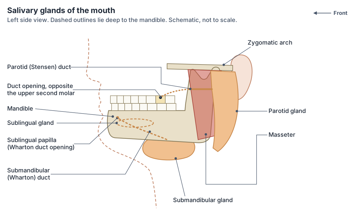
Major salivary glands
| Gland | Where it is | Duct and opening | Saliva | Share of daily saliva |
|---|---|---|---|---|
| Parotid (largest) | In front of the ear, over the masseter | Parotid (Stensen) duct: crosses masseter, pierces buccinator, opens opposite the upper second molar | Serous (watery, with amylase) | About 25 to 30% |
| Submandibular | Under the body of the mandible, wrapped around the back edge of mylohyoid | Submandibular (Wharton) duct, about 5 cm: opens on the sublingual papilla beside the lingual frenulum | Mixed serous and mucous | Up to about 70% |
| Sublingual (smallest) | Floor of the mouth under the tongue, deep to the sublingual fold | 8 to 20 small ducts on the sublingual fold; in some people also one larger duct that drains at the sublingual papilla | Mainly mucous | About 3 to 5% |
Shares are from Kenhub and TeachMeAnatomy and vary with stimulation. About 800 to 1,000 minor salivary glands in the lips, cheeks, palate and tongue add 1% or less.
Back of the mouth: arches, tonsils and the fauces
Two folds hang down on each side of the back of the mouth. The palatoglossal arch lies next to the base of the tongue, and the palatopharyngeal arch forms the upper and side margins of the fauces. The palatine tonsils are clusters of lymphoid tissue between the two arches. (OpenStax Anatomy and Physiology 2e, 23.3: The Mouth, Pharynx, and Esophagus.)
Each palatine tonsil sits in the tonsillar bed of the side wall of the oropharynx. Its inner surface is free and bulges into the throat, and its outer surface has a fibrous capsule. The tonsils drain to the jugulodigastric and upper deep cervical lymph nodes. (TeachMeAnatomy: The Tonsils and Adenoids (Waldeyer's Ring).)
Radiopaedia counts the oropharyngeal isthmus (fauces), the narrowed opening between the oral cavity and the throat, as part of the oropharynx. (Radiopaedia: Oral cavity.)
The oropharynx behind the mouth, the rest of the pharynx and the larynx. Throat anatomy.
Nerves, blood supply and lymph of the mouth
The trigeminal nerve carries sensation from the mouth. The maxillary division (V2) supplies the upper lip, the upper teeth with their gums and the palate. The mandibular division (V3) supplies the lower lip, the floor of the mouth, the lower teeth with their gums and general sensation from the front two-thirds of the tongue. V3 is also the motor nerve to the muscles of chewing, mylohyoid and tensor veli palatini. (TeachMeAnatomy: The Trigeminal Nerve (CN V).)
Most of the palate is supplied by the greater palatine artery, a branch of the maxillary artery, with the lesser palatine artery serving a small area at the back; the veins drain to the pterygoid venous plexus. (Radiopaedia: Hard palate.)
Lymph from the oral cavity drains to the submental, submandibular, retropharyngeal and deep cervical lymph nodes. (Radiopaedia: Oral cavity.)
Where the submental, submandibular and deep cervical nodes are. Lymph nodes of the head and neck.
What does the mouth do?
Eating starts in the mouth. The teeth and tongue break food up, saliva moistens it and salivary amylase begins to digest carbohydrate, and lingual lipase from the tongue's glands is secreted here although it acts later, in the stomach. In the voluntary first phase of swallowing, the tongue pushes the bolus up and back against the palate into the oropharynx. (OpenStax Anatomy and Physiology 2e, 23.3: The Mouth, Pharynx, and Esophagus.)
Kenhub lists further roles of the palate: with the tongue it creates the partial vacuum a baby uses for suckling, the soft palate is lowered during nose breathing, and the tongue touches the hard and soft palate to make palatal and velar speech sounds. Touching the soft palate or uvula triggers the gag reflex. (Kenhub: The palate.)
Cleft lip and palate, salivary stones and other clinical notes
A cleft lip forms when the medial nasal and maxillary prominences of the embryo fail to fuse, and a cleft palate when the palatal shelves fail to fuse in the midline. TeachMeAnatomy gives a prevalence of about 1 in 1,000 births. Problems include feeding difficulty, repeated ear infections and speech difficulty. (TeachMeAnatomy: The Palate.)
About 80% of salivary stones (sialoliths) form in the submandibular duct, helped by its length, its upward course and the nature of its saliva. (TeachMeAnatomy: The Submandibular Gland.)
A ranula is a mucus cyst in the floor of the mouth under the tongue. It is the most common disorder of the sublingual gland and usually follows damage to its delicate ducts. (TeachMeAnatomy: The Sublingual Gland.)
Gingivitis, inflammation of the gums caused by bacteria in the shallow pocket between tooth and gum, is the most common gum disease. It causes redness, swelling, tenderness and bleeding gums. (Kenhub: Gingiva.)
Looking at the uvula tests the vagus nerve. If the pharyngeal branch of the vagus is damaged on one side, the palatal arch on that side drops and the uvula deviates away from the affected side. (TeachMeAnatomy: The Vagus Nerve (CN X).)
Mouth anatomy in the 3D model
- Geniohyoid
- A narrow muscle above the mylohyoid in the floor of the mouth.
- Gingiva of lower jaw
- The gingiva (gum) of the lower jaw is the mucosa covering the alveolar process of the mandible and surrounding the necks of lower teeth.
- Gingiva of upper jaw
- The gingiva (gum) of the upper jaw is the mucosa covering the alveolar process of the maxilla and surrounding the necks of upper teeth.
- Hard palate
- The hard palate is the bony front part of the palate, which forms the roof of the mouth and the floor of the nasal cavity.
- Levator veli palatini
- A muscle of the soft palate.
- Lip
- The lips are two muscular, mobile folds that surround the opening of the mouth.
- Lower central secondary incisor tooth
- The permanent lower central incisor, the smallest tooth in the mouth, one on each side of the midline of the lower jaw.
- Lower first secondary molar tooth
- The permanent lower first molar, the largest lower tooth and the first permanent tooth to erupt.
- Lower first secondary premolar tooth
- The permanent lower first premolar, a canine-like tooth with one large and one small cusp.
- Lower lateral secondary incisor tooth
- The permanent lower lateral incisor, a chisel-shaped cutting tooth between the central incisor and the canine.
- Lower second secondary molar tooth
- The permanent lower second molar, a four-cusped grinding tooth at the back of the lower arch in this model.
- Lower second secondary premolar tooth
- The permanent lower second premolar, a crushing tooth in front of the first molar, often with three cusps.
- Lower secondary canine tooth
- The permanent lower canine, a pointed single-cusped tooth at the corner of the lower dental arch.
- Mylohyoid
- A flat muscle forming the muscular floor of the mouth.
- Palatine bone
- An L-shaped bone behind the maxilla.
- Palatine tonsil
- The palatine tonsils are the two masses of lymphoid tissue at the back of the mouth, one on each side of the oropharynx. They are what most people mean by the tonsils.
- Palatopharyngeus
- A muscle of the soft palate and pharynx that forms the posterior pillar of the fauces.
- Parotid gland
- The parotid gland is the salivary gland in front of and below each ear. It makes serous saliva, a watery fluid rich in enzymes.
- Soft palate
- The soft palate is the muscular back part of the roof of the mouth. Its free back edge carries the uvula.
- Sublingual gland
- The sublingual gland is the smallest of the three major salivary glands.
- Submandibular gland
- The submandibular gland is a major salivary gland located below the mandible.
- Tensor veli palatini
- A palatal muscle that tenses the soft palate.
- Tongue
- The tongue is a muscular organ of the mouth involved in taste, chewing, swallowing and speech.
- Upper central secondary incisor tooth
- The permanent upper central incisor, the largest and most prominent incisor, beside the midline of the upper jaw.
- Upper first secondary molar tooth
- The permanent upper first molar, a large three-rooted grinding tooth whose roots lie close to the maxillary sinus.
- Upper first secondary premolar tooth
- The permanent upper first premolar, a two-cusped tooth that usually has two roots.
- Upper lateral secondary incisor tooth
- The permanent upper lateral incisor, a smaller cutting tooth between the central incisor and the canine.
- Upper second secondary molar tooth
- The permanent upper second molar, a three-rooted grinding tooth at the back of the upper arch in this model.
- Upper second secondary premolar tooth
- The permanent upper second premolar, a two-cusped tooth that usually has one root.
- Upper secondary canine tooth
- The permanent upper canine (eye tooth), which has the longest root of any tooth.
- Uvular muscle
- A small paired muscle forming the core of the uvula.
Frequently asked questions
What are the parts of the mouth called?
The mouth (oral cavity) has the lips at the front, the cheeks at the sides, the hard and soft palate above with the uvula hanging from the soft palate, and the tongue on the floor. The teeth and gums divide it into the vestibule, between lips, cheeks and teeth, and the oral cavity proper behind the teeth. At the back, the fauces lead into the throat.
What is the roof of the mouth called?
The palate. Its front two-thirds is the hard palate, a plate of bone formed by the maxillae and palatine bones and covered by mucosa with ridges called palatine rugae. Its back third is the soft palate, a muscular fold that rises to close off the nose when you swallow and ends in the uvula.
What is the thing hanging at the back of the mouth?
The uvula, a small cone-shaped projection from the middle of the back edge of the soft palate. It contains the musculus uvulae, which shortens it. During swallowing the soft palate and uvula rise to block the nasopharynx so that food does not enter the nose. On each side of it hang the palatoglossal and palatopharyngeal arches, with a palatine tonsil between them.
What is the red part of the lips called?
The vermilion. It is a modified mucosa with a thin epithelium through which the rich blood supply shows. A pale line called the vermilion border separates it from the skin, and on the upper lip this border forms the Cupid's bow below the philtrum. Inside the mouth, the red line marks where the dry vermilion meets the wet oral lining.
What is the string under the tongue called?
The lingual frenulum, a fold of mucous membrane that tethers the underside of the tongue to the floor of the mouth. On each side of its base is a sublingual papilla, where the submandibular duct opens. If the frenulum is too short, the condition is called ankyloglossia, or tongue tie.
Where do the salivary glands open into the mouth?
The parotid duct opens on the inside of the cheek opposite the upper second molar. The submandibular duct opens on the sublingual papilla beside the lingual frenulum under the tongue. Each sublingual gland opens through 8 to 20 small ducts along the sublingual fold. Hundreds of minor salivary glands open directly through the lining of the lips, cheeks and palate.
How much saliva does the mouth make each day?
About 1 to 1.5 litres a day in an adult, according to both OpenStax and Kenhub. Most comes from the submandibular glands, up to about 70%, with about a quarter from the parotid glands and a few percent from the sublingual glands. Saliva is more than 99% water and also carries mucus, antibodies, lysozyme and the digestive enzyme amylase.
References
- Betts JG, et al. Anatomy and Physiology 2e, §23.3 The Mouth, Pharynx, and Esophagus. OpenStax, Rice University, 2022.
- TeachMeAnatomy. The Oral Cavity.
- TeachMeAnatomy. The Palate: Hard Palate, Soft Palate, Vasculature.
- TeachMeAnatomy. The Parotid Gland.
- TeachMeAnatomy. The Submandibular Gland.
- TeachMeAnatomy. The Sublingual Gland.
- TeachMeAnatomy. The Trigeminal Nerve (CN V).
- TeachMeAnatomy. Muscles of Facial Expression.
- TeachMeAnatomy. Lymphatics of the Head and Neck.
- TeachMeAnatomy. The Tonsils and Adenoids (Waldeyer's Ring).
- TeachMeAnatomy. The Vagus Nerve (CN X).
- Kenhub. Oral cavity: Anatomy, tongue muscles, nerves and vessels.
- Kenhub. Salivary glands: Overview, anatomy, clinical aspects.
- Kenhub. The palate.
- Kenhub. Hard palate: Anatomy, function and borders.
- Kenhub. Gingiva: Types, histology and clinical aspects.
- Dentalcare (Procter & Gamble). An Overview of Dental Anatomy: Anatomy of the Periodontium.
- Radiopaedia. Oral cavity (reference article).
- Radiopaedia. Hard palate (reference article).
- Radiopaedia. Soft palate (reference article).
- Wu WT, Chang KV, Naňka O, et al. Lip Sonoanatomy and Relevance to Aesthetic Filler Injections: A Pictorial Review. Journal of Cosmetic Dermatology 2025;24(4):e70164.
- Mitsuhashi N. et al. (2009). BodyParts3D: 3D structure database for anatomical concepts. Nucleic Acids Research 37, D782–D785. Data © The Database Center for Life Science, CC BY 4.0.
- Browne K., Schlehlein H. (2026). 3D Reference Organ Set for Male, v1.10 (Visible Human Male). Human Reference Atlas 3D Reference Object Library, HuBMAP / NIH. CC BY 4.0.
Related anatomy
- TongueTongue anatomy explained: parts and papillae of the dorsum, the underside with the frenulum and sublingual folds…
- TeethTooth anatomy with labeled diagrams and a 3D model: parts of a tooth, incisors, canines, premolars, molars, Universal…
- Throat anatomyThroat anatomy with a 3D model and labelled diagrams: the three parts of the pharynx, constrictor muscles, tonsils…
- Palatine bonePalatine bone (os palatinum) anatomy: horizontal and perpendicular plates, three processes, palatine foramina…
- Lymph nodes of the head and neckLymph node map of the head and neck: where neck lymph nodes are, submental and submandibular nodes, levels I to VI…
- Muscles of masticationThe muscles of mastication explained: masseter, temporalis, medial and lateral pterygoid, with labelled diagrams, the…
Explore the whole body in the interactive 3D human anatomy model, or browse the A–Z list of human structures.