

Where is the uterus? Location and position
The uterus lies in the female pelvis, behind (posterior to) the urinary bladder and in front of (anterior to) the rectum. Its broad, curved top, the fundus, is where the fallopian tubes connect, and its lowest part, the cervix, projects downwards into the vagina. (Ameer MA, Peterson DC. Anatomy, Abdomen and Pelvis: Uterus. StatPearls, 2025.)
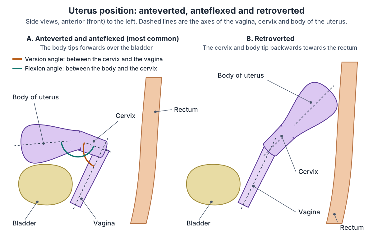
Its usual position is anteverted, tipped forwards relative to the vagina, and anteflexed, with the body bent forwards on the cervix, so it lies just behind and above the bladder. A uterus that is tipped backwards is more likely to prolapse into the vagina, particularly after damage to the pelvic floor. (TeachMeAnatomy: The Uterus.)
Radiologists describe the position with two angles. Version is the angle between the axis of the cervix and the axis of the vagina, so an anteverted uterus is tipped forwards relative to the vagina. Flexion is the angle between the axis of the body of the uterus and the axis of the cervix, so an anteflexed uterus has its body bent forwards on the cervix. Both angles change as the bladder and rectum fill. (Radiopaedia: Uterus.)
A retroverted (tilted) uterus, tipped backwards towards the rectum, is a normal variation found in about 1 in 5 women: Radiopaedia gives about 20% and Cleveland Clinic 20 to 25%. (Radiopaedia: Retroverted uterus.)
Peritoneum covers almost the whole uterus apart from the front of the cervix. It dips down in front of the uterus to form the vesicouterine pouch, between the uterus and bladder, and behind it to form the rectouterine pouch (pouch of Douglas), between the uterus and rectum. (Kenhub: Uterus.)
In the 3D model the uterus is highlighted in the middle of the pelvis with a uterine tube and an ovary on each side, held by the broad, round and ovarian ligaments. Turn the model to look from the side, or switch on the Urinary layer, to see the bladder in front of it.
The whole system: ovaries, tubes, uterus, vagina and vulva, with a sagittal section of the female pelvis. Female reproductive system.
Parts of the uterus: fundus, body, isthmus and cervix
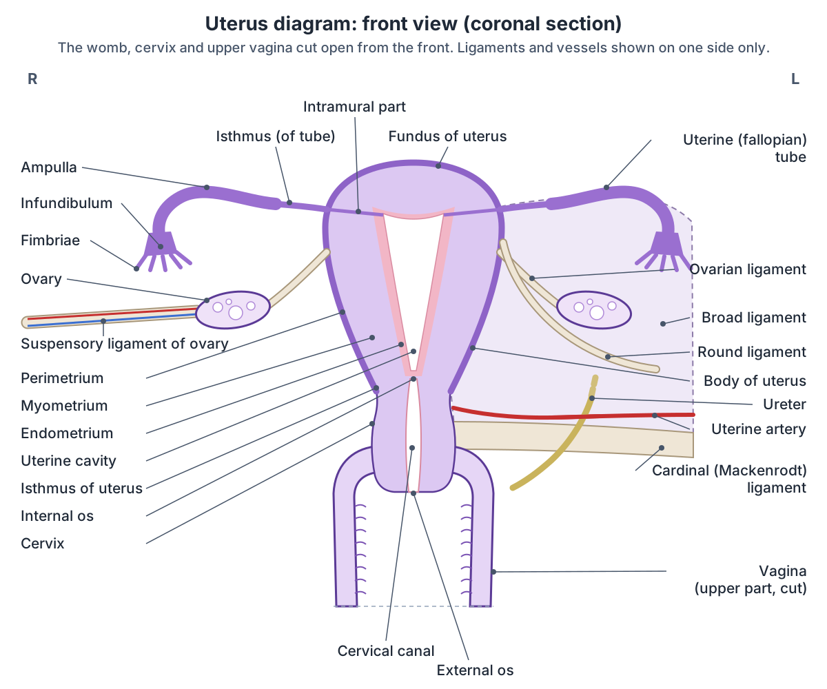
The uterus has four parts, from top to bottom: the fundus, a broad curved region where the fallopian tubes connect; the body (corpus), the main part below the level of the tubes; the isthmus, a narrowed segment at the lower uterine neck; and the cervix, which projects down into the vagina. (Ameer MA, Peterson DC. Anatomy, Abdomen and Pelvis: Uterus. StatPearls, 2025.)
OpenStax defines the fundus of the uterus as the part above the openings of the uterine tubes, the body as the middle section and the cervix as the narrow lower part that projects into the vagina. (OpenStax Anatomy and Physiology 2e, 27.2: Anatomy and Physiology of the Ovarian Reproductive System.)
The isthmus is a short narrowing between the body above and the cervix below, at the level of the internal os; Kenhub gives its length as about 1 cm. (Muckle CW. Clinical Anatomy of the Uterus, Fallopian Tubes, & Ovaries. Global Library of Women's Medicine.)
In an adult the body is about twice as long as the cervix (Radiopaedia gives a body to cervix ratio of 2:1), so the isthmus lies about two-thirds of the way down the uterus, not halfway. The cavity of the body is only a narrow slit, flattened from front to back. Seen from the front it is a triangle, with its base at the fundus between the openings of the two tubes and its apex at the internal os, where it joins the canal of the cervix. (Radiopaedia: Uterus.)
Cleveland Clinic describes the fundus as the uppermost and widest part of the uterus, the corpus as its main body and the isthmus as the part between the corpus and the cervix. (Cleveland Clinic: Uterus.)
In an adult who is not pregnant, the uterus is about 7.5 cm long, 5 cm wide at the top and 2.5 cm thick, and weighs about 30 to 40 g. (Radiopaedia: Uterus.)
The Global Library of Women's Medicine gives the same figures (7.5 cm long, 5 cm wide, 2.5 cm thick, 30 to 40 g), and Cleveland Clinic gives them in inches: about 3 inches long, 2 inches wide and 1 inch thick, about the size of a fist or a lemon. (Muckle CW. Clinical Anatomy of the Uterus, Fallopian Tubes, & Ovaries. Global Library of Women's Medicine.)
OpenStax gives a slightly smaller average of about 7 cm long and 5 cm wide outside pregnancy, while Kenhub gives about 8 cm, so most sources put the length at about 7 to 8 cm. (OpenStax Anatomy and Physiology 2e, 27.2: Anatomy and Physiology of the Ovarian Reproductive System.)
Parts of the uterus
| Part | Where it is | Key features |
|---|---|---|
| Fundus | Top of the uterus, above where the uterine tubes enter | Broad, curved and widest part; covered by peritoneum |
| Uterine horns (cornua) | Upper corners, where the tubes join | The round ligaments start here |
| Body (corpus) | Main part, below the level of the tubes | Holds the triangular, slit-like uterine cavity; usual site of implantation |
| Isthmus | Narrow junction between the body and the cervix | Slight constriction; the cavity narrows to the internal os |
| Cervix | Lowest part, projecting into the vagina | Endocervical canal from the internal os to the external os; its front lies below the peritoneum, while the back of the supravaginal part is covered by peritoneum (pouch of Douglas) |
From StatPearls, OpenStax, Radiopaedia and TeachMeAnatomy (see references).
Layers of the uterus wall: perimetrium, myometrium and endometrium
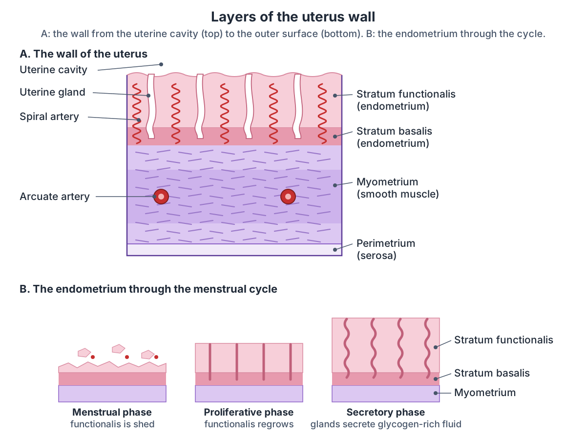
The wall of the uterus has three layers. The outer perimetrium is a serous membrane. The middle myometrium is a thick layer of smooth muscle that produces uterine contractions. The inner endometrium is the lining, with a deep stratum basalis next to the myometrium and a superficial stratum functionalis that grows and thickens in response to oestrogen and progesterone. (OpenStax Anatomy and Physiology 2e, 27.2: Anatomy and Physiology of the Ovarian Reproductive System.)
The stratum basalis changes little during the menstrual cycle and is not shed. The stratum functionalis is shed at menstruation and then regrows from the basalis. The perimetrium is a double layer of peritoneum continuous with the peritoneum of the abdomen, and in pregnancy the myometrium grows both by enlarging its cells (hypertrophy) and by making new ones (hyperplasia). (TeachMeAnatomy: The Uterus.)
Kenhub describes the myometrium as a complex of three smooth muscle layers, including a layer rich in blood vessels that plays a major role in labour, and the endometrium as lined by simple columnar epithelium. (Kenhub: Uterus.)
Spiral arteries, special branches of the uterine artery, supply the thickened stratum functionalis. The endometrium goes through a monthly cycle: in the menses phase the functionalis is shed, in the proliferative phase it regrows, and in the secretory phase its glands secrete a fluid rich in glycogen. Day 1 of the cycle is the first day of bleeding. (OpenStax Anatomy and Physiology 2e, 27.2: Anatomy and Physiology of the Ovarian Reproductive System.)
StatPearls describes the same endometrial phases, proliferative, secretory and menstrual, and counts the first day of heavy flow as day 1. (Thiyagarajan DK, Basit H, Jeanmonod R. Physiology, Menstrual Cycle. StatPearls, 2024.)
On MRI the wall shows three zones: the bright endometrium, a dark inner band of myometrium called the junctional zone, and the intermediate outer myometrium. The junctional zone is less distinct before the first period and in pregnancy. (Radiopaedia: Uterus.)
Cervix anatomy: the neck of the uterus
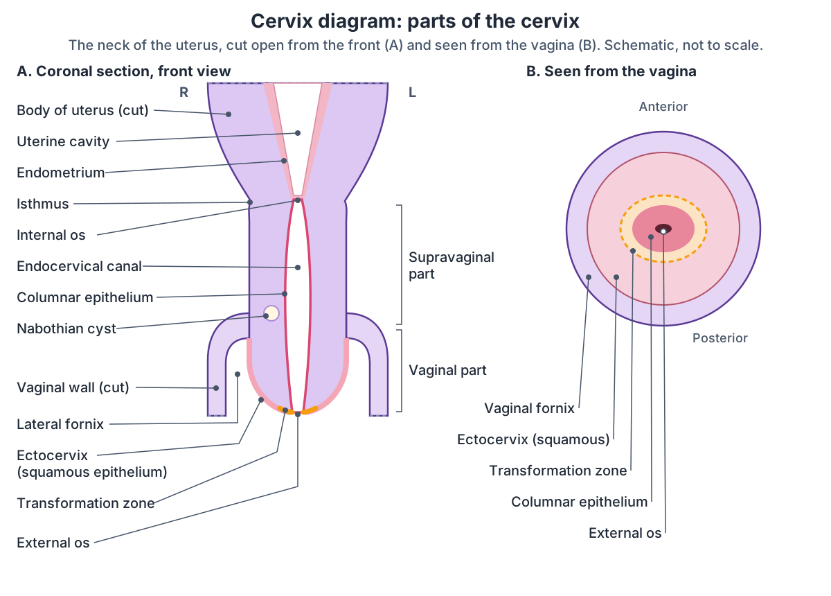
The cervix is the narrow lower part of the uterus. Its attachment to the vagina divides it into an upper supravaginal part and a lower vaginal part that projects into the vagina. Its canal, the endocervical canal, is spindle-shaped and flattened from front to back. It opens into the cavity of the body at the internal os and into the vagina at the external os. (Radiopaedia: Cervix.)
The cervix is about 2 to 3 cm long, and most of it protrudes into the vagina. Cleveland Clinic gives about an inch (2.5 cm) and notes that cervical size varies. (Muckle CW. Clinical Anatomy of the Uterus, Fallopian Tubes, & Ovaries. Global Library of Women's Medicine.)
The ectocervix, the part that bulges into the top of the vagina, is covered by non-keratinised stratified squamous epithelium. The endocervical canal is lined by mucus-secreting simple columnar epithelium. (TeachMeAnatomy: The Cervix.)
The two kinds of lining cells meet at the transformation zone. Cell changes often happen there, so clinicians look at it closely during cervical screening. (Cleveland Clinic: Cervix.)
Radiopaedia describes the same change, from columnar epithelium in the upper canal to stratified squamous epithelium close to the external os. Cervical glands that become blocked and swollen with retained mucus form small nabothian cysts. (Radiopaedia: Cervix.)
The cervix lets sperm into the uterus, particularly around ovulation. At other times tight closure and thick mucus in the canal make a physical and immunological barrier that helps keep the uterus sterile. (TeachMeAnatomy: The Cervix.)
Around ovulation the cervix releases thinner, less acidic mucus. In pregnancy it makes a mucus plug that seals the entry to the uterus, and in labour it softens and thins (effaces) and widens (dilates) so the baby can pass. (Cleveland Clinic: Cervix.)
Three pairs of ligaments attach to the cervix: the pubocervical ligaments in front, the transverse cervical (cardinal or Mackenrodt) ligaments at the sides and the uterosacral ligaments behind. (Radiopaedia: Cervix.)
The vaginal canal around the cervix: fornices, walls, layers and supports. Vagina anatomy.
Fallopian tube anatomy: the parts of the uterine tube
Each uterine (fallopian) tube is about 10 to 12 cm long. It runs between the two folds of the broad ligament from the upper corner of the uterus towards the ovary, and its outer end opens into the peritoneal cavity. (Radiopaedia: Fallopian tube.)
From the ovary towards the uterus, Radiopaedia lists five parts: finger-like fimbriae that drape over the ovary; the funnel-shaped infundibulum, from which they arise; the ampulla, the widest and longest part, more than half the length of the tube and the usual site of fertilisation; the isthmus, the narrowest part next to the uterus; and the interstitial (intramural) part inside the wall of the uterus. (Radiopaedia: Fallopian tube.)
Other sources count four parts. Cleveland Clinic lists the infundibulum, ampulla, isthmus and intramural part, with the fimbriae at the end of the tube, and gives each tube as 4 to 5 inches long. TeachMeAnatomy lists the fimbriae, infundibulum, ampulla and isthmus. (Cleveland Clinic: Fallopian Tubes.)
The tubes lie in the upper border of the broad ligament. Their lining has ciliated cells and non-ciliated secretory peg cells, which waft the egg towards the uterus and nourish it, and their smooth muscle contracts in waves (peristalsis), most strongly when oestrogen levels are high. Both the uterine and the ovarian arteries supply them. (TeachMeAnatomy: The Fallopian Tubes.)
OpenStax describes how the egg gets in: the wide infundibulum flares out with slender fimbriae, ciliated cells in the lining beat towards the uterus, and fertilisation often happens in the ampulla. (OpenStax Anatomy and Physiology 2e, 27.2: Anatomy and Physiology of the Ovarian Reproductive System.)
Parts of the uterine (fallopian) tube
| Part | Where it is | Key features |
|---|---|---|
| Fimbriae | Outer end, draped over the ovary | Finger-like projections that catch the egg |
| Infundibulum | Funnel-shaped outer end | Opens into the peritoneal cavity; the fimbriae arise from it |
| Ampulla | Middle, more than half the length | Widest and longest part; usual site of fertilisation |
| Isthmus | Next to the uterus | Narrowest part |
| Intramural (interstitial) part | Inside the wall of the uterus | Opens into the uterine cavity at its upper corner |
Radiopaedia counts five parts; Cleveland Clinic and TeachMeAnatomy count four. Each tube is about 10 to 12 cm long (Radiopaedia; Cleveland Clinic: 4 to 5 inches).
Ovary anatomy and location
The ovaries are paired oval organs about the size of an almond. OpenStax gives each a length of about 2 to 3 cm, while Radiopaedia gives typical measurements of 4 x 2 x 3 cm in women of reproductive age, so sources differ on their size. (OpenStax Anatomy and Physiology 2e, 27.2: Anatomy and Physiology of the Ovarian Reproductive System.)
Each ovary lies in the ovarian fossa, a shallow depression on the wall of the pelvis that Kenhub describes as bounded by the external iliac vein, the ureter and the uterine tube. (Kenhub: Ovaries.)
Three attachments hold each ovary. The mesovarium, a fold of peritoneum, joins it to the back of the broad ligament. The suspensory ligament of the ovary runs from the mesovarium to the pelvic wall and carries the ovarian vessels and nerves. The ligament of the ovary joins it to the uterus. (TeachMeAnatomy: The Ovaries.)
The surface of the ovary is a simple cuboidal (germinal) epithelium over a dense connective tissue capsule. Beneath it, the cortex holds the ovarian follicles, each with an oocyte, and the inner medulla is loose connective tissue with a rich supply of vessels and nerves. (TeachMeAnatomy: The Ovaries.)
The ovaries produce oocytes and, in response to the pituitary hormones LH and FSH, the sex steroids oestrogen and progesterone. Each ovarian artery arises directly from the abdominal aorta below the renal arteries, with a contribution from the uterine artery. The left ovarian vein drains into the left renal vein and the right into the inferior vena cava, and lymph drains to the para-aortic nodes. (TeachMeAnatomy: The Ovaries.)
Radiopaedia gives the same venous drainage and adds that the ovarian ligament is a continuation of the round ligament. (Radiopaedia: Ovary.)
Ligaments of the uterus, including the Mackenrodt ligament
The broad ligament is a flat sheet of peritoneum over the uterus, tubes and ovaries that reaches the side walls of the pelvis. Its parts are the mesometrium around the uterus, the mesosalpinx around the tube and the mesovarium attached to the ovary, and it contains the ovarian and uterine arteries. (TeachMeAnatomy: Ligaments of the Female Reproductive Tract.)
The round ligament is a remnant of the embryonic gubernaculum. It starts at the uterine horn, where the tube joins the uterus, passes through the inguinal canal and ends in the labium majus. (TeachMeAnatomy: Ligaments of the Female Reproductive Tract.)
OpenStax describes the round ligament in the same way, attaching near the uterine tubes and extending to the labia majora, and the uterosacral ligament as stabilising the uterus from behind by its connection from the cervix to the pelvic wall. (OpenStax Anatomy and Physiology 2e, 27.2: Anatomy and Physiology of the Ovarian Reproductive System.)
The cardinal ligament, also called the transverse cervical ligament or Mackenrodt ligament, runs from the side of the cervix and upper vagina to the side wall of the pelvis. Radiopaedia describes it as a condensation of connective tissue rather than a separate ligament. Its upper part carries the uterine artery and vein, and the ureter crosses it just below the uterine vessels, so pelvic surgery here, such as a hysterectomy, risks injuring the ureter or the uterine artery. With the uterosacral ligaments it suspends the uterus, cervix and upper vagina and helps prevent prolapse. (Radiopaedia: Cardinal ligament.)
TeachMeAnatomy places the cardinal ligament along the lower border of the broad ligament, and groups the support of the uterus into three levels: the broad and round ligaments above; the cardinal, pubocervical and uterosacral ligaments in the middle; and the pelvic floor (levator ani, perineal membrane and perineal body) below. (TeachMeAnatomy: Ligaments of the Female Reproductive Tract.)
StatPearls lists the same ligaments (utero-ovarian, round, broad, cardinal and uterosacral) and the same support from below by the pelvic diaphragm, the urogenital diaphragm and the perineal body. (Ameer MA, Peterson DC. Anatomy, Abdomen and Pelvis: Uterus. StatPearls, 2025.)
Levator ani, the perineal membrane and the perineal body: the lowest level of support. Pelvic floor.
Ligaments of the uterus and ovary
| Ligament | Runs from and to | Contains or does |
|---|---|---|
| Broad ligament | Sides of the uterus to the side walls of the pelvis | Peritoneal fold (mesometrium, mesosalpinx, mesovarium); contains the ovarian and uterine arteries |
| Round ligament of uterus | Uterine horn, through the inguinal canal, to the labium majus | Remnant of the gubernaculum; helps keep the uterus anteverted |
| Ovarian ligament (ligament of the ovary) | Ovary to the side of the uterus, just below the tube | Fibrous band inside the broad ligament |
| Suspensory ligament of ovary | Ovary to the pelvic wall | Carries the ovarian vessels and nerves |
| Cardinal (transverse cervical, Mackenrodt) ligament | Cervix and upper vagina to the side wall of the pelvis | Uterine artery and vein; with the uterosacral ligaments suspends the cervix and upper vagina |
| Uterosacral ligament | Cervix to the sacrum | Supports the cervix from behind |
| Pubocervical ligament | Cervix to the back of the pubic symphysis | Supports the cervix from the front |
From TeachMeAnatomy (uterus; ligaments of the female reproductive tract), Radiopaedia (cardinal ligament) and OpenStax.
Blood supply, lymph and nerves of the uterus
The main artery of the uterus is the uterine artery, a branch of the anterior division of the internal iliac artery. It runs medially in the base of the broad ligament to the side of the uterus and passes in front of the ureter on the way. It also gives branches to the vagina, the ovary and the uterine tube. (Radiopaedia: Uterine artery.)
TeachMeAnatomy gives the mnemonic 'water under the bridge' for this crossing: the ureter (the water) passes under the uterine artery (the bridge) just lateral to the internal os of the cervix. (TeachMeAnatomy: The Uterus.)
The uterine artery joins (anastomoses with) the uterine branches of the ovarian artery. Inside the wall, anterior and posterior arcuate arteries run around the uterus, and spiral arteries supply the endometrium; at menstruation the spiral arteries contract and the functional layer loses its blood supply. (Kenhub: Uterus.)
Blood drains to the uterine veins and on to the internal iliac veins. Lymph from the fundus drains to the para-aortic nodes, lymph from the body and cervix to the internal and external iliac nodes, and some lymph runs along the round ligament to the superficial inguinal nodes. (Radiopaedia: Uterus.)
Sympathetic fibres reach the uterus from the uterovaginal plexus, and parasympathetic fibres arrive in the pelvic splanchnic nerves, from the S2 to S4 segments of the spinal cord. (TeachMeAnatomy: The Uterus.)
Kenhub gives the same parasympathetic supply from the S2 to S4 spinal segments, gives the sympathetic supply from the T12 and L1 segments, and derives the nerves of the uterus from the inferior hypogastric plexus. (Kenhub: Uterus.)
A review of labour analgesia (Wong, 2010) explains that pain fibres from the uterus and cervix run with the sympathetic nerves through the pelvic and hypogastric plexuses and enter the spinal cord at T10 to L1. So the pain of the first stage of labour, from the dilating cervix and lower uterine segment, is referred to the skin supplied by T10 to L1, and pain relief has to block those segments. Pain from the stretched vagina and perineum later in labour travels in the pudendal nerves to S2 to S4. (Wong CA. Advances in labor analgesia. Int J Womens Health. 2010.)
What does the uterus do?
Cleveland Clinic lists four main jobs. 1. Menstrual cycles: the lining of the uterus is shed during a period. 2. Fertility: a fertilised egg usually implants in the uterus after conception. 3. Pregnancy: the uterus stretches as the fetus grows. 4. Labour and delivery: the uterus contracts regularly to help open the cervix. (Cleveland Clinic: Uterus.)
Two layers of the wall do most of this work: the stratum functionalis of the endometrium thickens under oestrogen and progesterone, and the smooth muscle of the myometrium produces the contractions. (OpenStax Anatomy and Physiology 2e, 27.2: Anatomy and Physiology of the Ovarian Reproductive System.)
The body of the uterus is where the early embryo (blastocyst) usually implants, and in pregnancy the myometrium enlarges to hold the growing fetus. (TeachMeAnatomy: The Uterus.)
How the uterus develops and changes with age
The uterus, the uterine tubes and part of the vagina develop from the paired Müllerian (paramesonephric) ducts. In a male embryo, secretions from the sustentacular (Sertoli) cells of the testis make these ducts break down. (OpenStax Anatomy and Physiology 2e, 27.3: Development of the Male and Female Reproductive Systems.)
Incomplete development of the ducts produces Müllerian anomalies, such as a uterine septum or a double uterus (uterus didelphys). (Ameer MA, Peterson DC. Anatomy, Abdomen and Pelvis: Uterus. StatPearls, 2025.)
Cleveland Clinic's list of uterine shapes present from birth includes bicornuate, arcuate, septate, unicornuate and didelphys (double) uteruses. (Cleveland Clinic: Uterus.)
The proportions of the uterus change through life. In a newborn the cervix is larger than the body. After puberty the body becomes the larger part, and in the reproductive years it is clearly bigger than the cervix. After the menopause the whole uterus becomes smaller and the body and cervix become closer in size. (Radiopaedia: Uterus.)
The uterus in the 3D model
The uterus, cervix, tubes, ovaries and ligaments in the 3D model come from the Visible Human Female data set of the Human Reference Atlas. The donor was 59 years old. (Herr BW et al. Specimen, biological structure, and spatial ontologies in support of a Human Reference Atlas. Sci Data. 2023.)
Because the uterus and ovaries shrink after the menopause, the model's uterus was enlarged 1.35 times and each ovary 1.75 times to typical adult size, and the colon was moved clear of them. The round ligaments and the uterine arteries are approximations, built along their textbook course from the uterine horn to the inguinal canal and from the side wall of the pelvis to the cervix. The cervix diagram and the wall-layers diagram on this page show detail the model does not: the epithelium of the cervix, the layers of the wall and the endometrium. The fundus, body, isthmus, cervix and the parts of each tube are separate parts: click one to read about it, or switch off Highlight topic to see the rest of the female body.
Clinical anatomy: fibroids, endometriosis, cancer and ectopic pregnancy
Fibroids are benign, oestrogen-dependent tumours of the myometrium. Most cause no symptoms, but large ones can cause heavy periods, pelvic pain or infertility. In endometriosis, endometrial tissue grows outside the uterus, most often on the ovaries and the uterine ligaments, and is linked to painful periods and infertility. (TeachMeAnatomy: The Uterus.)
In high-income countries endometrial carcinoma is the most common cancer of the female genital tract. It usually appears around or after the menopause and typically presents with abnormal bleeding, which needs to be checked by a clinician. (Crosbie EJ, Kitson SJ, McAlpine JN, et al. Endometrial cancer. Lancet. 2022.)
Other conditions of the uterus include adenomyosis, where endometrial tissue grows into the muscle wall; polyps of the lining; and uterine prolapse, when weakened supporting tissues let the uterus sag. (Cleveland Clinic: Uterus.)
Cervical cancer is linked to the human papillomavirus (HPV). HPV vaccination greatly reduces the risk, and regular screening checks the cervix for abnormal cells. (Cleveland Clinic: Cervix.)
An ectopic pregnancy in the uterine tube is a medical emergency, and surgical cutting of the tubes is one method of sterilisation. (TeachMeAnatomy: The Fallopian Tubes.)
How the female body differs from the male: pelvis, reproductive organs, breasts and urethra. Female anatomy.
Uterus in the 3D model
- Broad ligament of uterus
- The broad ligament is a double fold of peritoneum that hangs from the sides of the uterus to the side walls and floor of the pelvis.
- Cervix of uterus
- The cervix is the narrow, cylindrical lower part of the uterus that opens into the vagina.
- Ovarian ligament
- The ovarian ligament is a fibromuscular cord that joins the ovary to the uterus.
- Ovary
- The ovary is the female gonad, which produces oocytes and the hormones oestrogen and progesterone.
- Round ligament of uterus
- The round ligament is a fibromuscular cord from the uterus to the labium majus.
- Uterine tube
- The uterine tube is a muscular tube, about 10–12 cm long, that carries the oocyte from the ovary to the uterus.
- Uterus
- The uterus is a hollow, pear-shaped muscular organ in which a fetus develops.
Frequently asked questions
What are the 4 parts of the uterus?
From top to bottom: the fundus, the rounded top above where the fallopian tubes join; the body (corpus), the main part, which holds the triangular uterine cavity; the isthmus, a short narrowing; and the cervix, the neck that projects into the vagina. The cervical canal joins the uterine cavity at the internal os and opens into the vagina at the external os.
What are the 3 layers of the uterus?
The outer perimetrium is a serous layer of peritoneum. The middle myometrium is thick smooth muscle that contracts during periods and labour. The inner endometrium is the lining: its deep stratum basalis stays in place, while the superficial stratum functionalis thickens each cycle under oestrogen and progesterone and is shed at menstruation.
Where is the uterus located?
The uterus lies in the centre of the female pelvis, between the urinary bladder in front and the rectum behind. It usually tips forwards over the bladder (anteverted and anteflexed), and its position shifts as the bladder and rectum fill. The cervix projects into the top of the vagina. Peritoneum dips between the uterus and bladder (vesicouterine pouch) and between the uterus and rectum (pouch of Douglas).
How big is the uterus?
Outside pregnancy the adult uterus is about 7 to 8 cm long, 5 cm wide at the top and 2.5 cm thick, roughly the size of a fist, and weighs about 30 to 40 g. Sources differ slightly: OpenStax gives 7 cm by 5 cm and Kenhub about 8 cm long. The uterus grows greatly in pregnancy and shrinks in old age.
How long is the cervix?
The cervix is about 2 to 3 cm long, roughly an inch, though its size varies, and most of it projects into the top of the vagina. Its canal runs from the internal os, where it meets the uterine cavity, to the external os in the vagina. The canal is lined by mucus-secreting columnar cells, while the part in the vagina is covered by squamous cells.
What are the parts of the fallopian tube?
From the ovary to the uterus: the fimbriae, finger-like fringes that catch the egg; the funnel-shaped infundibulum; the ampulla, the widest and longest part, where fertilisation usually happens; the isthmus, the narrow part next to the uterus; and the intramural part inside the uterine wall. Each tube is about 10 to 12 cm long.
What is the Mackenrodt ligament?
The Mackenrodt ligament is another name for the cardinal or transverse cervical ligament. It runs from the side of the cervix and upper vagina to the side wall of the pelvis and carries the uterine artery and vein. With the uterosacral ligaments it suspends the cervix and upper vagina and helps prevent prolapse.
What is a tilted (retroverted) uterus?
A retroverted or tilted uterus tips backwards towards the rectum instead of forwards over the bladder. It is a normal variation seen in about 1 in 5 women. It usually causes no symptoms, although Radiopaedia notes it is sometimes linked with painful sex or painful periods. TeachMeAnatomy notes that, because it sits directly above the vagina, it is more likely to prolapse when pressure in the abdomen rises.
References
- OpenStax. Anatomy and Physiology 2e, §27.2 Anatomy and Physiology of the Ovarian Reproductive System. Rice University, 2022.
- OpenStax. Anatomy and Physiology 2e, §27.3 Development of the Male and Female Reproductive Systems. Rice University, 2022.
- Ameer MA, Peterson DC. Anatomy, Abdomen and Pelvis: Uterus. StatPearls. Treasure Island (FL): StatPearls Publishing; 2025.
- Thiyagarajan DK, Basit H, Jeanmonod R. Physiology, Menstrual Cycle. StatPearls. Treasure Island (FL): StatPearls Publishing; 2024.
- TeachMeAnatomy. The Uterus.
- TeachMeAnatomy. The Fallopian Tubes.
- TeachMeAnatomy. The Ovaries.
- TeachMeAnatomy. Ligaments of the Female Reproductive Tract.
- TeachMeAnatomy. The Cervix.
- Uterus. Radiopaedia reference article.
- Fallopian tube. Radiopaedia reference article.
- Cervix. Radiopaedia reference article.
- Ovary. Radiopaedia reference article.
- Cardinal ligament. Radiopaedia reference article.
- Uterine artery. Radiopaedia reference article.
- Retroverted uterus. Radiopaedia reference article.
- Uterus: anatomy, blood supply, histology, functions. Kenhub anatomy library.
- Ovaries: anatomy and embryology. Kenhub anatomy library.
- Cleveland Clinic. Uterus: Anatomy, Function, Size, Position and Conditions. Reviewed 2026.
- Cleveland Clinic. Cervix: Anatomy, Function, Changes and Conditions. Reviewed 2026.
- Cleveland Clinic. Fallopian Tubes: Location, Anatomy and Function. Reviewed 2026.
- Muckle CW. Clinical Anatomy of the Uterus, Fallopian Tubes, and Ovaries. Global Library of Women's Medicine, volume 1, chapter 2.
- Crosbie EJ, Kitson SJ, McAlpine JN, Mukhopadhyay A, Powell ME, Singh N. Endometrial cancer. Lancet. 2022.
- Wong CA. Advances in labor analgesia. Int J Womens Health. 2010;1:139-154.
- Herr BW, Hardi J, Quardokus EM, et al. Specimen, biological structure, and spatial ontologies in support of a Human Reference Atlas. Sci Data. 2023;10:171.
- Mitsuhashi N. et al. (2009). BodyParts3D: 3D structure database for anatomical concepts. Nucleic Acids Research 37, D782–D785. Data © The Database Center for Life Science, CC BY 4.0.
- Browne K., Schlehlein H. 3D Reference Organ Set for United, Female (Visible Human Female). Human Reference Atlas 3D Reference Object Library, HuBMAP / NIH. Data from the NLM Visible Human Project. CC BY 4.0.
Related anatomy
- Female reproductive systemLabelled female reproductive system diagrams: ovaries, uterine tubes, uterus and cervix, vagina and rugae, vulva and…
- VaginaVagina anatomy with a labeled vagina diagram and 3D model: the vaginal canal and its length, walls, fornices, rugae…
- VulvaVulva anatomy with labelled diagrams: mons pubis, labia, clitoris (glans, body, crura, bulbs), vestibule, hymen…
- Cervix of uterusCervix of uterus: The cervix is the narrow, cylindrical lower part of the uterus that opens into the vagina. See where…
- OvaryOvary: The ovary is the female gonad, which produces oocytes and the hormones oestrogen and progesterone. See where it…
- Pelvic floor and perineumPerineum and pelvic floor anatomy with labelled diagrams and a 3D model: levator ani, coccygeus, perineal body, both…
Explore the whole body in the interactive 3D human anatomy model, or browse the A–Z list of human structures.