

How many baby teeth are there? The 20 primary teeth
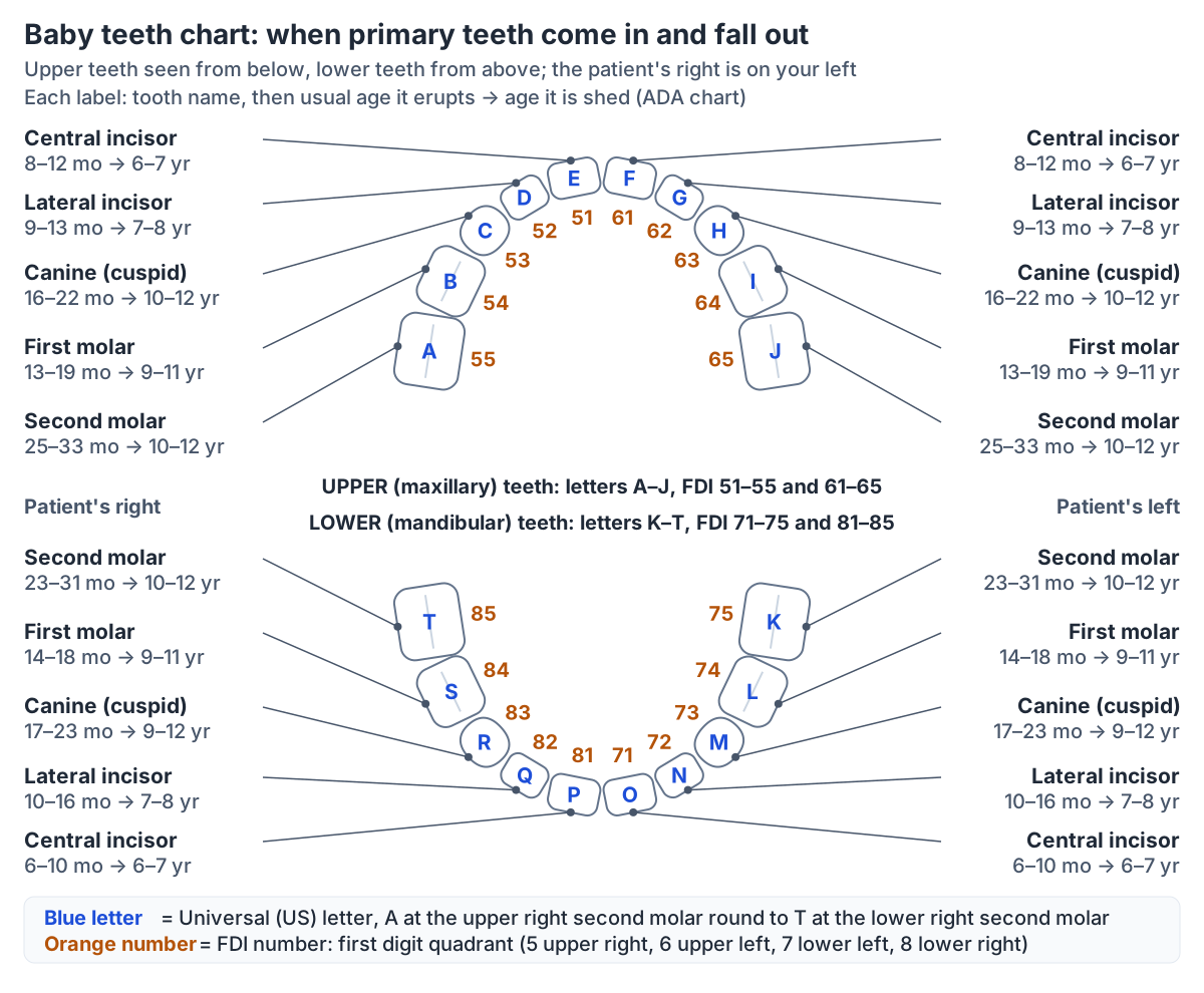
There are 20 baby teeth, also called primary or deciduous teeth. Each arch holds two central incisors, two lateral incisors, two canines and four molars, so every quarter of the mouth (quadrant) has five teeth. The primary dentition has no premolars and no third molars. (Dentalcare: Primary Dentition.)
TeachMeAnatomy gives the same count: 10 teeth in each arch, with two incisors (central and lateral), a canine and two molars per quadrant, making 8 incisors, 4 canines and 8 molars in total. (TeachMeAnatomy: Child and Adult Dentition.)
These teeth are already there before they can be seen. The American Dental Association notes that babies are born with all 20 primary teeth already in their jaws, and that by about age 3 children typically have 10 top teeth and 10 bottom teeth. (American Dental Association (MouthHealthy): Baby Teeth.)
Usually none of them is visible at birth: Kenhub, which also uses the name milk teeth, notes that no deciduous teeth are present in the mouth at birth and that all twenty are present by the time a child turns three. The rare exceptions are natal teeth, covered below. (Kenhub: Deciduous teeth.)
Like all teeth, baby teeth have a crown above the gum line and one or more roots in the jaw, an inner pulp cavity with nerves and blood vessels, dentin around it, enamel over the crown and cementum over the root. (OpenStax Anatomy and Physiology 2e, 23.3: The Mouth, Pharynx, and Esophagus.)
The parts of a tooth, the types of teeth and how adult teeth are built, with a 3D model of the permanent teeth. Tooth anatomy.
Baby teeth chart: when each tooth comes in and falls out
The American Dental Association (ADA) chart gives an age range for every primary tooth. Upper teeth come in at 8 to 12 months (central incisors), 9 to 13 months (lateral incisors), 16 to 22 months (canines), 13 to 19 months (first molars) and 25 to 33 months (second molars). Lower teeth come in at 6 to 10, 10 to 16, 17 to 23, 14 to 18 and 23 to 31 months for the same teeth. (American Dental Association (MouthHealthy): Primary Tooth Development chart (2012).)
The same chart gives the ages at which they are shed: 6 to 7 years for the central incisors, 7 to 8 years for the lateral incisors, 9 to 11 years for the first molars and 10 to 12 years for the second molars. Upper canines are shed at 10 to 12 years and lower canines at 9 to 12 years. (American Dental Association (MouthHealthy): Primary Tooth Development chart (2012).)
Cleveland Clinic's tooth eruption chart lists the same eruption and shedding ranges for all five upper and five lower tooth types, and adds that the complete set of primary teeth is in the mouth from about 2½ to 3 years until about 6 to 7 years of age. (Cleveland Clinic: Teething and Tooth Eruption Chart.)
Not every reference agrees. The American Academy of Pediatric Dentistry (AAPD) uses an older table adapted from Logan and Kronfeld (1933), with somewhat earlier eruption ranges: 5 to 8 months for the lower central incisors, 11 to 18 months for the first molars and 20 to 30 months for the second molars. It warns that many otherwise normal infants do not follow the schedule strictly. (American Academy of Pediatric Dentistry: Dental Growth and Development (after Logan and Kronfeld, 1933).)
Smaller differences exist too: the Dentalcare dental anatomy course, which presents its table as average ages, gives 22 to 31 months for the lower second molar, one month earlier than the ADA. (Dentalcare: Primary Dentition.)
Baby teeth chart: letters, numbers and ages for each primary tooth
| Tooth | Universal letters | FDI numbers | Comes in (ADA) | Comes in (AAPD) | Falls out (ADA) | Falls out (AAPD) |
|---|---|---|---|---|---|---|
| Upper central incisor | E, F | 51, 61 | 8–12 months | 6–10 months | 6–7 years | 7–8 years |
| Upper lateral incisor | D, G | 52, 62 | 9–13 months | 8–12 months | 7–8 years | 8–9 years |
| Upper canine (cuspid) | C, H | 53, 63 | 16–22 months | 16–20 months | 10–12 years | 11–12 years |
| Upper first molar | B, I | 54, 64 | 13–19 months | 11–18 months | 9–11 years | 9–11 years |
| Upper second molar | A, J | 55, 65 | 25–33 months | 20–30 months | 10–12 years | 9–12 years |
| Lower central incisor | P, O | 81, 71 | 6–10 months | 5–8 months | 6–7 years | 6–7 years |
| Lower lateral incisor | Q, N | 82, 72 | 10–16 months | 7–10 months | 7–8 years | 7–8 years |
| Lower canine (cuspid) | R, M | 83, 73 | 17–23 months | 16–20 months | 9–12 years | 9–11 years |
| Lower first molar | S, L | 84, 74 | 14–18 months | 11–18 months | 9–11 years | 10–12 years |
| Lower second molar | T, K | 85, 75 | 23–31 months | 20–30 months | 10–12 years | 11–13 years |
Letters and numbers are given for the patient's right, then left. ADA: American Dental Association chart (2012), which matches the Cleveland Clinic table. AAPD: American Academy of Pediatric Dentistry table, adapted from Logan and Kronfeld (1933). Dentalcare gives 22–31 months for the lower second molar. Individual children vary.
When do babies get teeth? When teething starts
The first baby teeth usually come through the gums at about 6 months of age. OpenStax gives this as the age at which the 20 deciduous teeth first begin to appear. (OpenStax Anatomy and Physiology 2e, 23.3: The Mouth, Pharynx, and Esophagus.)
TeachMeAnatomy gives the same start: the primary teeth begin to erupt at 6 months of age, and the permanent teeth begin to replace them at about 6 years. (TeachMeAnatomy: Child and Adult Dentition.)
The ADA describes a range rather than a single date: the first teeth typically come through between 6 and 12 months, the exact timing varies between children, and the full set of baby teeth is usually in place by age 3. (American Dental Association (MouthHealthy): Teething.)
The NHS likewise says most babies start teething at around 6 months, that the central incisors are usually the first to come through, and that most children have all their milk teeth by the time they are 2 to 3 years old. (NHS: Baby teething symptoms.)
Radiopaedia names the first tooth: the deciduous teeth start erupting at six months with the lower central incisor. (Radiopaedia: Teeth.)
The AAPD table gives 5 to 8 months for the lower central incisors and 6 to 10 months for the upper ones, so a first tooth a little before 6 months is within the published range. (American Academy of Pediatric Dentistry: Dental Growth and Development (after Logan and Kronfeld, 1933).)
The AAPD describes the primary dentition stage as beginning with the eruption of the first tooth, usually at about 6 months, and as complete from about 2 to 6 years of age, when all the primary teeth are in the mouth. (American Academy of Pediatric Dentistry: Management of the Developing Dentition and Occlusion in Pediatric Dentistry (2024).)
What order do baby teeth come in?
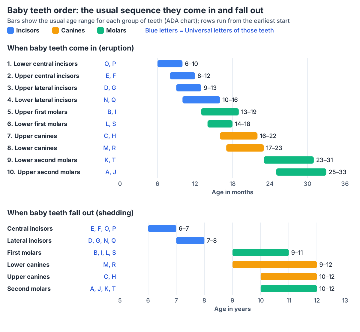
Baby teeth come in from the front of the mouth backwards, with one exception: the canines. The NHS gives the usual order as central incisors at around 6 months, lateral incisors at around 8 months, first molars at around 12 months, canines at around 18 months and second molars at around 24 months. Bottom teeth often come through before the matching top teeth. (NHS: Baby teething symptoms.)
Cleveland Clinic adds that the first two teeth are usually the two bottom central incisors, followed by the top four front teeth, and that teeth usually come in left-right pairs. Lower teeth often come in first, but on its chart the upper lateral incisors, first molars and canines start slightly earlier than the lower ones. (Cleveland Clinic: Teething and Tooth Eruption Chart.)
Reading the ADA ranges in order of their earliest age gives the same sequence: lower central incisors (6 to 10 months), upper central incisors (8 to 12), upper lateral incisors (9 to 13), lower lateral incisors (10 to 16), first molars (13 to 19 upper, 14 to 18 lower), canines (16 to 22 upper, 17 to 23 lower) and finally the second molars (23 to 31 lower, 25 to 33 upper). (American Dental Association (MouthHealthy): Primary Tooth Development chart (2012).)
So the first molars usually arrive before the canines, even though the canines sit in front of them. The AAPD table shows the same order: first molars at 11 to 18 months and canines at 16 to 20 months. (American Academy of Pediatric Dentistry: Dental Growth and Development (after Logan and Kronfeld, 1933).)
When do baby teeth fall out, and how many do you lose?
All 20 baby teeth normally fall out. The ADA describes losing baby teeth as a natural part of growing up and says the adult teeth start coming in at around age 6. (American Dental Association (MouthHealthy): Baby Teeth.)
The ADA's eruption chart page adds that the primary teeth fall out (shed) at various times throughout childhood. (American Dental Association (MouthHealthy): Eruption Charts.)
Shedding follows roughly the same front-to-back order as eruption: central incisors at 6 to 7 years, lateral incisors at 7 to 8 years, first molars at 9 to 11 years, then the canines and second molars at 9 to 12 years. Kenhub's table of loss ages matches these ADA ranges for every tooth. (Kenhub: Deciduous teeth.)
The AAPD table again differs in detail. It gives 7 to 8 years for the upper central incisors, places the lower canines (9 to 11 years) before the lower first molars (10 to 12 years), and extends the lower second molars to 11 to 13 years. (American Academy of Pediatric Dentistry: Dental Growth and Development (after Logan and Kronfeld, 1933).)
The years when baby and adult teeth are in the mouth together are called the mixed dentition. The AAPD places it from about age 6 to 11, when primary and permanent teeth are both present in the mouth. (American Academy of Pediatric Dentistry: Management of the Developing Dentition and Occlusion in Pediatric Dentistry (2024).)
OpenStax gives a slightly wider window, describing the 20 deciduous teeth as being replaced by 32 permanent teeth between approximately age 6 and 12. (OpenStax Anatomy and Physiology 2e, 23.3: The Mouth, Pharynx, and Esophagus.)
A baby tooth with no permanent successor usually resorbs too, but later than usual, and some stay in the mouth for many years. In a review of root resorption, the shedding of baby teeth and the arrival of their successors were found to be closely matched between the left and right sides of the mouth, which suggests the two events are coupled. (Harokopakis-Hajishengallis E. Physiologic root resorption in primary teeth: molecular and histological events. J Oral Sci. 2007.)
Which permanent teeth replace the baby teeth?
Each baby incisor and canine is replaced by a permanent tooth of the same type. The baby molars are replaced by the permanent premolars, and the permanent molars come in behind them. Dentalcare gives 6 to 7 years for the first permanent molars, the earliest range in its table of permanent teeth. (Dentalcare: Permanent Dentition.)
Dentalcare puts it the same way in its account of tooth types: there are no premolars in the primary dentition, and the premolars replace the primary molars when they are shed. (Dentalcare: Types of Teeth and their Functions.)
The first permanent molar therefore replaces no baby tooth. The AAPD table gives it an eruption age of 5½ to 7 years and ranks it first in the order of permanent eruption, followed by the lower central incisors. (American Academy of Pediatric Dentistry: Dental Growth and Development (after Logan and Kronfeld, 1933).)
A University of Baghdad dental lecture makes the contrast explicit: the first primary tooth to erupt is a lower incisor, while the first permanent tooth to erupt is the lower first molar. (University of Baghdad College of Dentistry: Morphologic differences between primary and permanent teeth (Pediatric Dentistry lecture 1).)
All 32 permanent teeth with their names, Universal numbers 1 to 32 and FDI numbers. Tooth chart: adult teeth names and numbers.
Which permanent tooth replaces each baby tooth
| Baby tooth | Replaced by | Successor comes in (Dentalcare) | Successor comes in (AAPD) |
|---|---|---|---|
| Central incisor | Permanent central incisor | 7–8 years | Upper 7–8, lower 6–7 years |
| Lateral incisor | Permanent lateral incisor | 8–9 years | Upper 8–9, lower 7–8 years |
| Canine | Permanent canine | Upper 11–12, lower 9–10 years | Upper 11–12, lower 9–11 years |
| First molar | First premolar | Upper 10–11, lower 10–12 years | Upper 10–11, lower 10–12 years |
| Second molar | Second premolar | Upper 10–12, lower 11–12 years | Upper 10–12, lower 11–13 years |
| None | First permanent molar (behind the baby teeth) | 6–7 years | 5.5–7 years |
| None | Second permanent molar | 11–13 years | 12–14 years |
Permanent molars have no baby predecessor; third molars (wisdom teeth) come later still. Sources: Dentalcare Permanent Dentition and Types of Teeth; AAPD Dental Growth and Development.
Baby teeth letters and numbers: Universal A to T, FDI 51 to 85 and Palmer
In the Universal system used in the United States, baby teeth are lettered rather than numbered. The dentist looks into the open mouth and goes clockwise from the upper right: A is the upper right second molar, E and F are the upper central incisors, J is the upper left second molar, K is the lower left second molar, O and P are the lower central incisors and T is the lower right second molar. (Radiopaedia: American Dental Association Universal Numbering System.)
The MSD Manual lists the same pairs: upper second molars A and J, upper first molars B and I, upper canines C and H, upper lateral incisors D and G, upper central incisors E and F, lower second molars T and K, lower first molars S and L, lower canines R and M, lower lateral incisors Q and N, and lower central incisors P and O. (MSD Manual Consumer Version: Identifying the Primary (Baby) Teeth.)
The international FDI system gives every tooth two digits. For baby teeth the first digit (the quadrant) runs from 5 to 8: 5 upper right, 6 upper left, 7 lower left and 8 lower right. The second digit counts from the midline, from 1 (central incisor) to 5 (second molar), because there are no premolars. So 82 is the lower right baby lateral incisor. (Radiopaedia: FDI World Dental Federation notation.)
Kenhub describes the same two-digit scheme for deciduous teeth, with quadrants 5 to 8 and the teeth numbered 1 to 5 from the midline. (Kenhub: Deciduous teeth.)
Palmer notation, used mainly in the United Kingdom, letters the baby teeth A to E from the midline in each quadrant (A central incisor, B lateral incisor, C canine, D first molar, E second molar) and adds a bracket symbol to show the quadrant. (Radiopaedia: Palmer notation.)
TeachMeAnatomy likewise refers to the five primary teeth in each quadrant by the letters A, B, C, D and E. So the letter C means a canine in Palmer notation but the upper right canine only in the Universal system: always check which system a chart uses. (TeachMeAnatomy: Child and Adult Dentition.)
Baby teeth letters and numbers in the three dental notation systems
| Tooth type | Universal (US) | FDI (international) | Palmer (UK) |
|---|---|---|---|
| Central incisor | E, F (upper); O, P (lower) | 51, 61, 71, 81 | A in each quadrant |
| Lateral incisor | D, G (upper); N, Q (lower) | 52, 62, 72, 82 | B in each quadrant |
| Canine | C, H (upper); M, R (lower) | 53, 63, 73, 83 | C in each quadrant |
| First molar | B, I (upper); L, S (lower) | 54, 64, 74, 84 | D in each quadrant |
| Second molar | A, J (upper); K, T (lower) | 55, 65, 75, 85 | E in each quadrant |
FDI quadrants for baby teeth: 5 upper right, 6 upper left, 7 lower left, 8 lower right. In Palmer notation a bracket symbol shows the quadrant. Sources: Radiopaedia, MSD Manual, Kenhub, TeachMeAnatomy.
Baby skull teeth: the permanent teeth growing under the baby teeth
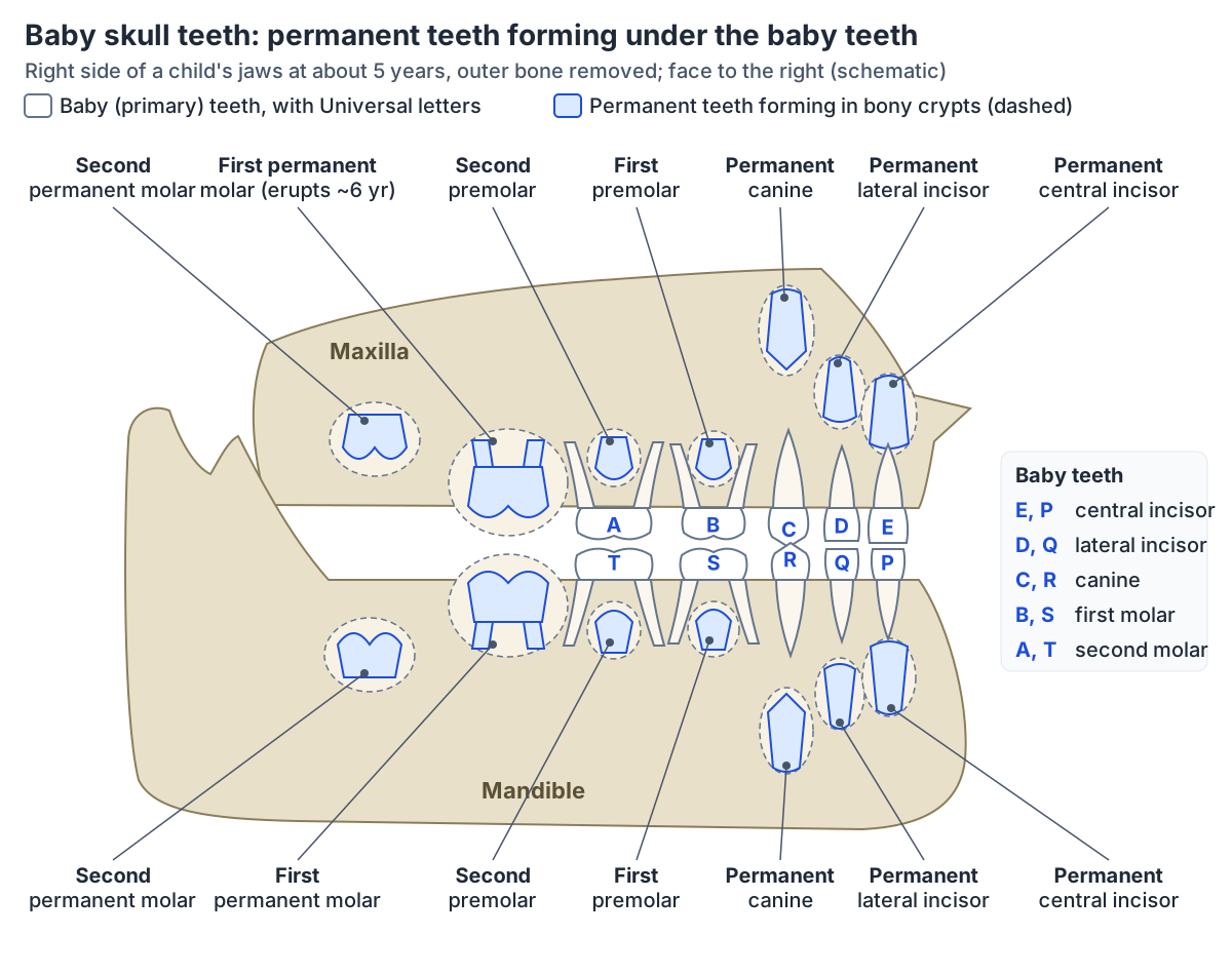
Pictures of a child's skull with the outer bone cut away seem to show two sets of teeth, and that is what they are. By about age 3, when all the primary teeth have erupted, all the permanent teeth except the third molars are already at some stage of development inside the jaws. (Dentalcare: Primary Dentition.)
Tooth formation starts long before birth. In the AAPD table the hard tissue of every baby tooth begins to calcify in the fourth month of fetal life, and the first permanent molar begins to calcify at birth. (American Academy of Pediatric Dentistry: Dental Growth and Development (after Logan and Kronfeld, 1933).)
A paediatric dentistry lecture from Al-Mustaqbal University gives about 14 weeks in utero for the start of calcification of the central incisors, with the lateral incisors and canines a little later, and notes that calcification of the baby tooth crowns is still incomplete at birth. It also states that some calcification of the first permanent molars is always present at birth. (Al-Mustaqbal University College: Early development and calcification of the anterior primary teeth (Pediatric Dentistry lecture 4).)
The permanent front teeth form on the tongue side of their baby predecessors and towards their root tips. A review of root resorption describes the completed crown of a permanent incisor as lying lingual to the apical third of the baby tooth's root. In the molar region the developing permanent teeth also start on the tongue side and later move under the divergent roots of the baby molars. (Harokopakis-Hajishengallis E. Physiologic root resorption in primary teeth: molecular and histological events. J Oral Sci. 2007.)
Under the baby molars the premolars develop between the roots. The roots of the primary molars are relatively long, slender and flared, and this flaring leaves room between them for the developing premolar crowns. (Al-Mustaqbal University College: Early development and calcification of the anterior primary teeth (Pediatric Dentistry lecture 4).)
Each developing tooth sits in a dental follicle, the connective tissue sac around a growing tooth and root. On X-rays of a young patient, follicles show as dark (lucent) areas with a thin bright rim. (Radiopaedia rPoster: The tooth, the whole tooth, and nothing but the tooth (2023).)
The bones of the skull, including the maxilla and mandible that hold the developing teeth. Skull bones.
Do baby teeth have roots?
Yes. Every baby tooth has at least one root anchoring it in the jaw. The roots finish forming between about 18 months and 3 years of age, and the complete primary dentition is then in the mouth from about 2 to about 6 years while the permanent teeth form in the jaws below. (University of Baghdad College of Dentistry: Morphologic differences between primary and permanent teeth (Pediatric Dentistry lecture 1).)
The AAPD table gives the ages at which each baby tooth is fully formed: 18 to 24 months for the incisors, 24 to 30 months for the first molars, 30 to 39 months for the canines and 36 months for the second molars. (American Academy of Pediatric Dentistry: Dental Growth and Development (after Logan and Kronfeld, 1933).)
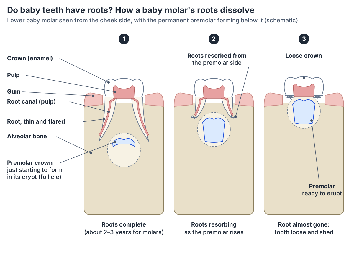
The upper baby molars have three roots, two on the cheek side and one on the palate side, and the lower baby molars have two, one mesial and one distal. Compared with the crown, the roots of the baby molars are longer and more slender than those of permanent teeth, and they flare outwards towards their tips. (University of Baghdad College of Dentistry: Morphologic differences between primary and permanent teeth (Pediatric Dentistry lecture 1).)
Baby incisors and canines have one root each. The Al-Mustaqbal lecture describes the root of the upper baby central incisor as cone-shaped with tapered sides and the canine's as long, slender and tapering, and the three roots of the upper first baby molar as long, slender and widely spread. (Al-Mustaqbal University College: Early development and calcification of the anterior primary teeth (Pediatric Dentistry lecture 4).)
A micro-CT study of baby molar roots describes them as longer, thinner, curved and divergent, and its measurements cover the mesiobuccal, distobuccal and palatal roots of the upper molars and the mesial and distal roots of the lower molars. (Vidi B et al. Description of the root anatomy of the primary molars using high resolution micro-CT. Front Dent Med. 2024.)
The reason people think baby teeth have no roots is that a shed baby tooth usually has little or none left. By the time it falls out, most of the root has been resorbed, as the next section explains. (Harokopakis-Hajishengallis E. Physiologic root resorption in primary teeth: molecular and histological events. J Oral Sci. 2007.)
The lower jaw bone whose alveolar part holds the roots of the lower teeth. Mandible (lower jaw).
How baby teeth fall out: root resorption
Baby teeth loosen because their roots are dissolved. Root resorption is a normal (physiological) event in the life of a primary tooth, carried out by cells called odontoclasts. The pressure of the erupting permanent tooth is thought to help start it, and the dental follicle of the permanent tooth appears to control it, but a successor is not essential: baby teeth without one also resorb, only later. (Harokopakis-Hajishengallis E. Physiologic root resorption in primary teeth: molecular and histological events. J Oral Sci. 2007.)
Resorption begins on the part of the root nearest the permanent tooth. The bud of a permanent front tooth lies on the tongue side of its baby predecessor and towards the root tip, so resorption of baby incisors and canines starts on the tongue side of the apical third of the root, and it continues as the permanent tooth moves closer to the surface until the baby tooth is loose and falls out. In the baby molars resorption usually begins on the inner surfaces of the roots, next to the bone between them. (University of Baghdad College of Dentistry: Morphologic differences between primary and permanent teeth (Pediatric Dentistry lecture 1).)
A micro-CT study of baby molars describes the same pattern: resorption appears on the root surfaces closest to the permanent tooth, the lingual surface of single-rooted teeth and the inner surfaces of the furcation of the molars. (Vidi B et al. Description of the root anatomy of the primary molars using high resolution micro-CT. Front Dent Med. 2024.)
The process is not continuous. Periods of active resorption alternate with rest and with repair, when new cementum or bone is laid down on the root. This may explain why a wobbly tooth seems to loosen and then tighten again before it finally falls out. Late in the process the inside of the crown can be resorbed too, leaving a thin shell that breaks off in pieces. (Harokopakis-Hajishengallis E. Physiologic root resorption in primary teeth: molecular and histological events. J Oral Sci. 2007.)
Sometimes the sequence goes wrong. If the permanent lower incisors do not move far enough towards the lip before they emerge, the roots of the baby incisors resorb incompletely or late, and the permanent incisors can erupt on the tongue side of baby incisors that are still in the mouth. (Harokopakis-Hajishengallis E. Physiologic root resorption in primary teeth: molecular and histological events. J Oral Sci. 2007.)
In ankylosis the cementum of the root fuses directly to the bone and the periodontal ligament is replaced by bone, so the tooth can no longer erupt. It stays at the same level while the neighbouring teeth keep erupting in a growing child, so it seems to sink (infraocclusion). Ankylosis occurs in both dentitions, but baby molars are the teeth most often affected. (American Academy of Pediatric Dentistry: Management of the Developing Dentition and Occlusion in Pediatric Dentistry (2024).)
Baby teeth on an X-ray
On dental X-rays and CT, enamel is the brightest (most radiopaque) part of a tooth. Dentin and cementum are less radiopaque and cannot be told apart, the pulp chamber and root canal are dark (lucent) in the middle of the tooth, and the periodontal ligament is a thin dark line between the root and the lamina dura, the cortical bone that lines the socket. (Radiopaedia: Teeth.)
The usual X-ray for looking at a child's whole set of teeth is the orthopantomogram (OPG), a panoramic single image of the mandible, maxilla and teeth. Its uses include following the growth and development of children's teeth (position, shape and angle) and checking for extra (supernumerary) teeth or missing tooth germs. (Radiopaedia: Orthopantomography.)
Radiopaedia's normal panoramic X-ray of a 6-year-old shows the start of the change of teeth: the first permanent molars and the lower central incisors have erupted, and the third molars have not yet begun to develop. (Radiopaedia case: Normal orthopantomography, mixed dentition stage.)
The permanent teeth still inside the jaws show up because each sits in its dental follicle. In a young patient, rounded dark (lucent) areas with thin sclerotic (bright) rims around the developing teeth and roots are follicles, not infection; the rim is lost if infection is present. (Radiopaedia rPoster: The tooth, the whole tooth, and nothing but the tooth (2023).)
X-rays understate the pulp of baby teeth: a Baghdad dental lecture notes that radiographs do not show the full extent of the pulp horns into the cusps, and that the pulp horns of baby teeth lie closer to the outer surface than those of permanent teeth. (University of Baghdad College of Dentistry: Morphologic differences between primary and permanent teeth (Pediatric Dentistry lecture 1).)
Where a baby tooth is ankylosed, an X-ray of the tooth may show loss of the periodontal ligament, external resorption of the root and its replacement by bone, although a two-dimensional film has limits. (American Academy of Pediatric Dentistry: Management of the Developing Dentition and Occlusion in Pediatric Dentistry (2024).)
The upper jaw bone and its alveolar process, which holds the upper teeth and their developing successors. Maxilla (upper jaw).
Babies born with teeth: natal and neonatal teeth
A few babies have a tooth at birth. A natal tooth is one present in the mouth at birth; a neonatal tooth comes through within the first 30 days of life. Most are baby teeth that have erupted early rather than extra teeth. (Szymczak A et al. Incidence of natal and neonatal teeth: a 15-year retrospective study from the Greater Poland voivodeship. BMC Oral Health. 2025.)
A 2023 meta-analysis of 23 prevalence studies estimated that natal teeth occur in about 34.6 per 10,000 newborns (about 1 in 289) and neonatal teeth in about 4.5 per 10,000 (about 1 in 2,212). For natal teeth the estimates ranged from about 11 per 10,000 in Asia to about 75 per 10,000 in North America, and none of the included studies met every quality criterion. The authors note that these teeth often need prompt professional care. (Vitali FC et al. Worldwide prevalence of natal and neonatal teeth: systematic review and meta-analysis. J Am Dent Assoc. 2023.)
Individual studies vary much more, from about 1 in 1,000 to 1 in 30,000 depending on the type of study. A case-control study from Poland cites the same 1 in 289 and 1 in 2,212 figures and reports a natal to neonatal ratio between 3:1 and 1:1.2 in the literature. (Krasuska-Sławińska E et al. Natal and neonatal teeth in newborns and infants: a case-control study. Sci Rep. 2025.)
In the Greater Poland region, National Health Fund records for 2007 to 2022 identified 471 cases, 0.08% of live births, and 93.8% of them were natal rather than neonatal teeth. The recorded incidence rose from 0.039% in 2007 to 0.134% in 2022. The same paper's review notes that most natal and neonatal teeth appear in the front of the lower jaw, and that about 1 to 10% are supernumerary (extra) teeth. (Szymczak A et al. Incidence of natal and neonatal teeth: a 15-year retrospective study from the Greater Poland voivodeship. BMC Oral Health. 2025.)
The case-control study likewise describes most natal teeth as prematurely erupted baby teeth, most commonly the lower central incisors, with 5 to 10% supernumerary. Every natal or neonatal tooth in its 52 children was a lower central incisor, and some had defective (hypoplastic) enamel. (Krasuska-Sławińska E et al. Natal and neonatal teeth in newborns and infants: a case-control study. Sci Rep. 2025.)
Natal teeth may be very loose; in one published case the extracted teeth had crowns but no roots. The same report lists possible complications such as injury to the tongue and poor feeding, and other names for these teeth, including congenital, fetal and predeciduous teeth. (Newadkar UR et al. Natal and neonatal teeth: terminologies with diverse superstitions. J Family Med Prim Care. 2016.)
Damage to the baby's tongue or the lining of the mouth from repeated rubbing against these teeth is called Riga-Fede disease. They can also make feeding difficult and carry a risk of aspiration (the tooth being inhaled). (Szymczak A et al. Incidence of natal and neonatal teeth: a 15-year retrospective study from the Greater Poland voivodeship. BMC Oral Health. 2025.)
Because these teeth often need prompt care, a tooth in a newborn's mouth should be checked by a doctor or dentist. (Vitali FC et al. Worldwide prevalence of natal and neonatal teeth: systematic review and meta-analysis. J Am Dent Assoc. 2023.)
Baby teeth vs permanent teeth
Baby teeth are not simply small adult teeth. Their crowns are shorter and narrower at the neck (cervical constriction), their enamel and dentin are thinner, and they are lighter in colour. Their pulp is larger relative to the crown, and the pulp horns lie closer to the outer surface. (University of Baghdad College of Dentistry: Morphologic differences between primary and permanent teeth (Pediatric Dentistry lecture 1).)
The Al-Mustaqbal lecture adds that the crowns of baby teeth are wider for their length, the cervical ridge of enamel is much more prominent, the roots of the front teeth are narrow and long compared with the crown, and baby teeth are usually lighter in colour than permanent teeth. (Al-Mustaqbal University College: Early development and calcification of the anterior primary teeth (Pediatric Dentistry lecture 4).)
The two sets also differ in number: the 20 deciduous teeth are replaced by 32 permanent teeth. The third molar in each set of three permanent molars is the wisdom tooth; it erupts late, and it is not uncommon for it to fail to erupt at all. (OpenStax Anatomy and Physiology 2e, 23.3: The Mouth, Pharynx, and Esophagus.)
The biggest difference is their fate. In bone, resorption goes on all through life, but among teeth only the primary dentition undergoes normal resorption: the roots of baby teeth dissolve, while the roots of permanent teeth do not. (Harokopakis-Hajishengallis E. Physiologic root resorption in primary teeth: molecular and histological events. J Oral Sci. 2007.)
Baby teeth vs permanent teeth
| Feature | Baby (primary) teeth | Permanent teeth |
|---|---|---|
| Number | 20 | 32, including 4 third molars (wisdom teeth) |
| Types in each quadrant | 2 incisors, 1 canine, 2 molars | 2 incisors, 1 canine, 2 premolars, 3 molars |
| First to come in | Lower central incisor, about 6 months | First permanent molar, about 6 years |
| Colour | Lighter (whiter) | Darker |
| Enamel and dentin | Thinner | Thicker |
| Crown | Shorter, constricted at the neck, with a prominent cervical ridge | Longer, less constricted |
| Pulp | Larger relative to the crown; pulp horns closer to the surface | Smaller relative to the crown |
| Roots | Long and slender relative to the crown; molar roots flare to make room for the premolar | Relatively shorter and stouter; not resorbed in normal life |
| Fate | Roots resorbed, tooth shed between about 6 and 12 years | Not shed naturally (roots are not resorbed) |
Sources: OpenStax 23.3, Dentalcare, University of Baghdad and Al-Mustaqbal University dental lectures, Radiopaedia, Harokopakis-Hajishengallis (2007).
Why baby teeth matter: chewing, speech and space for adult teeth
Although they are shed, baby teeth do several jobs. The ADA calls them nature's braces because they help set the spacing for the adult teeth, and notes that they are needed for chewing and help children speak clearly. (American Dental Association (MouthHealthy): Baby Teeth.)
Cleveland Clinic lists the same roles: baby teeth reserve space for their permanent counterparts, give the face its usual shape, help the development of clear speech and help children eat well. (Cleveland Clinic: Teething and Tooth Eruption Chart.)
Spacing is normal in a baby's mouth. Gaps between the front baby teeth are common, and the most marked, called primate spaces, lie on the midline side of the upper canines and on the far side of the lower canines. Spacing in the primary dentition is regarded as a sign of favourable development of the permanent bite. (Vegesna M et al. Occlusal characteristics and spacing in primary dentition: a gender comparative cross-sectional study. Int Sch Res Notices. 2014.)
When a baby tooth is lost early, through decay, infection or injury, the AAPD notes that the arch may lose length and the space for the permanent successor can shrink, which can lead to crowding, rotated teeth or teeth erupting in the wrong place. (American Academy of Pediatric Dentistry: Management of the Developing Dentition and Occlusion in Pediatric Dentistry (2024).)
The lips, palate, gums, tongue and salivary glands around the teeth. Mouth anatomy.
Frequently asked questions
When do babies start teething?
Most babies get their first tooth at about 6 months. The American Dental Association gives a usual range of 6 to 12 months, and the American Academy of Pediatric Dentistry table starts the lower central incisors at 5 to 8 months, so timing varies. The first tooth is usually a lower central incisor, and the full set of 20 baby teeth is usually in by about age 3.
What teeth do babies get first?
The first teeth are usually the two lower central incisors, the bottom front teeth, at about 6 to 10 months. Next come the upper central incisors at 8 to 12 months, then the upper and lower lateral incisors. The first molars arrive at about 13 to 19 months, before the canines at 16 to 23 months, and the second molars come in last, between about 23 and 33 months.
What order do baby teeth come in?
The usual order is: lower central incisors, upper central incisors, upper lateral incisors, lower lateral incisors, first molars, canines and finally second molars. Teeth tend to come in left-right pairs, and the lower central incisors usually come before the upper ones. The canines usually arrive after the first molars even though they sit in front of them. The ranges overlap, so a different order is common and usually normal.
How many baby teeth do you lose?
Children normally lose all 20 baby teeth: 8 incisors, 4 canines and 8 molars. Each incisor and canine is replaced by the same type of permanent tooth and each baby molar by a premolar. The 12 permanent molars, including the wisdom teeth, do not replace any baby tooth; they come in behind them, so an adult ends up with 32 teeth.
When do baby teeth fall out?
Baby teeth usually start to fall out at about 6 to 7 years, beginning with the central incisors. The lateral incisors follow at 7 to 8 years, the first molars at 9 to 11 years, and the canines and second molars at about 9 to 12 years. Some charts extend the last baby molars to about 13 years. Timing varies between children.
Do baby teeth have roots?
Yes. Each baby tooth has one root (incisors and canines) or two to three roots (molars), which finish forming between about 18 months and 3 years. As the permanent tooth below moves up, the root is gradually resorbed by cells called odontoclasts. By the time the tooth falls out little or no root is left, which is why shed baby teeth look rootless.
Can babies be born with teeth?
Yes, but rarely. A tooth present at birth is a natal tooth and one that comes through in the first 30 days is a neonatal tooth. A 2023 meta-analysis estimated about 1 in 289 newborns have natal teeth and about 1 in 2,212 neonatal teeth. Most are early-erupting lower central incisors, often loose and with little root.
What letters are used for baby teeth?
In the US Universal system baby teeth are lettered A to T, clockwise as the dentist looks into the mouth: A is the upper right second molar, J the upper left second molar, K the lower left second molar and T the lower right second molar. The FDI system numbers them 51 to 55, 61 to 65, 71 to 75 and 81 to 85.
References
- American Dental Association. Primary Tooth Development (eruption chart). MouthHealthy, 2012.
- American Dental Association. Eruption Charts. MouthHealthy.
- American Dental Association. Baby Teeth. MouthHealthy.
- American Dental Association. Teething. MouthHealthy, updated April 2026.
- American Academy of Pediatric Dentistry. Dental Growth and Development. Reference Manual resource section; adapted from Logan WHG, Kronfeld R. J Am Dent Assoc 1933;20(3):379-427.
- American Academy of Pediatric Dentistry. Management of the developing dentition and occlusion in pediatric dentistry. The Reference Manual of Pediatric Dentistry. Chicago, IL: AAPD; 2025:497-515 (latest revision 2024).
- Cleveland Clinic. Teething (Teething Syndrome): Symptoms and Tooth Eruption Chart. Updated 24 May 2023.
- Dentalcare (Procter & Gamble). An Overview of Dental Anatomy: Primary Dentition. CE500.
- Dentalcare (Procter & Gamble). An Overview of Dental Anatomy: Permanent Dentition. CE500.
- Dentalcare (Procter & Gamble). An Overview of Dental Anatomy: Types of Teeth and their Functions. CE500.
- NHS. Baby teething symptoms. Reviewed 14 May 2026.
- OpenStax. Anatomy and Physiology 2e, §23.3 The Mouth, Pharynx, and Esophagus. Rice University, 2022.
- McKernon S. Child and Adult Dentition. TeachMeAnatomy, updated 2025.
- Sieroslawska A. Deciduous teeth. Kenhub, reviewed 2023.
- Deng F, et al. American Dental Association Universal Numbering System. Radiopaedia reference article.
- Smith H, Al Kabbani A, Weerakkody Y, et al. FDI World Dental Federation notation. Radiopaedia reference article.
- Deng F. Palmer notation. Radiopaedia reference article.
- Knipe H, Foster T, et al. Teeth. Radiopaedia reference article.
- Klaassen K, Ryan P, Arora A, et al. Orthopantomography. Radiopaedia reference article.
- Uribe SE. Normal orthopantomography mixed dentition stage. Radiopaedia case, 2016.
- Oo A, Spence C. The tooth, the whole tooth, and nothing but the tooth. Radiopaedia rPoster, 2023.
- MSD Manual Consumer Version. Identifying the Primary (Baby) Teeth. Merck & Co.
- Harokopakis-Hajishengallis E. Physiologic root resorption in primary teeth: molecular and histological events. J Oral Sci. 2007;49(1):1-12.
- Yaseen HN. Morphologic differences between primary and permanent teeth. Pediatric Dentistry lecture 1, College of Dentistry, University of Baghdad, 2021.
- Al-Falluji ML. Early development and calcification of the anterior primary teeth. Pediatric Dentistry lecture 4, Al-Mustaqbal University College.
- Vitali FC, Santos PS, Massignan C, et al. Worldwide prevalence of natal and neonatal teeth: systematic review and meta-analysis. J Am Dent Assoc. 2023;154(10):910-921.e4.
- Szymczak A, Suchodolski M, Forszt D, et al. Incidence of natal and neonatal teeth: a 15-year retrospective study from the Greater Poland voivodeship. BMC Oral Health. 2025.
- Krasuska-Sławińska E, Turska-Szybka A, Pham A, et al. Natal and neonatal teeth in newborns and infants: a case-control study. Sci Rep. 2025.
- Newadkar UR, Chaudhari L, Khalekar YK. Natal and neonatal teeth: terminologies with diverse superstitions. J Family Med Prim Care. 2016;5(1):184-185.
- Vidi B, Gil-Valcarcel AM, Obispo-Diaz C, et al. Description of the root anatomy of the primary molars using high resolution computed microtomography (Micro-CT): an analysis of three-dimensional root canal system. Front Dent Med. 2024;5:1522414.
- Vegesna M, Chandrasekhar R, Chandrappa V. Occlusal characteristics and spacing in primary dentition: a gender comparative cross-sectional study. Int Sch Res Notices. 2014;2014:512680.
About this page
Drafted with AI assistance from the sources listed above, checked against them and reviewed by Dr Shaista Andleeb; last reviewed on 3 November 2026. The editorial policy explains how sources are chosen and how errors are corrected. To report a mistake, use the contact page.
Related anatomy
- Tooth chartTooth number chart for all 32 adult teeth: every name with its Universal, FDI and Palmer number, labelled diagrams…
- TeethTooth anatomy with labeled diagrams and a 3D model: crown, root, enamel, dentin, pulp and cementum, incisors, canines…
- Mouth anatomyMouth anatomy with a 3D model and labelled diagrams: lips, cheeks, hard and soft palate, uvula, tongue, teeth, gums…
- Skull bonesSkull anatomy with labelled diagrams and a 3D model: the 8 cranial and 14 facial bones, sutures, fontanelles, the…
- MandibleMandible anatomy with a labelled diagram: body, ramus, angle, condylar and coronoid processes, foramina, muscle…
- MaxillaMaxilla bone anatomy: the body and four processes, palatine and alveolar processes, maxillary sinus, articulations, Le…
Explore the whole body in the interactive 3D human anatomy model, or browse the A–Z list of human structures.