

What is a dermatome?
A dermatome is the area of skin supplied by the sensory axons of one segmental spinal nerve root. Its partner on the motor side is the myotome: the group of muscle fibres supplied by the motor axons of the same root. Both terms are defined this way in the international standards used to examine people after spinal cord injury. (Kirshblum SC et al. International standards for neurological classification of spinal cord injury (revised 2011). J Spinal Cord Med. 2011.)
The skin of a dermatome is supplied through the posterior (dorsal) root of the spinal nerve. The cell bodies of these sensory neurons do not sit in the spinal cord's grey matter but in the spinal (dorsal root) ganglion just outside it. (Kenhub: Spinal nerves.)
There are 31 pairs of spinal nerves: 8 cervical (C1 to C8), 12 thoracic, 5 lumbar, 5 sacral and 1 coccygeal. Each is named for the level of the spinal cord where it emerges. (OpenStax Anatomy and Physiology 2e, 13.4: The Peripheral Nervous System.)
C1 is the exception: it has no associated dermatome, and in many people the C1 nerve is absent. That is why dermatome maps start at C2. (Kenhub: Dermatomes.)
Healthline therefore counts 30 dermatomes for the 31 pairs of spinal nerves, one fewer than the number of nerves. (Healthline: What Are Dermatomes?.)
Cleveland Clinic adds that many people lack a C1 spinal nerve, and that where one is present its small skin area lies at the centre of the back of the head. Dermatomes cover the whole body except the face, which is supplied by nerves that do not pass through the spinal cord. (Cleveland Clinic: Dermatomes.)
Dorsal and ventral roots, the dorsal root ganglion, rami and the tracts that carry skin sensation upward. Spinal cord anatomy.
Dermatome map: front and back of the body
A dermatome map is a chart of all the dermatomes on the body surface. Kenhub groups it by region: the head, face and neck belong to the trigeminal nerve (CN V) and spinal nerves C2 to C4, the upper limbs to C5 to T2, the thorax and abdomen to T3 to T12, and the lower limbs and genitalia to L1 to S5. (Kenhub: Dermatomes.)
The trunk dermatomes form horizontal bands. On the limbs the pattern changes: the upper limb dermatomes run lengthways down the arm from C5 to T1, while the lower limb dermatomes start at the hip, shift medially towards the knee and then spiral down to the ankle and the top of the foot. (Lecturio: Dermatomes.)
On the chest this creates a jump. Because C5 to T1 are carried out into the arm, C4 lies directly next to T2 on the Foerster map, a little below the clavicle. (Kenhub: Dermatomes.)
The two maps on this page follow that Foerster-type pattern and mark the key sensory points of the International Standards for Neurological Classification of Spinal Cord Injury (ISNCSCI) as dots. They are schematic: every map is a simplification, as the section on why maps differ explains. (Kirshblum SC et al. International standards for neurological classification of spinal cord injury (revised 2011). J Spinal Cord Med. 2011.)
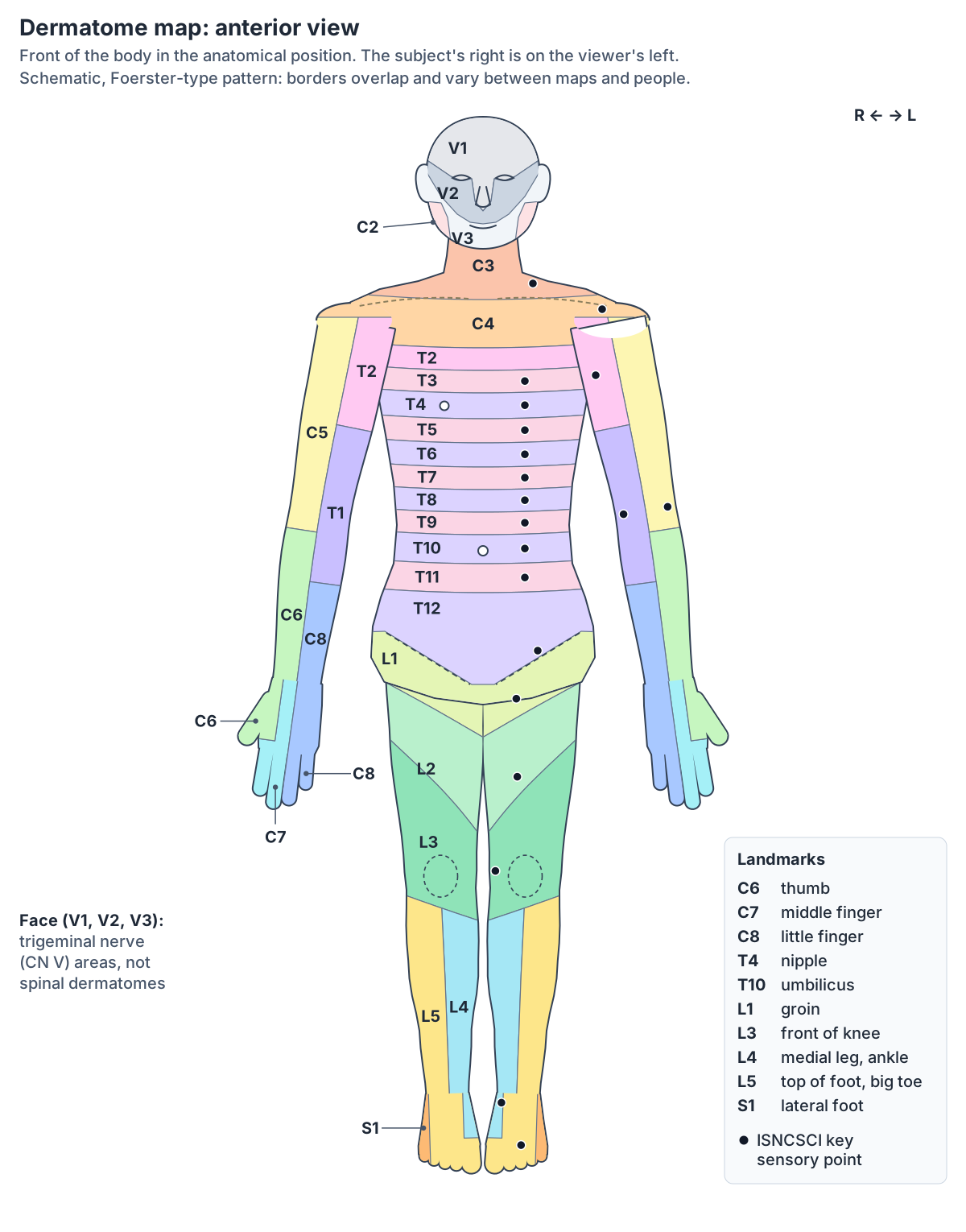
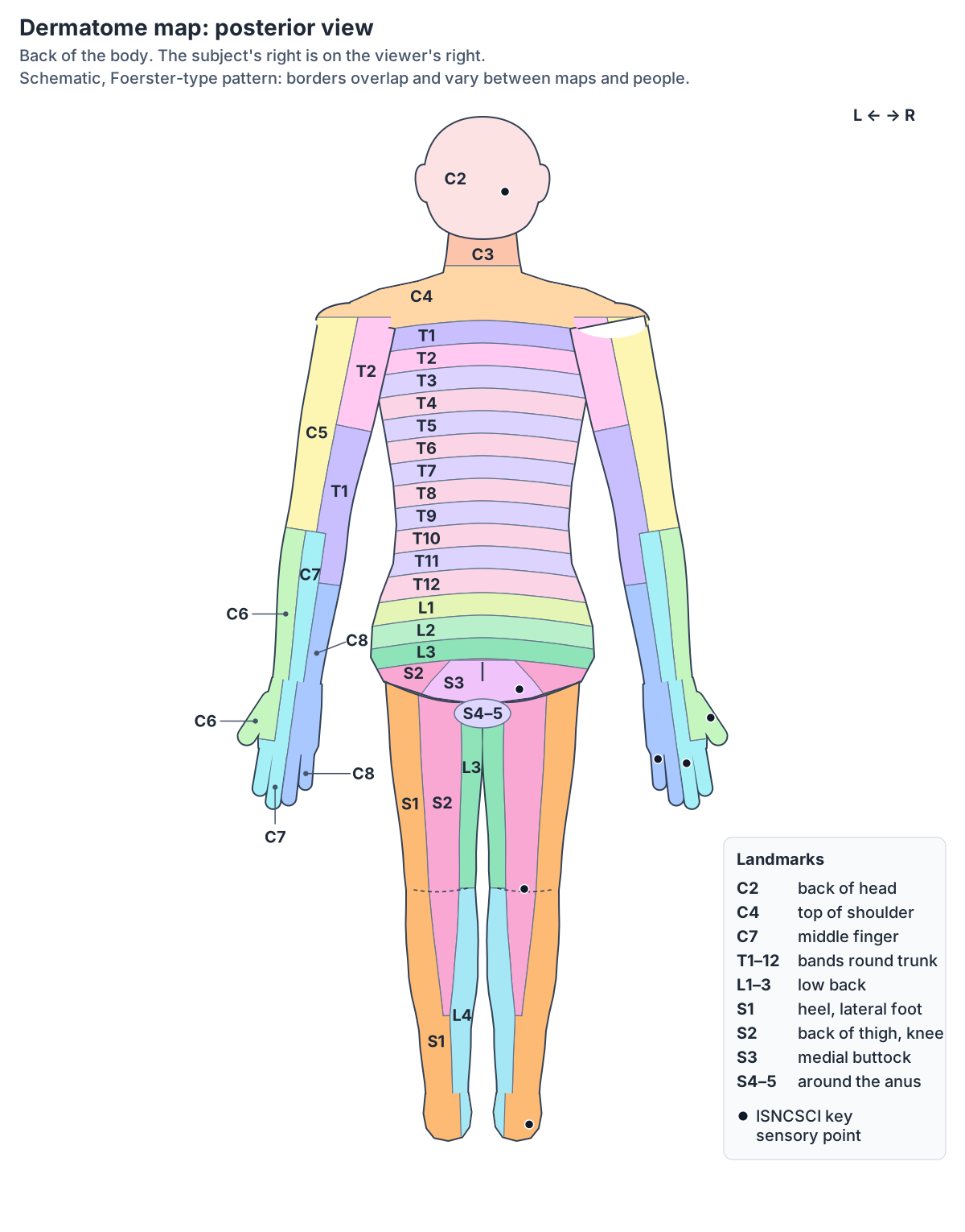
Dermatome levels by region (dermatome chart)
Head and neck: C2 covers the back and top of the head, C3 the front of the neck, the upper back of the neck and the supraclavicular fossa, and C4 the shoulder, the skin below the clavicle and the back of the lower neck. The skin of the face belongs to the three divisions of the trigeminal nerve: V1 the forehead and nose, V2 the cheekbones and upper jaw, and V3 the lower jaw including the skin in front of the ear. (Kenhub: Dermatomes.)
Upper limb: C5 spreads over the lateral arm, C6 the radial side of the forearm and the thumb, C7 the middle of the back of the forearm and the middle finger, C8 the ulnar side of the forearm and hand and the little finger, T1 the medial forearm and lower arm, and T2 the medial upper arm into the axilla. (Kenhub: Dermatomes.)
Thorax and abdomen: T3 lies at the level of the lower axilla, T4 at the nipple, T5 just below it, T6 at the xiphoid process, T7 to T9 evenly spaced down to T10 at the umbilicus, and T11 and T12 spaced between the umbilicus and the top of the pelvis. (Kenhub: Dermatomes.)
Lower limb and perineum: L1 wraps from the back beside the L1 vertebra round to the groin, L2 covers the front of the thigh below the inguinal region, L3 runs down the medial thigh, L4 curves from the lateral thigh to the medial leg and foot, and L5 covers the anterolateral leg and the top and sole of the foot. S1 runs down the posterolateral thigh and leg to the lateral malleolus, S2 from the buttock down the back of the thigh, S3 covers the medial buttock and perineum, and S4 and S5 the perineum and the skin around the anus. (Kenhub: Dermatomes.)
Cleveland Clinic summarises the same pattern by spinal region: cervical dermatomes for the head, neck, shoulders, arms and hands; thoracic dermatomes, which are mostly on the trunk, with T1 entirely on the arm; lumbar dermatomes for the hips, legs and feet; and sacral and coccygeal dermatomes for the buttocks, the back of the legs and the genitals. (Cleveland Clinic: Dermatomes.)
Why the face is mapped by the trigeminal nerve's V1, V2 and V3 rather than by spinal dermatomes. Cranial nerves and nerve anatomy of the face.
Dermatome chart: every level, its skin area and its key sensory point
| Level | Skin area (Kenhub) | ISNCSCI key sensory point |
|---|---|---|
| C2 | Back and top of the head | About 1 cm lateral to the external occipital protuberance (or about 3 cm behind the ear) |
| C3 | Front of the neck, back of the upper neck and the supraclavicular fossa | Supraclavicular fossa, midclavicular line |
| C4 | Shoulder, skin below the clavicle and back of the lower neck | Top of the acromioclavicular joint |
| C5 | Lateral side of the arm | Lateral (radial) side of the antecubital fossa |
| C6 | Radial side of the forearm and the thumb | Back of the thumb, proximal phalanx |
| C7 | Middle of the back of the forearm and the middle finger | Back of the middle finger, proximal phalanx |
| C8 | Ulnar side of the forearm and hand and the little finger | Back of the little finger, proximal phalanx |
| T1 | Medial forearm and lower arm | Medial (ulnar) side of the antecubital fossa |
| T2 | Medial upper arm, continuing into the axilla | Apex of the axilla |
| T3 | Chest and back at the level of the lower axilla | Third intercostal space, midclavicular line |
| T4 | Chest at the level of the nipple | Fourth intercostal space at the nipple line |
| T5 to T9 | Bands between the nipple and the umbilicus; T6 at the xiphoid process | Fifth to ninth intercostal spaces (T6 at the xiphisternum) |
| T10 | Abdomen at the level of the umbilicus | Tenth intercostal space, level of the umbilicus |
| T11 | Between T10 and T12 | Eleventh intercostal space |
| T12 | Lower abdomen just above the pelvic girdle | Midpoint of the inguinal ligament |
| L1 | From the back beside the L1 vertebra round to the groin | Halfway between the T12 and L2 points |
| L2 | Front of the thigh below the inguinal region | Front of the thigh, halfway between the inguinal ligament and the medial femoral condyle |
| L3 | Lower thigh and medial knee region | Medial femoral condyle, just above the knee |
| L4 | From the lateral thigh across the knee to the medial leg and foot | Medial malleolus |
| L5 | Anterolateral leg, top and sole of the foot, middle toes | Top of the foot at the third metatarsophalangeal joint |
| S1 | Posterolateral thigh and leg to the lateral malleolus; little toe | Lateral heel (calcaneus) |
| S2 | Buttock to the back of the thigh, the popliteal fossa and the leg | Midpoint of the popliteal fossa |
| S3 | Medial buttock and perineum | Ischial tuberosity or infragluteal fold |
| S4 to S5 | Perineum, genitals and the skin around the anus | Perianal skin within 1 cm of the anal margin |
Skin areas paraphrase Kenhub's region descriptions (which mix features of the Foerster and Keegan and Garrett maps); key points are from the ISNCSCI standards (Kirshblum et al. 2011). C1 usually has no dermatome.
Key dermatome landmarks: C6 thumb, T4 nipple, T10 umbilicus, L4 and S1
A few landmarks make the whole map easy to rebuild. The thumb is C6, the middle finger C7 and the little finger C8. On the trunk the nipple is T4 and the umbilicus T10. In the leg the ankle and medial leg are L4, the big toe and top of the foot L5, and the little toe and lateral side of the foot S1. (Kenhub: Dermatomes.)
The ISNCSCI key sensory points sit in the same places: the back of the thumb for C6, the fourth intercostal space at the nipple line for T4 and the tenth intercostal space at the level of the umbilicus for T10. In the lower limb the standards test L4 over the medial malleolus, L5 on the top of the foot at the third metatarsophalangeal joint and S1 on the lateral heel. L3 is tested on the medial femoral condyle, just above the knee, and L1 halfway between the T12 point on the inguinal ligament and the L2 point on the front of the thigh. (Kirshblum SC et al. International standards for neurological classification of spinal cord injury (revised 2011). J Spinal Cord Med. 2011.)
Sources disagree about the knee. Kenhub's landmark list puts the knee at L3 and the ankle at L4, although its detailed map carries L4 across the knee to the medial leg. Healthline places the area of the knee in L4, together with the front of the thigh and calf and the inside of the ankle. The ISNCSCI points sit just above the knee (L3) and at the inner ankle (L4), so the front of the knee lies near the L3/L4 border. (Healthline: What Are Dermatomes?.)
The axilla is T2, and the T1 point lies on the medial side of the elbow crease. The C5 point lies on the lateral side of the same crease, so C5 and T1 are tested at opposite ends of one skin fold. (Kirshblum SC et al. International standards for neurological classification of spinal cord injury (revised 2011). J Spinal Cord Med. 2011.)
Why dermatomes form bands: development
Dermatomes go back to the third week of development. The paraxial mesoderm beside the neural tube splits into paired blocks called somites. TeachMeAnatomy describes 44 pairs forming, of which 13 break down, leaving 31 that match the 31 pairs of spinal nerves. (TeachMeAnatomy: Dermatomes.)
Kenhub gives a slightly different count: about 42 to 44 pairs of somites, made up of 4 occipital, 8 cervical, 12 thoracic, 5 lumbar, 5 sacral and 8 to 10 coccygeal segments. (Kenhub: Dermatomes.)
Each somite divides into a sclerotome, which forms the vertebrae and ribs, a myotome, which forms skeletal muscle, and a dermatome, which forms the connective tissue and dermis of the skin. (Kenhub: Dermatomes.)
As the limbs grow, the dermis of the limb buds is stretched and carried down the limb. That is why the trunk shows simple bands while the limbs show long, twisted strips. (TeachMeAnatomy: Dermatomes.)
The epidermis, dermis and hypodermis that each dermatome supplies with sensory nerve endings. Skin layers.
Dermatome vs myotome
A dermatome is skin; a myotome is muscle. Kenhub defines the myotome as the group of muscles supplied by the ventral (anterior) root of a single spinal nerve, the motor equivalent of a dermatome. Both overlap with their neighbours, and a single muscle may receive fibres from more than one level. (Kenhub: Myotomes.)
Because most muscles get input from several spinal levels, myotomes are tested through movements of a joint or muscle group rather than single muscles. Testing dermatomes and myotomes together is a core part of the neurological examination. (Elsevier Student Hub: Dermatomes and Myotomes.)
The ISNCSCI exam uses one key muscle group per level, such as the wrist extensors for C6, the elbow extensors for C7, the knee extensors for L3, the ankle dorsiflexors for L4 and the ankle plantar flexors for S1. (Kirshblum SC et al. International standards for neurological classification of spinal cord injury (revised 2011). J Spinal Cord Med. 2011.)
Comparing the dermatome and the myotome of one level helps separate a purely sensory lesion from one that also causes weakness at that level. (Lecturio: Dermatomes.)
Dermatomes vs myotomes: key muscles and reflexes by level
| Level | ISNCSCI key muscle group | Wider movement range (Kenhub) | Tendon reflex (Kenhub) |
|---|---|---|---|
| C5 | Elbow flexors (biceps, brachialis) | Shoulder abduction (C5); shoulder flexion (C5 to C6) | None listed |
| C6 | Wrist extensors | Supination (C6); wrist flexion and extension (C6 to C7) | Biceps reflex |
| C7 | Elbow extensors (triceps) | Elbow extension (C6 to C7) | Triceps reflex |
| C8 | Finger flexors (middle finger) | Finger flexion and extension (C7 to C8) | None listed |
| T1 | Little finger abductors | Finger abduction and adduction (T1) | None listed |
| L2 | Hip flexors (iliopsoas) | Hip flexion (L1 to L3) | None listed |
| L3 | Knee extensors (quadriceps) | Knee extension (L3 to L4) | Patellar reflex (L3 to L4) |
| L4 | Ankle dorsiflexors (tibialis anterior) | Ankle dorsiflexion (L4 to S1) | None listed |
| L5 | Long toe extensors | Toe extension at the MTP joints (L5) | None listed |
| S1 | Ankle plantar flexors (gastrocnemius, soleus) | Ankle plantarflexion (S1 to S2) | Achilles reflex (S1 to S2) |
The single-level key muscles come from the ISNCSCI standards; Kenhub's wider ranges show that most movements depend on more than one root, which is why one myotome is tested through a movement rather than one muscle.
Dermatomes vs peripheral nerve territories
A dermatome is not the same as the skin area of a named nerve such as the median or femoral nerve. The limb nerves come out of plexuses that mix fibres from several spinal nerves, so a peripheral nerve's territory covers parts of several dermatomes. (Kenhub: Dermatomes.)
Clinicians use both maps, together with where any pins and needles are felt, to decide whether a problem lies at the spinal nerve root or further out in a peripheral nerve. (TeachMeAnatomy: Dermatomes.)
In the trunk the two maps line up more closely. Spinal nerves T2 to T11 do not join a plexus; they run between the ribs as the intercostal nerves. (OpenStax Anatomy and Physiology 2e, 13.4: The Peripheral Nervous System.)
Brachial plexus and the skin territories of the axillary, radial, median and ulnar nerves. Nerves of the upper limb.
Why dermatome maps differ (Foerster, Keegan and Garrett, evidence-based maps)
Textbooks mainly reproduce two maps. Foerster's map (1933) correlates better with clinical practice, while Keegan and Garrett's map (1948) leans towards a developmental view. They differ in several places: C2/C3 versus C3 for the ear and jaw region, C5 versus C4 over the shoulder, T1 versus T2 in the upper pectoral region, and the lower limb, which Keegan and Garrett draw as a spiral and Foerster as more segmental. (Kenhub: Dermatomes.)
TeachMeAnatomy notes that Foerster's map is the one most commonly used and the one used in the ASIA scale for spinal cord injury, and that it shows the medial arm supplied by T1 to T3, matching where the pain of angina or a heart attack is felt. Keegan and Garrett's map reflects the segmental progression of limb development. (TeachMeAnatomy: Dermatomes.)
The maps differ because they were made in different ways. Head and Campbell (1900) mapped the rash and tenderness of 450 people with shingles; Sherrington worked on monkeys; Foerster studied people whose dorsal roots had been cut; and Keegan and Garrett tested pin-scratch sensation in patients with a single compressed root from a disc prolapse. (WikiMSK: Dermatomes.)
In 2008 Lee and colleagues reviewed the published evidence and produced a composite, evidence-based map. They left out Keegan and Garrett's data on methodological grounds, found more variability in the lumbosacral than the cervical dermatomes and showed little overlap at the midline. Their map uses broader, chunk-like areas instead of thin stripes. (WikiMSK: Dermatomes.)
Two further facts explain why no map is exact. Neighbouring dermatomes overlap, so usually two or three adjacent posterior roots must be affected before an area becomes completely numb, and dermatomes vary considerably between individuals. (Kenhub: Dermatomes.)
Overlap stops at the axial lines. Both classic maps are arranged around these lines, across which neighbouring dermatomes do not overlap. (TeachMeAnatomy: Dermatomes.)
Shingles and dermatomes
Shingles (herpes zoster) shows dermatomes on the skin. After chickenpox, the varicella-zoster virus stays dormant in the dorsal root ganglia. When it reactivates, people with shingles develop a rash in one or two adjacent dermatomes, and the rash usually does not cross the midline. (CDC: Clinical Overview of Shingles (Herpes Zoster).)
The rash most often appears along a thoracic dermatome on the trunk or on the face. It usually heals in 2 to 4 weeks. Risk rises sharply after 50 years of age, and complications include postherpetic neuralgia, a nerve pain that can outlast the rash. (CDC: Clinical Overview of Shingles (Herpes Zoster).)
Kenhub likewise notes that shingles most often affects people over 50 and commonly involves the thoracic and lumbar dermatomes. It gives three to five weeks for the illness as a whole and notes that postherpetic neuralgia can persist for years. (Kenhub: Dermatomes.)
Radiculopathy, sciatica and dermatomal pain
Kenhub separates two ideas. Radicular pain is radiating pain from irritation of a single posterior root or its ganglion, often from spinal stenosis or a herniated disc. Radiculopathy is objective sensory and/or motor loss in a dermatome or myotome because conduction along the nerve root is impaired. The two often occur together. Sciatica is lumbar radicular pain that runs down the back of the thigh and leg along the sciatic nerve. (Kenhub: Dermatomes.)
Pain, numbness or tingling in a dermatomal distribution may suggest nerve root compression, for example from disc herniation or foraminal stenosis, but the pattern has to be read together with myotomes, reflexes and imaging. (Lecturio: Dermatomes.)
Pain maps alone are a weak guide. In a study of 93 patients with MRI-confirmed L5 or S1 radiculopathy, pain patterns overlapped published dermatomes by only about 50% to 80%, and clinicians who tried to name the root from pain drawings were right about as often as chance (54.4% to 54.8%). (Albert HB et al. Where do patients with MRI-confirmed single-level radiculopathy experience pain? Chiropr Man Therap. 2019.)
In spinal cord injury, light touch (cotton wool) and pinprick are tested separately in each dermatome, because the two sensations travel in different parts of the spinal cord. (TeachMeAnatomy: Dermatomes.)
Lumbar and sacral plexuses, the sciatic nerve and the skin territories of the leg. Nerves of the lower limb.
Referred pain and dermatomes
Pain from an internal organ is often felt in the skin of a dermatome rather than in the organ. This is referred pain. The most widely accepted explanation is that visceral sensory fibres enter the same level of the spinal cord as the somatic sensory fibres from that skin area, although no definitive mechanism has been established. (OpenStax Anatomy and Physiology 2e, 15.2: Autonomic Reflexes and Homeostasis.)
The classic example is the heart: strong sensations from it are felt as pain in the left shoulder and left arm. A ruptured spleen can cause pain in the left shoulder and neck because blood irritates the diaphragm above it. (OpenStax Anatomy and Physiology 2e, 15.2: Autonomic Reflexes and Homeostasis.)
Visceral sensory and motor pathways, and the spinal levels of sympathetic outflow. Autonomic nervous system.
Dermatomes and the spine in the 3D model
The 3D viewer highlights the vertebrae from the atlas (C1) to the fifth lumbar vertebra, the sacrum and the intervertebral discs, viewed from behind. Spinal nerves C1 to C7 leave the vertebral canal above the vertebra of the same number, C8 leaves below C7, and the remaining nerves mostly leave through the intervertebral foramen below their vertebra. (Kenhub: Spinal nerves.)
The spinal cord itself tapers into the conus medullaris below about the L1/L2 level, and the lower nerve roots continue down the canal as the cauda equina. Lumbar and sacral roots therefore travel some distance inside the canal before they leave it, which is why a disc lower in the spine can press on them. (Kenhub: Spinal nerves.)
The BodyParts3D model includes only the top of the spinal cord's central canal (also highlighted) and no spinal nerves or dermatomes, so use the model to find the vertebral levels and the two maps above to see where each nerve's skin area lies.
The cervical, thoracic and lumbar vertebrae, discs and intervertebral foramina in 3D. Spine anatomy.
Dermatomes in the 3D model
- Atlas
- The first cervical vertebra (C1), a ring-shaped bone that supports the skull.
- Axis
- The second cervical vertebra (C2), distinguished by a tooth-like upward projection called the dens.
- Central canal of spinal cord
- The central canal is a narrow, CSF-filled channel running through the length of the spinal cord.
- Eighth thoracic vertebra
- The eighth of the twelve thoracic vertebrae, labelled T8, which articulate with ribs.
- Eleventh thoracic vertebra
- The eleventh of the twelve thoracic vertebrae, labelled T11, which articulate with ribs.
- Fifth cervical vertebra
- The fifth of the seven cervical (neck) vertebrae, labelled C5.
- Fifth lumbar vertebra
- The fifth of the five lumbar vertebrae, labelled L5, in the lower back.
- Fifth thoracic vertebra
- The fifth of the twelve thoracic vertebrae, labelled T5, which articulate with ribs.
- First lumbar vertebra
- The first of the five lumbar vertebrae, labelled L1, in the lower back.
- First thoracic vertebra
- The first of the twelve thoracic vertebrae, labelled T1, which articulate with ribs.
- Fourth cervical vertebra
- The fourth of the seven cervical (neck) vertebrae, labelled C4.
- Fourth lumbar vertebra
- The fourth of the five lumbar vertebrae, labelled L4, in the lower back.
- Fourth thoracic vertebra
- The fourth of the twelve thoracic vertebrae, labelled T4, which articulate with ribs.
- Intervertebral disk
- A fibrocartilage cushion between two adjacent vertebral bodies, consisting of a gel-like nucleus pulposus inside a ring-like annulus fibrosus.
- Intervertebral disk of fifth lumbar vertebra
- The intervertebral disc between L5 and S1 (the L5–S1 disc), a fibrocartilage pad with a gel-like nucleus pulposus inside a ring-like anulus fibrosus.
- Intervertebral disk of fourth lumbar vertebra
- The intervertebral disc between L4 and L5 (the L4–L5 disc), a fibrocartilage pad with a gel-like nucleus pulposus inside a ring-like anulus fibrosus.
- Ninth thoracic vertebra
- The ninth of the twelve thoracic vertebrae, labelled T9, which articulate with ribs.
- Sacrum
- A triangular bone of five fused sacral vertebrae.
- Second lumbar vertebra
- The second of the five lumbar vertebrae, labelled L2, in the lower back.
- Second thoracic vertebra
- The second of the twelve thoracic vertebrae, labelled T2, which articulate with ribs.
- Seventh cervical vertebra
- The seventh of the seven cervical (neck) vertebrae, labelled C7.
- Seventh thoracic vertebra
- The seventh of the twelve thoracic vertebrae, labelled T7, which articulate with ribs.
- Sixth cervical vertebra
- The sixth of the seven cervical (neck) vertebrae, labelled C6.
- Sixth thoracic vertebra
- The sixth of the twelve thoracic vertebrae, labelled T6, which articulate with ribs.
- Tenth thoracic vertebra
- The tenth of the twelve thoracic vertebrae, labelled T10, which articulate with ribs.
- Third cervical vertebra
- The third of the seven cervical (neck) vertebrae, labelled C3.
- Third lumbar vertebra
- The third of the five lumbar vertebrae, labelled L3, in the lower back.
- Third thoracic vertebra
- The third of the twelve thoracic vertebrae, labelled T3, which articulate with ribs.
- Twelfth thoracic vertebra
- The twelfth of the twelve thoracic vertebrae, labelled T12, which articulate with ribs.
Frequently asked questions
What is a dermatome?
A dermatome is the area of skin supplied by the sensory fibres of one spinal nerve root. The sensory nerve cell bodies sit in the dorsal root ganglion. Dermatomes are named after their spinal nerve, from C2 at the back of the head to S4 and S5 around the anus, and they are mapped to work out which root or spinal cord level is affected when sensation changes.
How many dermatomes are there?
There are 31 pairs of spinal nerves but usually 30 dermatomes on each side, because C1 normally has no dermatome. The face is not part of the dermatome map: it is supplied by the trigeminal nerve (CN V) through its three divisions, V1, V2 and V3. Coccygeal skin near the tailbone is sometimes counted as a final small area.
What is the difference between a dermatome and a myotome?
A dermatome is the skin supplied by the sensory (dorsal) root of one spinal nerve; a myotome is the group of muscles supplied by the motor (ventral) root of the same nerve. Dermatomes are tested with light touch and pinprick, myotomes with movements such as wrist extension for C6 or ankle dorsiflexion for L4.
Which dermatome is the thumb, the nipple and the belly button?
The thumb is C6, tested on the back of its proximal phalanx. The nipple lies in T4, at the fourth intercostal space. The belly button (umbilicus) lies in T10. Other useful landmarks are the middle finger (C7), the little finger (C8), the groin (L1), the medial ankle (L4), the top of the foot (L5) and the lateral foot and heel (S1).
Which dermatome covers the knee?
The front of the knee lies near the border of L3 and L4. Kenhub's landmark list gives the knee as L3, while Healthline places the area of the knee in L4. The clinical test points sit just above the knee on the medial femoral condyle (L3) and at the medial malleolus of the ankle (L4). The back of the knee, the popliteal fossa, is S2.
Why do dermatome maps look different in different books?
Each map was made differently: Head mapped shingles rashes, Foerster cut dorsal roots, and Keegan and Garrett tested patients with disc prolapse. Foerster's 1933 map is used in the ASIA spinal injury exam, Keegan and Garrett's 1948 map shows long spiral bands, and Lee's 2008 evidence-based map uses broader areas. Overlap and individual variation also blur the borders.
Does shingles follow a dermatome?
Yes. The varicella-zoster virus lies dormant in the dorsal root ganglia after chickenpox. When it reactivates, the shingles rash appears in one or two adjacent dermatomes, usually on one side without crossing the midline. It is most common along a thoracic dermatome on the trunk or on the face, and risk rises sharply after age 50.
References
- Kenhub. Dermatomes.
- Kenhub. Myotomes.
- Kenhub. Spinal nerves.
- TeachMeAnatomy. Dermatomes.
- Kirshblum SC, Burns SP, Biering-Sorensen F, et al. International standards for neurological classification of spinal cord injury (revised 2011). J Spinal Cord Med. 2011;34(6):535-546.
- Albert HB, Hansen JK, Søgaard H, Kent P. Where do patients with MRI-confirmed single-level radiculopathy experience pain, and what is the clinical interpretability of these pain patterns? Chiropr Man Therap. 2019;27:50.
- OpenStax. Anatomy and Physiology 2e, §13.4 The Peripheral Nervous System. Rice University, 2022.
- OpenStax. Anatomy and Physiology 2e, §15.2 Autonomic Reflexes and Homeostasis. Rice University, 2022.
- Cleveland Clinic. Dermatomes.
- Healthline. What Are Dermatomes? Medically reviewed by Susan W. Lee, DO, 2024.
- Centers for Disease Control and Prevention. Clinical Overview of Shingles (Herpes Zoster).
- WikiMSK. Dermatomes.
- Lecturio. Dermatomes.
- Elsevier Student Hub. Dermatomes and Myotomes.
- TeachMeAnatomy. The Trigeminal Nerve (CN V).
- Mitsuhashi N. et al. (2009). BodyParts3D: 3D structure database for anatomical concepts. Nucleic Acids Research 37, D782–D785. Data © The Database Center for Life Science, CC BY 4.0.
Related anatomy
- Spinal cordSpinal cord anatomy with labelled diagrams: cross-section, grey matter horns, spinothalamic and dorsal column tracts…
- Vertebral columnSpine anatomy with a 3D model and labelled diagrams: cervical, thoracic and lumbar vertebrae, parts of a vertebra…
- Upper limb nervesBrachial plexus diagram with roots, trunks, cords and branches; axillary, musculocutaneous, median, ulnar and radial…
- Lower limb nervesLower limb nerves explained: lumbar and sacral plexus diagram, femoral, saphenous, sciatic, tibial, common peroneal…
- Cranial nervesThe 12 cranial nerves in order with labelled diagrams, plus nerve anatomy of the face: facial nerve branches…
Explore the whole body in the interactive 3D human anatomy model, or browse the A–Z list of human structures.