

Where is the temple on your head?
The temple, or temporal region, is the area on the side of the head bounded above and behind by the temporal lines, in front by the lateral margin of the orbit and below by the zygomatic arch. It overlies the temporal fossa and contains the pterion. In front and above it borders the frontal region, above it the parietal region, and behind and below the occipital and mastoid regions. (IMAIOS e-Anatomy: Temporal region.)
The lower edge of the temple is the zygomatic arch, the bony arch on the side of the skull that runs from the cheek to just above the ear canal. It is formed by two processes: the temporal process of the zygomatic bone in front and the zygomatic process of the temporal bone behind. Above the arch is the shallow temporal fossa; below the arch and deep to the ramus of the mandible is the infratemporal fossa. (OpenStax Anatomy and Physiology 2e, 7.2: The Skull.)
Bones of the temple
The floor of the temporal fossa, the bony base of the temple, is formed by four bones: the back of the frontal bone, the front lower corner of the parietal bone, the lateral surface of the greater wing of the sphenoid, and the squamous part of the temporal bone. Their junction is the pterion. The front border of the fossa is the back of the frontal process of the zygomatic bone and the zygomatic process of the frontal bone, and the lower border is the zygomatic arch and the infratemporal crest of the greater wing of the sphenoid. (Kenhub: Temporal fossa.)
Radiopaedia divides the floor into an upper part, formed by the frontal and parietal bones, and a lower part, formed by the greater wing of the sphenoid and the squamous temporal bone. (Radiopaedia: Temporal fossa.)
The squamous, petrous, tympanic and mastoid parts of the temporal bone. Temporal bone.
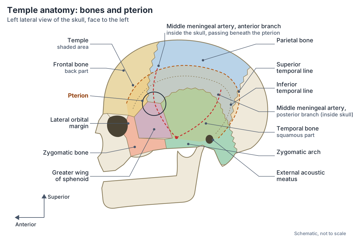
Bones of the temple region
| Bone | Part at the temple | Notes |
|---|---|---|
| Frontal bone | Back part of the bone, behind its zygomatic process | The zygomatic process forms part of the front border of the temporal fossa |
| Parietal bone | Front lower corner | Forms the upper part of the floor of the temporal fossa |
| Sphenoid bone | Lateral surface of the greater wing | Its infratemporal crest forms part of the lower border of the fossa |
| Temporal bone | Squamous part | Its zygomatic process forms the back of the zygomatic arch |
| Zygomatic bone | Frontal and temporal processes | Frontal process forms the front border; temporal process forms the front of the zygomatic arch |
The first four bones meet at the pterion. Compiled from Kenhub, Radiopaedia and OpenStax.
What is the pterion?
The pterion is the H-shaped meeting of sutures on the side of the skull where four bones join: the greater wing of the sphenoid, the squamous part of the temporal bone, the frontal bone and the parietal bone. It lies at the front end of the squamous suture. A clinical rule places it two fingers' breadth above the zygomatic arch and a thumb's breadth behind the frontal process of the zygomatic bone. (Radiopaedia: Pterion.)
OpenStax describes the pterion as the weakest part of the skull, with a major branch of an artery that supplies the skull and the coverings of the brain lying immediately deep to it on the inside. (OpenStax Anatomy and Physiology 2e, 7.2: The Skull.)
The pterion is covered by the scalp and by the origin of temporalis with its fascia, so it cannot be seen or felt through the skin. An osteometric study of 100 pterions placed its centre about 3.5 cm behind and 1.5 cm above the frontozygomatic suture and found the sphenoparietal type in 89%, the frontotemporal and stellate types in 4% each and the epipteric type in 3%. (Aggarwal N et al. Analysis of the Variations in the Morphology, Topography of the Pterion: An Osteometric Study. Asian J Neurosurg 2023.)
A 2025 meta-analysis of 79 studies (18,694 skull sides) found the sphenoparietal type in 78.5% of sides, the epipteric type in 8.4%, the frontotemporal type in 5.7% and the stellate type in 4.3%. On average the pterion lay 38.7 mm above the midpoint of the zygomatic arch and 31.3 mm from the frontozygomatic suture. Sex and side did not significantly change which type was present. (Triantafyllou G et al. Unveiling pterion variability: a meta-analytic approach. Surg Radiol Anat 2025.)
All 22 bones of the skull, the sutures and the fontanelles. Skull bones.
Types of pterion
| Type | How the bones meet | Pooled frequency (2025 meta-analysis, 18,694 sides) | Frequency in a 2023 study of 100 sides |
|---|---|---|---|
| Sphenoparietal | Greater wing of the sphenoid meets the parietal bone, forming an H | 78.5% | 89% |
| Epipteric | A small extra (sutural) bone sits at the junction | 8.4% | 3% |
| Frontotemporal | Frontal bone touches the squamous temporal bone directly | 5.7% | 4% |
| Stellate | All four bones meet at one point, forming a K | 4.3% | 4% |
Murphy's classification as defined by Aggarwal and colleagues (2023); pooled frequencies from Triantafyllou and colleagues (2025).
The temporal fossa: borders and contents
The superior temporal line, an arching ridge on the side of the skull, forms the upper and back margin of the temporal fossa and gives attachment to the temporal fascia. Below it, the inferior temporal line marks the highest point of the temporalis muscle. The temporal fascia forms the roof of the fossa, and below, deep to the zygomatic arch, the fossa opens into the infratemporal fossa, a route by which infection can spread. (Kenhub: Temporal fossa.)
Kenhub lists the contents of the temporal fossa as temporalis; the superficial temporal artery and vein, the middle temporal artery and the anterior, middle and posterior deep temporal arteries with their veins; and four nerves: the deep temporal nerves and auriculotemporal nerve from the mandibular nerve, the zygomaticotemporal nerve from the maxillary nerve, and the temporal branches of the facial nerve. (Kenhub: Temporal fossa.)
Radiopaedia gives the same picture in brief: the fossa contains mainly the temporalis muscle with the deep temporal arteries and nerves, the superficial temporal artery and the zygomaticotemporal nerve; the temporal fascia attaches to the superior temporal line and the muscle to the inferior temporal line. (Radiopaedia: Temporal fossa.)
Layers of the temple from skin to bone
Deep to the skin and subcutaneous tissue of the temple is the superficial temporal fascia, also called the temporoparietal fascia. At the superior temporal line it continues upwards as the galea aponeurotica of the scalp, and at the level of the zygoma it continues downwards as the superficial musculoaponeurotic system (SMAS) of the face. A flap of this fascia can be raised on the superficial temporal vessels. (Fagan J. Temporalis Muscle Flap. Open Access Atlas of Otolaryngology, Head and Neck Operative Surgery (LibreTexts).)
Beneath it lies the deep temporal fascia, a downward continuation of the pericranium, which covers temporalis from the superior temporal line. Below the level of the upper orbital margin it splits into superficial and deep layers that attach to the outer and inner margins of the zygomatic arch, with the superficial temporal fat pad between them. (Fagan J. Temporalis Muscle Flap. Open Access Atlas of Otolaryngology, Head and Neck Operative Surgery (LibreTexts).)
The temporal branch of the facial nerve, which surgeons often call the frontal branch, runs in the plane deep to the SMAS and superficial to the superficial layer of the deep temporal fascia. Its course is estimated with Pitanguy's line, from a point 0.5 cm below the tragus towards the eyebrow, passing about 1.5 cm above its lateral end. (Schleicher W, Feldman M, Rhodes J. Review of Facial Nerve Anatomy: Trauma to the Temporal Region. Eplasty 2013.)
Inside the skull, the lower part of the temple wall (the greater wing of the sphenoid and the temporal bone) forms the lateral part of the middle cranial fossa, which supports the temporal lobe of the brain. The middle meningeal artery enters this fossa through the foramen spinosum together with the middle meningeal vein and a meningeal branch of the mandibular nerve. (TeachMeAnatomy: The Middle Cranial Fossa.)
The five layers of the scalp, which continue into the layers of the temple. Scalp.
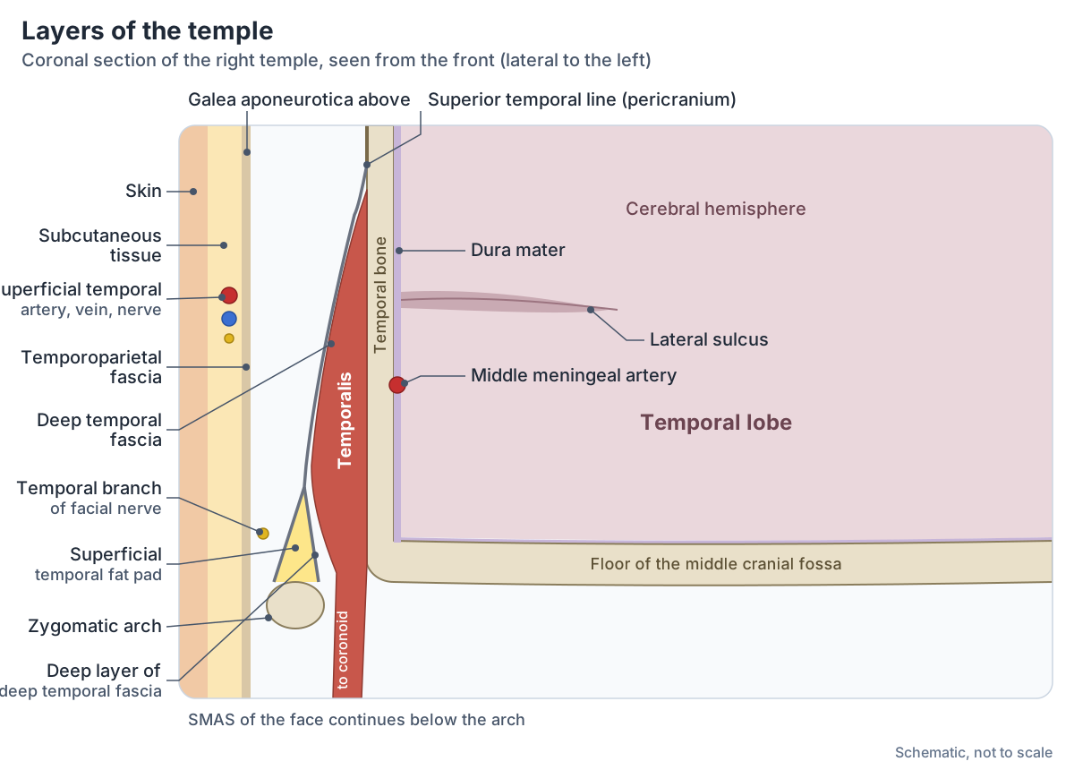
Layers of the temple from skin to brain
| Layer | What it is | Structures in or on it |
|---|---|---|
| Skin and subcutaneous tissue | Outermost layer of the temple | Sensory branches of the auriculotemporal and zygomaticotemporal nerves |
| Temporoparietal fascia (superficial temporal fascia) | Continuous with the galea aponeurotica above and the SMAS below | Superficial temporal vessels |
| Plane above the deep temporal fascia | Deep to the SMAS, superficial to the deep temporal fascia | Temporal (frontal) branch of the facial nerve |
| Deep temporal fascia | Attached to the superior temporal line; splits into two layers that attach to the zygomatic arch | Superficial temporal fat pad between its two layers |
| Temporalis muscle | Fan-shaped muscle filling the temporal fossa | Deep temporal nerves and arteries |
| Bone | Frontal, parietal, sphenoid and temporal bones, thin at the pterion | Pericranium on its outer surface |
| Dura mater | Outer covering of the brain, against the inner skull | Middle meningeal artery and its anterior branch |
| Brain | Temporal lobe in the middle cranial fossa | Lateral sulcus separates it from the frontal and parietal lobes |
Compiled from Fagan (Open Access Atlas of Otolaryngology), Schleicher and colleagues, Kenhub, Radiopaedia, TeachMeAnatomy and OpenStax.
Temporalis: the muscle of the temple
Temporalis arises from the whole floor of the temporal fossa below the temporal line and from the temporal fascia. Its anterior and posterior fibres converge on a narrow tendon that runs medial to the zygomatic arch and inserts on the apex and medial surface of the coronoid process of the mandible. (Kenhub: Temporalis muscle.)
The anterior fibres elevate the mandible and the posterior fibres pull it backwards (retrusion), so the muscle closes the mouth and, acting on one side, helps with side-to-side chewing. It is supplied by the anterior, middle and posterior deep temporal nerves from the mandibular nerve (V3) and by the deep temporal branches of the maxillary artery, with help from the middle temporal branch of the superficial temporal artery. (Kenhub: Temporalis muscle.)
In surgical descriptions temporalis is a fan-shaped muscle that fills the temporal fossa and inserts on the coronoid process and the front border of the ramus of the mandible. (Fagan J. Temporalis Muscle Flap. Open Access Atlas of Otolaryngology, Head and Neck Operative Surgery (LibreTexts).)
Temporalis, masseter and the pterygoids, with origins, insertions and actions. Muscles of mastication.
Superficial temporal artery and the temple pulse
The superficial temporal artery is one of the two terminal branches of the external carotid artery. It arises within the parotid gland, passes between its deep and superficial lobes and runs up over the zygomatic arch. Its branches are the frontal and parietal terminal branches, the transverse facial artery, the zygomatico-orbital artery, the middle temporal artery, auricular branches and a parotid branch. (Radiopaedia: Superficial temporal artery.)
It arises near the neck of the mandible and climbs in front of the tragus of the ear, crossing the zygomatic process with its vein and the auriculotemporal nerve. Where it crosses the zygomatic process, just in front of the ear, its pulse can be felt. It supplies the skin and muscles of the side of the face and scalp, the parotid gland and the temporomandibular joint. (Kenhub: Superficial temporal artery.)
The common, internal and external carotid arteries and their branches. Carotid arteries.
The middle meningeal artery under the pterion
The middle meningeal artery is the main blood supply of the dura mater around the brain. It arises from the first part of the maxillary artery, a terminal branch of the external carotid, and enters the middle cranial fossa through the foramen spinosum. There it divides: the anterior division runs over the greater wing of the sphenoid, often in a groove in the bone, and passes under the pterion, while the posterior division runs back over the squamous temporal bone. (Radiopaedia: Middle meningeal artery.)
This is why a blow to the temple can be serious. An extradural (epidural) haemorrhage is bleeding between the skull and the dura, usually after trauma strong enough to fracture the skull. Radiopaedia reports that about 75% are caused by a torn middle meningeal artery and about 75% have a skull fracture, that the temporoparietal region is the most common site (about 60%), and that the classic lucid interval is seen in fewer than 20% of cases. (Radiopaedia: Extradural haemorrhage.)
Because the pterion marks the position of the anterior branch of the middle meningeal artery, it is used as a landmark for burr holes to drain an extradural haematoma. The pterional approach is also popular in neurosurgery, partly because its skin incision lies behind the hairline. (Aggarwal N et al. Analysis of the Variations in the Morphology, Topography of the Pterion: An Osteometric Study. Asian J Neurosurg 2023.)
Nerves of the temple
Sensation from the skin of the temple is carried by branches of the trigeminal nerve: the zygomaticotemporal nerve from the maxillary division and the auriculotemporal nerve from the mandibular division. Further forward, the supraorbital and supratrochlear nerves from the ophthalmic division supply the forehead and front of the scalp. (TeachMeAnatomy: The Scalp.)
The facial nerve supplies the muscles of the temple region. It leaves the skull through the stylomastoid foramen, runs into the parotid gland and divides into five branches. The temporal branch crosses the temple to supply frontalis, orbicularis oculi and corrugator supercilii. (TeachMeAnatomy: The Facial Nerve (CN VII).)
In the Open Access Atlas of Otolaryngology, the frontal (temporal) branches are described as crossing the zygomatic arch and running over the superficial temporal fat pad, then passing deep to orbicularis oculi just lateral to the orbital rim. Temporalis itself is supplied separately, by the deep temporal nerves from the mandibular nerve. (Fagan J. Temporalis Muscle Flap. Open Access Atlas of Otolaryngology, Head and Neck Operative Surgery (LibreTexts).)
The trigeminal and facial nerves and the other ten cranial nerves. Cranial nerves.
The anatomy behind temple pain
Tension in temporalis is a common source of pain over the temple. Kenhub lists tooth and jaw misalignment, trauma, long periods of immobilisation, tooth grinding (bruxism) and long dental procedures with the mouth held open as causes. (Kenhub: Temporalis muscle.)
Giant cell arteritis is an inflammation of medium and large arteries that most often involves branches of the superficial temporal artery. It usually affects people over 50, with a peak between about 70 and 80, and is about twice as common in women. Headache is the most common symptom, with tenderness over the scalp or temporal artery and pain in the jaw on chewing; visual disturbance occurs in about 15%. Temporal artery biopsy is the reference test, and ultrasound may show a halo sign around the artery. (Radiopaedia: Giant cell arteritis.)
Pain from the jaw joint can also be felt around the temple. Cleveland Clinic lists headaches, jaw and face pain and clicking or locking of the jaw among the symptoms of temporomandibular joint disorders. Persistent or severe temple pain needs assessment by a clinician; this page describes anatomy only. (Cleveland Clinic: TMJ Disorders.)
The jaw joint just below the temple: disc, ligaments and movements. Temporomandibular joint (TMJ).
The temple in the 3D model
The 3D viewer highlights the frontal, parietal, temporal, sphenoid and zygomatic bones of the BodyParts3D model, an adult male. Turn to the left lateral view and look just above the zygomatic arch, behind the orbit: the pterion is where the greater wing of the sphenoid reaches up between the frontal, parietal and temporal bones. The model's skull has no separate sutures, and it does not include temporalis, the superficial temporal or middle meningeal arteries or the facial nerve, so use the diagrams for those layers. (Mitsuhashi N et al. BodyParts3D: 3D structure database for anatomical concepts. Nucleic Acids Res 2009.)
An overview of the whole head: skull, brain, face, sense organs, vessels and nerves. Head anatomy.
Temple in the 3D model
- Frontal bone
- The bone of the forehead and the roof of the orbits.
- Parietal bone
- A paired, curved bone forming the roof and sides of the skull.
- Sphenoid bone
- A butterfly-shaped bone at the base of the skull.
- Temporal bone
- A paired bone forming part of the side and base of the skull.
- Zygomatic bone
- The cheekbone.
Frequently asked questions
Where is your temple located?
The temple is on each side of the head between the outer corner of the eye and the ear. It is bounded below by the zygomatic arch, in front by the outer edge of the orbit, and above and behind by the temporal lines of the skull. It covers the temporal fossa, a shallow hollow filled by the temporalis muscle.
What bones make up the temple?
Four bones form the floor of the temple: the frontal bone, the parietal bone, the greater wing of the sphenoid and the squamous part of the temporal bone. They meet at the pterion. The zygomatic bone forms the front border, and the zygomatic arch, made by the zygomatic and temporal bones, forms the lower border.
What is the pterion?
The pterion is the H-shaped junction on the side of the skull where the frontal, parietal, sphenoid and temporal bones meet. It lies about two fingers' breadth above the zygomatic arch and a thumb's breadth behind the frontal process of the zygomatic bone. In most people the sphenoid meets the parietal bone here (the sphenoparietal type).
Why is the temple a weak spot of the skull?
The bone at the pterion is thin and is described as the weakest part of the skull. The anterior branch of the middle meningeal artery runs just inside it. A fracture here can tear the artery, so blood collects between the skull and the dura mater as an extradural haematoma, which can compress the brain and needs urgent treatment.
What artery is in the temple?
The superficial temporal artery, a terminal branch of the external carotid artery, runs up in front of the ear and across the temple, where its pulse can be felt over the zygomatic arch. It divides into frontal and parietal branches. Deeper, the deep temporal arteries supply temporalis, and inside the skull the middle meningeal artery runs beneath the pterion.
What muscle is in the temple?
Temporalis is the muscle of the temple. It is fan-shaped, arises from the floor of the temporal fossa and the temporal fascia, and passes deep to the zygomatic arch to insert on the coronoid process of the mandible. It closes the jaw and pulls it backwards, and it is supplied by the deep temporal nerves from the mandibular nerve.
What nerve supplies the skin of the temple?
The skin of the temple is supplied by two branches of the trigeminal nerve: the zygomaticotemporal nerve from the maxillary division and the auriculotemporal nerve from the mandibular division. The temporal branch of the facial nerve also crosses the temple, but it is a motor nerve to frontalis, orbicularis oculi and corrugator supercilii.
References
- Betts JG, et al. Anatomy and Physiology 2e, §7.2 The Skull. OpenStax, Rice University, 2022.
- Betts JG, et al. Anatomy and Physiology 2e, §13.2 The Central Nervous System. OpenStax, Rice University, 2022.
- IMAIOS e-Anatomy. Temporal region. Accessed 2026-10-24.
- Kenhub. Temporal fossa. Accessed 2026-10-24.
- Radiopaedia. Temporal fossa. Accessed 2026-10-24.
- Radiopaedia. Pterion. Accessed 2026-10-24.
- Aggarwal N, Kaur N, Patra A, Gupta M. Analysis of the Variations in the Morphology, Topography of the Pterion, and Their Implications in Neurosurgery: An Osteometric Study. Asian J Neurosurg. 2023;18(3):581-586.
- Triantafyllou G, Karangeli N, Duparc F, et al. Unveiling pterion variability: a meta-analytic approach to enhance neurosurgical precision. Surg Radiol Anat. 2025;47(1):66.
- Fagan J. Temporalis Muscle Flap. In: Open Access Atlas of Otolaryngology, Head and Neck Operative Surgery. University of Cape Town (LibreTexts).
- Schleicher W, Feldman M, Rhodes J. Review of Facial Nerve Anatomy: Trauma to the Temporal Region. Eplasty. 2013;13:ic54.
- Kenhub. Temporalis muscle. Accessed 2026-10-24.
- Radiopaedia. Superficial temporal artery. Accessed 2026-10-24.
- Kenhub. Superficial temporal artery. Accessed 2026-10-24.
- Radiopaedia. Middle meningeal artery. Accessed 2026-10-24.
- Radiopaedia. Extradural haemorrhage. Accessed 2026-10-24.
- TeachMeAnatomy. The Middle Cranial Fossa. Accessed 2026-10-24.
- TeachMeAnatomy. The Scalp. Accessed 2026-10-24.
- TeachMeAnatomy. The Facial Nerve (CN VII). Accessed 2026-10-24.
- Radiopaedia. Giant cell arteritis. Accessed 2026-10-24.
- Cleveland Clinic. TMJ Disorders. Accessed 2026-10-24.
- Mitsuhashi N, et al. BodyParts3D: 3D structure database for anatomical concepts. Nucleic Acids Res. 2009;37:D782-D785.
- Mitsuhashi N. et al. (2009). BodyParts3D: 3D structure database for anatomical concepts. Nucleic Acids Research 37, D782–D785. Data © The Database Center for Life Science, CC BY 4.0.
Related anatomy
- Skull bonesSkull anatomy with labelled diagrams and a 3D model: the 8 cranial and 14 facial bones, sutures, fontanelles, the…
- Temporal boneThe temporal bone (os temporale) explained: petrous part and inner ear, mastoid process, zygomatic and styloid…
- Sphenoid boneThe sphenoid bone (os sphenoidale): sella turcica and hypophyseal fossa, greater and lesser wings, pterygoid process…
- Muscles of masticationThe muscles of mastication explained: masseter, temporalis, medial and lateral pterygoid, with labelled diagrams, the…
- Temporomandibular joint (TMJ)TMJ anatomy: the condyle, fossa, articular disc, capsule and ligaments of the temporomandibular joint and how it…
- Head anatomyHead anatomy guide: skull, brain, face, sense organs and ENT structures, head and neck muscles, vessels, nerves and…
Explore the whole body in the interactive 3D human anatomy model, or browse the A–Z list of human structures.