

What is the temporomandibular joint (TMJ)?
The temporomandibular joint is the synovial joint between the mandible and the temporal bone. It lies in front of the tragus of the ear, and its surfaces are the mandibular fossa and articular tubercle of the squamous temporal bone above and the head of the mandible below. The bones never touch each other directly, because an articular disc lies between them. (TeachMeAnatomy: The Temporomandibular Joint.)
Everyone has two temporomandibular joints, one on each side of the jaw. You can feel them by placing your fingers just in front of your ears and opening your mouth. TMJ is the name of the joint itself; temporomandibular disorders (TMD) are the conditions that cause pain or dysfunction in it. (National Institute of Dental and Craniofacial Research: TMD (Temporomandibular Disorders).)
The TMJ is classified as a ginglymoarthrodial joint, a name that combines ginglymus (a hinge joint) and arthrodia (a gliding joint). The lower part of the joint hinges as the jaw opens and closes, while the upper part lets the condyle glide forwards and backwards between the fossa and the articular tubercle. (Elsevier Complete Anatomy: Temporomandibular joint.)
Bones of the TMJ: condyle, mandibular fossa and articular eminence
The upper part of the joint is formed by the temporal bone. The mandibular fossa is the deep, oval depression on the outer base of the skull, and the articular tubercle (articular eminence) is the smooth ridge immediately in front of it. The lower part is the condylar process of the mandible, the back projection of the ramus, which is topped by the oval condyle. The more anterior projection of the ramus is the flattened coronoid process. (OpenStax Anatomy and Physiology 2e, 7.2: The Skull.)
Unlike most synovial joints, whose surfaces are covered with hyaline cartilage, the TMJ surfaces are covered by fibrocartilage. Elsevier's anatomy notes point out that fibrocartilage is better able to withstand high forces, which suits a joint that carries chewing loads. (Elsevier Complete Anatomy: Temporomandibular joint.)
A small bony lip at the back of the fossa, the postglenoid tubercle, works with the strong lateral ligament to make posterior dislocation of the jaw very rare: a large force is needed to overcome them. (TeachMeAnatomy: The Temporomandibular Joint.)
The squamous, petrous, tympanic and mastoid parts, including the mandibular fossa. Temporal bone.
Parts of the temporomandibular joint
| Part | What it is | Role |
|---|---|---|
| Condyle (head of the mandible) | Rounded top of the condylar process of the mandible | Lower joint surface; rotates and glides under the disc |
| Mandibular fossa | Oval hollow on the underside of the squamous temporal bone, in front of the ear canal | Upper joint surface when the mouth is closed |
| Articular eminence (articular tubercle) | Smooth ridge of temporal bone just in front of the fossa | Upper joint surface during wide opening |
| Articular disc | Biconcave plate of fibrocartilage with anterior band, intermediate zone and posterior band | Separates the bones completely and divides the joint into two spaces |
| Retrodiscal tissue (bilaminar zone) | Loose, vascular, richly innervated tissue behind the disc with an upper and a lower lamina | Limits slipping and excessive rotation of the disc |
| Joint capsule | Fibrous sleeve lined by synovial membrane | Loose above the disc for gliding, tight below it so the disc moves with the condyle |
| Lateral (temporomandibular) ligament | Thickened lateral part of the capsule | Main stabilising ligament; resists posterior displacement |
Compiled from Radiopaedia, Kenhub, TeachMeAnatomy and OpenStax.
The TMJ articular disc
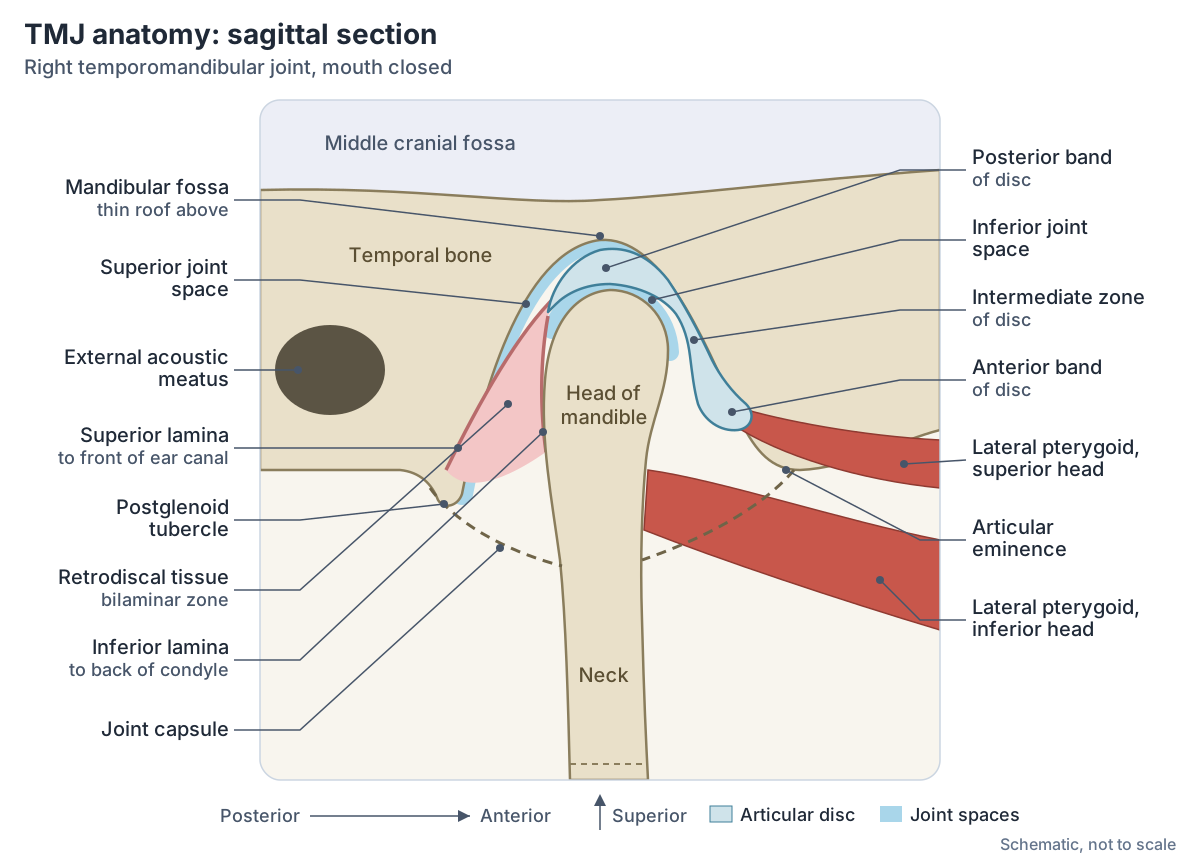
The articular disc is a biconcave plate of fibrocartilage, thin in the centre and thick around its edge. In side view it looks like a bow tie: a thick anterior band and a thick posterior band joined by a thin intermediate zone. It is tilted forwards by 10 to 30 degrees from the horizontal, attaches in front to the joint capsule, and is tied at its sides to the medial and lateral poles of the condyle. (Radiopaedia: Temporomandibular joint.)
With the mouth closed, the junction between the posterior band and the tissue behind it sits near the 11 or 12 o'clock position on top of the condyle. Radiologists call the disc anteriorly displaced when this junction lies further forwards, or when the posterior band is more than 10 degrees from the vertical line through the condyle. (Oliveira LRLB et al. Temporomandibular joint: from anatomy to internal derangement. Radiol Bras 2023.)
The disc is attached to the inner surface of the capsule and separates the joint surfaces completely. It stabilises the condyle, reduces friction and may help lubrication. The articular tissues and the dense part of the disc have no nerve supply. (Kenhub: Temporomandibular joint.)
Retrodiscal tissue (bilaminar zone)
Behind the disc, the posterior band splits into two layers: an upper fibroelastic lamina and a lower fibrous lamina. Between them is the loose, richly innervated, vascular retrodiscal (interlaminar) tissue, also called the bilaminar zone. It is best seen on MRI with the mouth open. (Radiopaedia: Temporomandibular joint.)
The upper layer, the temporal lamina, inserts into the front of the external auditory canal, and the lower layer inserts into the back of the condyle. Together they stop the disc slipping or rotating too far. (Oliveira LRLB et al. Temporomandibular joint: from anatomy to internal derangement. Radiol Bras 2023.)
The external acoustic meatus and middle ear that lie just behind the TMJ. Ear anatomy.
Joint capsule and the two joint spaces
The fibrous capsule arises from the borders of the mandibular fossa, encloses the articular tubercle, and attaches below to the neck of the mandible, just above the pterygoid fovea. It is loose, which allows the mandible to move forwards a long way without tearing capsular fibres. (Kenhub: Temporomandibular joint.)
Radiopaedia describes the capsule as loose above the disc, reaching the edges of the articular surface of the fossa so that the condyle can translate forwards, and tight below the disc, anchoring it to the condylar neck so that disc and condyle move together. (Radiopaedia: Temporomandibular joint.)
The disc divides the joint into two synovial spaces, each lined by its own synovial membrane. The upper (discotemporal) space, between the disc and the temporal bone, allows gliding: protrusion, retraction and side-to-side movement. The lower (discomandibular) space, between the disc and the condyle, allows rotation for opening and closing. (Radiopaedia: Temporomandibular joint.)
TMJ ligaments: lateral, sphenomandibular and stylomandibular
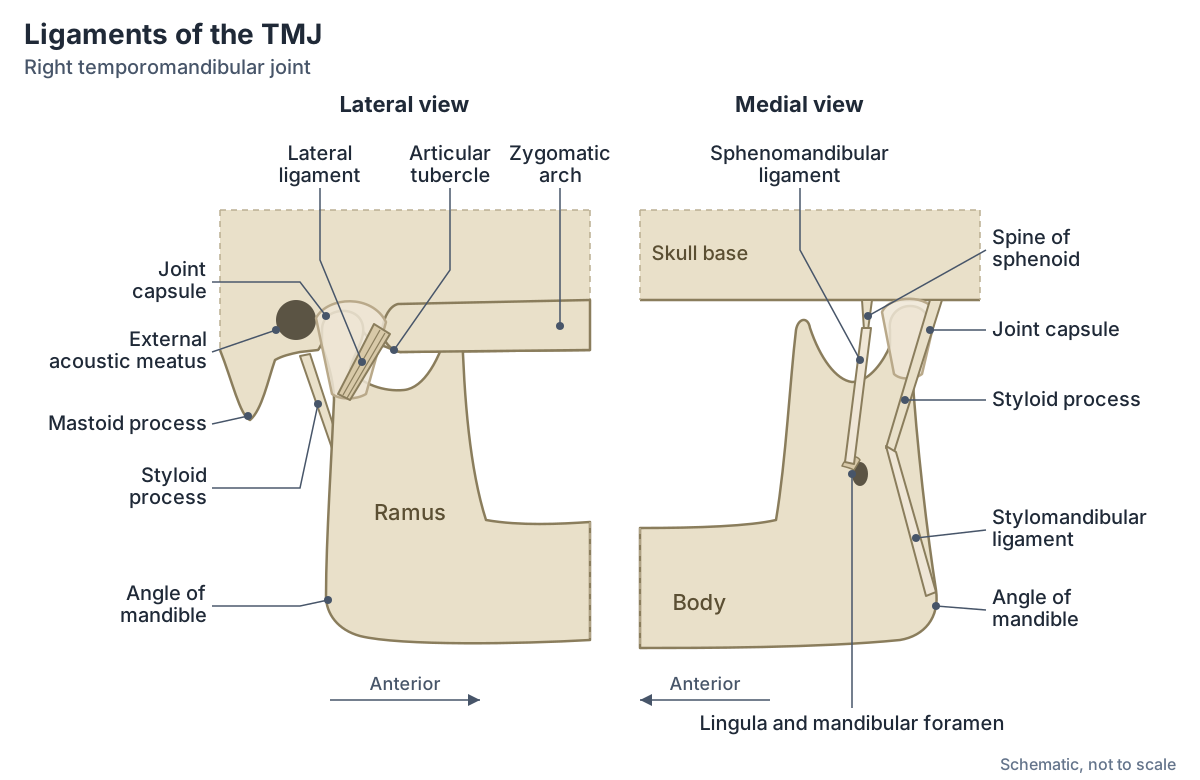
The lateral ligament, also called the temporomandibular ligament, is an intrinsic ligament: a thickened part of the capsule that strengthens the joint on its lateral side. Together with the postglenoid tubercle it prevents posterior dislocation. Kenhub describes the two other ligaments as extrinsic and says they give no significant support to the joint, although the sphenomandibular ligament acts as a swinging hinge for the mandible. (Kenhub: Temporomandibular joint.)
The sphenomandibular ligament descends from the spine of the sphenoid bone to the lingula on the inner surface of the ramus. It is pierced by the nerve to mylohyoid, and the auriculotemporal nerve and inferior alveolar vessels pass between it and the neck of the mandible. Radiopaedia calls it, with the muscles of mastication, the primary passive support of the mandible. (Radiopaedia: Sphenomandibular ligament.)
The stylomandibular ligament runs from the styloid process to the angle of the mandible. TeachMeAnatomy describes it as a thickening of the fascia of the parotid gland which, with the facial muscles, supports the weight of the jaw. The same page traces the lateral ligament from the articular tubercle to the neck of the mandible. (TeachMeAnatomy: The Temporomandibular Joint.)
Ligaments of the TMJ
| Ligament | Attachments | Type | Notes |
|---|---|---|---|
| Lateral (temporomandibular) ligament | Articular tubercle to the neck of the mandible | Intrinsic (part of the capsule) | Strengthens the joint laterally; with the postglenoid tubercle prevents posterior dislocation |
| Sphenomandibular ligament | Spine of the sphenoid to the lingula on the inner surface of the ramus | Extrinsic | Pierced by the nerve to mylohyoid; inferior alveolar vessels and auriculotemporal nerve pass between it and the mandibular neck |
| Stylomandibular ligament | Styloid process of the temporal bone to the angle of the mandible | Extrinsic | A thickening of the parotid fascia; adds little support |
Sources: Kenhub, TeachMeAnatomy and Radiopaedia. Small discal (collateral) ligaments also tie the disc to the medial and lateral poles of the condyle.
How the TMJ moves: hinge and glide
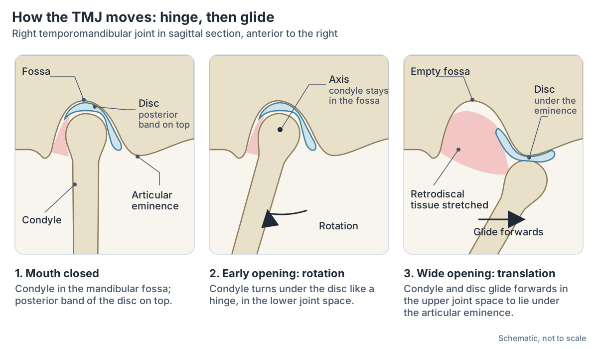
The mandible can be depressed, elevated, moved forwards and backwards, and moved slightly from side to side. IMAIOS explains that small openings, such as in ordinary speech, use only the lower joint, which works like a hinge. Wider opening also brings in the upper joint, where the disc and condyle glide forwards onto the articular tubercle. (IMAIOS e-Anatomy: Temporomandibular joint.)
The axis of rotation lies a little below the centre of the condyle, at the level of the insertion of the temporomandibular ligament. When the mouth opens fully, the condyle moves forwards with the disc and comes to lie beneath the articular eminence. (Radiopaedia: Temporomandibular joint.)
In real chewing and talking the two kinds of movement are combined rather than separate: rotation and translation usually happen at the same time. Rotation in the lower space can occur around axes in all three planes. (Kenhub: Temporomandibular joint.)
Side-to-side grinding is not a separate movement of its own. It is produced by alternating protrusion and retraction of the mandible on each side. (TeachMeAnatomy: The Temporomandibular Joint.)
How wide should your mouth open?
Maximal mouth opening is measured between the edges of the upper and lower incisors. In a study of 894 healthy Indian adults aged 21 to 70, the average was 51.3 mm in men and 44.3 mm in women, and opening became smaller with age in both sexes. (Khare N et al. Normal Mouth Opening in an Adult Indian Population. J Maxillofac Oral Surg 2012.)
A 2025 study of 400 Saudi adults found an average of 45.7 mm (47.9 mm in men and 44.3 mm in women) and reviewed studies of other populations, most of which report averages between about 40 and 55 mm. The authors note that trismus, restricted mouth opening, is commonly defined as a maximal opening of less than 35 mm. (Almohaimeed B, Ikram R. The association between maximum mouth opening and sociodemographic factors among Saudi adults. Front Oral Health 2025.)
Muscles that move the jaw at the TMJ
The TMJ is moved by the muscles of mastication and the hyoid muscles. Elevation, the strong closing movement, is produced by temporalis, masseter and the medial pterygoid. Depression is mostly due to gravity, helped by the digastric, geniohyoid and mylohyoid against resistance. Protrusion is by the lateral pterygoid, assisted by the medial pterygoid, and retraction by the posterior fibres of temporalis. (TeachMeAnatomy: The Temporomandibular Joint.)
The lateral pterygoid has a special relationship with the joint. Its upper fibres insert into the capsule and disc and its lower fibres into the pterygoid fovea on the condylar neck, so it pulls the disc and condyle forwards. Radiopaedia notes that the exact roles of the masticatory muscles in TMJ movement, and the lateral pterygoid's part in dysfunction, are still debated. (Radiopaedia: Temporomandibular joint.)
Masseter, temporalis and the medial and lateral pterygoids in detail. Muscles of mastication.
Jaw movements at the TMJ
| Movement | Joint space | Main muscles |
|---|---|---|
| Depression (opening) | Lower space (rotation), then upper space (glide) for wide opening | Gravity; lateral pterygoid; digastric, geniohyoid and mylohyoid against resistance |
| Elevation (closing) | Lower space (rotation); upper space as the condyle glides back | Temporalis, masseter, medial pterygoid |
| Protrusion (jaw forwards) | Upper space | Lateral pterygoid, helped by medial pterygoid |
| Retraction (jaw backwards) | Upper space | Posterior fibres of temporalis |
| Side-to-side (grinding) | Upper space (translation) | Alternating protrusion and retraction of each side |
Based on TeachMeAnatomy, Kenhub and Radiopaedia. Kenhub lists additional helpers for retraction and side-to-side movement.
Blood and nerve supply of the TMJ
The TMJ receives blood mainly from branches of the external carotid artery, especially the superficial temporal artery, with contributions from the deep auricular, ascending pharyngeal and maxillary arteries. Its nerves are the auriculotemporal and masseteric branches of the mandibular nerve (V3), the third division of the trigeminal nerve. (TeachMeAnatomy: The Temporomandibular Joint.)
Kenhub gives a slightly different emphasis: it names the deep auricular artery, a branch of the maxillary artery, as the main supplier, with the superficial temporal and anterior tympanic arteries, and lists the deep temporal nerves as a minor nerve supply. Veins drain to the superficial temporal and maxillary veins. (Kenhub: Temporomandibular joint.)
The trigeminal nerve and its mandibular division, which supplies the TMJ and the chewing muscles. Cranial nerves.
TMJ disorders (TMD) and jaw clicking
Temporomandibular disorders are a group of more than 30 conditions that cause pain and dysfunction in the jaw joint and the muscles that move it. The US National Institute of Dental and Craniofacial Research estimates that TMD affects about 5% of US adults and is twice as common in women as in men, especially women aged 35 to 44. Clicking or popping without pain is common and normal. (National Institute of Dental and Craniofacial Research: TMD (Temporomandibular Disorders).)
Symptoms include jaw and face pain, headaches, stiffness or locking of the jaw, and clicking, popping or grinding sounds. Cleveland Clinic notes that TMJ disorders most often begin between the ages of 20 and 40 and that women are about twice as likely to develop them. (Cleveland Clinic: TMJ Disorders.)
A 2023 review gives TMD figures of up to 28% of the general population and names internal derangement, an abnormal relationship between the disc and the condyle, as the main cause of TMJ dysfunction; its signs include joint pain, deviation of the jaw on opening and clicks. The disc can be displaced forwards, backwards, medially or laterally, with recapture (it returns to its place as the mouth opens) or without recapture. Small anterior displacements are seen in up to 33% of people with no symptoms, while displacement without recapture is often linked with a deformed disc, joint effusion or perforation. (Oliveira LRLB et al. Temporomandibular joint: from anatomy to internal derangement. Radiol Bras 2023.)
TMJ dislocation
In a TMJ dislocation the condyle loses contact with the mandibular fossa. Anterior dislocation is by far the most common: the condyle slides forwards over the articular eminence, either through an exaggerated, often recurrent, normal forward movement or through forceful opening of the mouth. Radiopaedia reports that up to 7% of the population dislocate the jaw at some point, most often between 20 and 40 years of age. Cranial dislocation is uncommon: an upward blow fractures the mandibular (glenoid) fossa and the condyle enters the middle cranial fossa. (Radiopaedia: Temporomandibular joint dislocation.)
A dislocated jaw can follow a big yawn, a large bite or a blow to the side of the face, and the person cannot close the mouth. The facial and auriculotemporal nerves run close to the joint and can be damaged in high-energy injuries. Posterior dislocation is very rare because of the postglenoid tubercle and the lateral ligament. (TeachMeAnatomy: The Temporomandibular Joint.)
The TMJ in the 3D model
The 3D viewer highlights the mandible, the temporal bone, the sphenoid and the zygomatic bone in the BodyParts3D model of an adult male. Turn to the left lateral view to find the condylar process sitting in the mandibular fossa just in front of the external acoustic meatus, with the articular eminence in front of it and the coronoid process passing under the zygomatic arch. Look from below to see the spine of the sphenoid, where the sphenomandibular ligament begins. The model has no articular disc, capsule, ligaments or masticatory muscles, so use the diagrams on this page for those. (Mitsuhashi N et al. BodyParts3D: 3D structure database for anatomical concepts. Nucleic Acids Res 2009.)
The lower jaw bone, its condylar and coronoid processes, ramus and foramina. Mandible.
Temporomandibular joint (TMJ) in the 3D model
- Mandible
- The lower jaw bone.
- Sphenoid bone
- A butterfly-shaped bone at the base of the skull.
- Temporal bone
- A paired bone forming part of the side and base of the skull.
- Zygomatic bone
- The cheekbone.
Frequently asked questions
What type of joint is the TMJ?
The TMJ is a synovial joint, classified as ginglymoarthrodial because it combines a hinge (ginglymus) with a gliding joint (arthrodia). It is atypical: its surfaces are covered with fibrocartilage instead of hyaline cartilage, and a fibrocartilage disc divides it into an upper gliding compartment and a lower hinge compartment.
Where is the temporomandibular joint located?
Each TMJ lies just in front of the tragus of the ear, where the condyle of the mandible fits into the mandibular fossa of the temporal bone. You can feel it move by placing a fingertip in front of the ear and opening and closing your mouth. There are two TMJs, one on each side of the jaw.
What are the 3 ligaments of the TMJ?
The three TMJ ligaments are the lateral (temporomandibular) ligament, which is a thickening of the capsule and the main stabiliser, and two extrinsic ligaments: the sphenomandibular ligament, from the spine of the sphenoid to the lingula of the mandible, and the stylomandibular ligament, from the styloid process to the angle of the mandible.
What does the TMJ disc do?
The articular disc is a bow-tie shaped plate of fibrocartilage between the condyle and the temporal bone. It separates the bones completely, divides the joint into upper and lower spaces, stabilises the condyle and reduces friction. It moves forwards with the condyle when the mouth opens wide, held back by the elastic bilaminar tissue behind it.
Why does my jaw click?
Clicking without pain is common and usually normal. One cause is a disc that sits slightly forwards and returns to its normal place on the condyle as the mouth opens, called disc displacement with recapture (reduction). Small displacements are seen in up to a third of people without symptoms. Painful clicking, locking or limited opening are signs of a temporomandibular disorder that a clinician should assess.
How wide should your mouth be able to open?
Healthy adults typically open about 40 to 55 mm between the edges of the upper and lower incisors. Studies find slightly larger openings in men than women, and opening decreases with age. Restricted opening, called trismus, is commonly defined as less than 35 mm.
Is TMJ the joint or the disorder?
TMJ is the name of the joint, the temporomandibular joint. The conditions that cause jaw pain, clicking or locking are temporomandibular disorders, shortened to TMD, although people often say they have TMJ when they mean TMD. TMD includes more than 30 conditions affecting the joint and the chewing muscles.
References
- Betts JG, et al. Anatomy and Physiology 2e, §7.2 The Skull. OpenStax, Rice University, 2022.
- TeachMeAnatomy. The Temporomandibular Joint. Accessed 2026-10-23.
- Kenhub. Temporomandibular joint. Accessed 2026-10-23.
- Radiopaedia. Temporomandibular joint. Accessed 2026-10-23.
- Radiopaedia. Sphenomandibular ligament. Accessed 2026-10-23.
- Radiopaedia. Temporomandibular joint dislocation. Accessed 2026-10-23.
- Elsevier Complete Anatomy. Temporomandibular joint. Accessed 2026-10-23.
- IMAIOS e-Anatomy. Temporomandibular joint. Accessed 2026-10-23.
- Oliveira LRLB, Alves IS, Vieira APF, Passos UL, Leite CC, Gebrim ES. Temporomandibular joint: from anatomy to internal derangement. Radiol Bras. 2023;56(2).
- National Institute of Dental and Craniofacial Research. TMD (Temporomandibular Disorders). Accessed 2026-10-23.
- Cleveland Clinic. TMJ Disorders. Accessed 2026-10-23.
- Khare N, Patil SB, Kale SM, et al. Normal Mouth Opening in an Adult Indian Population. J Maxillofac Oral Surg. 2012.
- Almohaimeed B, Ikram R. The association between maximum mouth opening and sociodemographic factors among Saudi adults: a cross-sectional study. Front Oral Health. 2025;6.
- Mitsuhashi N, et al. BodyParts3D: 3D structure database for anatomical concepts. Nucleic Acids Res. 2009;37:D782-D785.
- Mitsuhashi N. et al. (2009). BodyParts3D: 3D structure database for anatomical concepts. Nucleic Acids Research 37, D782–D785. Data © The Database Center for Life Science, CC BY 4.0.
Related anatomy
- MandibleMandible anatomy with a labelled diagram: body, ramus, angle, condylar and coronoid processes, foramina, muscle…
- Temporal boneThe temporal bone (os temporale) explained: petrous part and inner ear, mastoid process, zygomatic and styloid…
- Muscles of masticationThe muscles of mastication explained: masseter, temporalis, medial and lateral pterygoid, with labelled diagrams, the…
- TempleTemple anatomy: where the temple is, the four bones that meet at the pterion, temporalis, the temporal arteries and…
- Skull bonesSkull anatomy with labelled diagrams and a 3D model: the 8 cranial and 14 facial bones, sutures, fontanelles, the…
- Head anatomyHead anatomy guide: skull, brain, face, sense organs and ENT structures, head and neck muscles, vessels, nerves and…
Explore the whole body in the interactive 3D human anatomy model, or browse the A–Z list of human structures.