

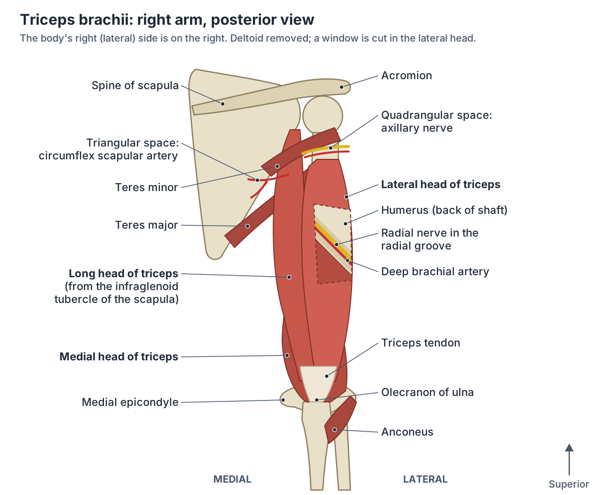
Where is the triceps brachii located?
The triceps brachii lies in the posterior compartment of the upper arm, the back of the arm between the shoulder and the elbow. TeachMeAnatomy describes the posterior compartment as containing the triceps brachii with its three heads, and its blood as coming from the profunda brachii (deep brachial) artery. (TeachMeAnatomy: Muscles of the Upper Arm.)
OpenStax names two extensors of the forearm at the elbow, triceps brachii and the small anconeus, and describes the triceps as the posterior muscle that extends the forearm, as during a punch. (OpenStax Anatomy and Physiology 2e, 11.5: Muscles of the Pectoral Girdle and Upper Limbs.)
The name gives the count. In muscle names, OpenStax explains, the prefix bi- means two origins and tri- means three, so triceps brachii is the three-headed muscle of the arm, and biceps brachii, in front of it, has two heads. (OpenStax Anatomy and Physiology 2e, 11.2: Naming Skeletal Muscles.)
All the muscles of the upper arm together: biceps, brachialis, coracobrachialis, triceps and the deltoid. Arm muscles.
The three heads of the triceps and their origins
The long head arises from the infraglenoid tubercle of the scapula, a rough area just below the glenoid cavity of the shoulder joint. It is the only head that starts on the scapula, so it is the only one that crosses the shoulder as well as the elbow. (University of Washington Musculoskeletal Atlas: Triceps brachii.)
Elsevier's Complete Anatomy adds that the long head arises by a flat tendon and that its upper end passes between teres major and teres minor. Kenhub notes that its attachment blends with the capsule of the shoulder joint. (Elsevier Complete Anatomy: Long head of triceps brachii.)
The lateral head arises from a narrow ridge on the back of the humerus just above the radial groove, the shallow channel that winds round the back of the bone. The medial head has a broad origin along the back of the humerus below the radial groove, and also from the medial and lateral intermuscular septa. (Kenhub: Triceps brachii muscle.)
The medial head is the deep head: it lies under the long and lateral heads, and the long head lies medial to the lateral head. Elsevier gives the lateral head an extra origin from the lateral intermuscular septum and notes that its upper end lies under the deltoid. (Elsevier Complete Anatomy: Lateral head of triceps brachii.)
Radiopaedia gives the same map and notes that the medial head lies largely deep to the lateral head, with the radial nerve and the profunda brachii vessels running in the radial groove between them. (Radiopaedia: Triceps brachii.)
Variations in the form of the triceps are uncommon. Radiopaedia lists a fourth head arising from the medial humerus. (Radiopaedia: Triceps brachii.)
A 2023 review of variants around the back of the elbow notes that this head lies superficial to the medial head and that only five cases have been described. A related variant is the dorsoepitrochlearis, a slip of latissimus dorsi found in about 1.6 to 10% of people, which may end on the triceps, the brachial fascia or the olecranon. Very rarely, about 1 in 1,940 people in one series, a sesamoid bone (patella cubiti) sits in the triceps tendon. (Kunc V et al. Anatomical variants complicating the posterior approaches towards the elbow joint. Surg Radiol Anat. 2023.)
The arm bone in 3D: the radial groove, surgical neck and the olecranon fossa. Humerus.
Where does the triceps insert?
All three heads join one tendon that inserts on the upper end of the olecranon, the point of the elbow on the ulna, and on the deep fascia of the forearm. The University of Washington atlas gives the insertion as the proximal end of the olecranon process of the ulna and the fascia of the forearm. (University of Washington Musculoskeletal Atlas: Triceps brachii.)
Kenhub places the common tendon on the back of the upper surface of the olecranon, with a band of fibres that passes laterally over anconeus to blend with the antebrachial (forearm) fascia. (Kenhub: Triceps brachii muscle.)
The medial head attaches a little differently. Radiopaedia says it inserts partly on the olecranon, partly into the deep surface of the common tendon and partly into the back of the elbow joint capsule. (Radiopaedia: Triceps brachii.)
Elsevier notes that the fibres reaching the capsule are sometimes described as a separate small muscle, articularis cubiti (subanconeus). (Elsevier Complete Anatomy: Medial head of triceps brachii.)
The olecranon, the elbow joint capsule and the ligaments around the triceps insertion. Elbow anatomy.
Triceps brachii: origin, insertion, nerve, action and blood supply
| Head | Origin | Insertion | Nerve (roots) | Main actions | Blood supply |
|---|---|---|---|---|---|
| Long head | Infraglenoid tubercle of the scapula (flat tendon) | Olecranon of the ulna via the common tendon; forearm fascia | Radial nerve; axillary nerve branch reported in some people | Extends elbow (mainly against resistance); extends and adducts arm at shoulder; steadies humeral head | Deep brachial artery |
| Lateral head | Ridge on posterior humerus above the radial groove; lateral intermuscular septum | Olecranon via the common tendon; forearm fascia | Radial nerve | Extends elbow (mainly against resistance) | Deep brachial and posterior circumflex humeral arteries |
| Medial head | Posterior humerus below the radial groove; medial and lateral intermuscular septa | Olecranon, deep surface of the tendon, posterior elbow capsule | Radial nerve | Extends elbow in every extension; retracts (pulls back) the elbow capsule | Deep brachial and superior ulnar collateral arteries |
| Whole muscle | Scapula (one head) and humerus (two heads) | Upper end of the olecranon of the ulna | Radial nerve, roots C6, C7, C8 | Chief extensor of the elbow; antagonist of biceps | Mainly the deep brachial (profunda brachii) artery |
Compiled from the University of Washington atlas, Kenhub, Radiopaedia, TeachMeAnatomy and Elsevier Complete Anatomy. Root values per head differ between sources, so roots are given for the whole muscle.
Nerve and blood supply of the triceps
The triceps is supplied by the radial nerve, with fibres from spinal roots C6, C7 and C8. The radial nerve, which comes from the posterior cord of the brachial plexus, supplies all three heads. (University of Washington Musculoskeletal Atlas: Triceps brachii.)
Each head gets its own branch. In 30 dissected arms, Mombellet and colleagues always found at least four motor branches: one to the long head, one to the lateral head and two to the medial head. All of them left the radial nerve between the lower edge of teres major and the radial groove, before the nerve reached the groove. (Mombellet M et al. Radial nerve branches to the triceps brachii: cadaveric analysis and surgical implications for neurotization. Surg Radiol Anat. 2026.)
TeachMeAnatomy describes the radial nerve leaving the axilla through the triangular interval and running down in the radial groove of the humerus, accompanied by the deep brachial artery. (TeachMeAnatomy: The Radial Nerve.)
Does the long head get a branch from the axillary nerve? TeachMeAnatomy says that in some people it does, and Radiopaedia cites a 2017 cadaver study by Erhardt and Futterman. A 2025 review calls axillary supply of the long head the most frequently observed variation, although textbooks describe the radial nerve as the only motor supply. (Mohel HK et al. Variable Innervation of the Triceps Brachii: Anatomical and Clinical Perspectives in a Narrative Review. Cureus. 2025.)
Not every study finds it: the 2026 dissection of 30 arms found no axillary nerve branch to the triceps at all. So the radial nerve is the textbook answer, with an axillary nerve contribution to the long head reported in some people. (Mombellet M et al. Radial nerve branches to the triceps brachii: cadaveric analysis and surgical implications for neurotization. Surg Radiol Anat. 2026.)
Kenhub and Elsevier both assign one root to each head, but other sources assign them differently, so this guide gives C6 to C8 for the muscle as a whole. (Kenhub: Triceps brachii muscle.)
The blood supply comes mainly from the deep brachial (profunda brachii) artery. Kenhub and Elsevier add the superior ulnar collateral artery, and both list the posterior circumflex humeral artery as an extra supply to the lateral head. (Elsevier Complete Anatomy: Lateral head of triceps brachii.)
The brachial plexus and the full course of the radial and axillary nerves. Nerves of the arm.
What does the triceps do?
The triceps is the chief extensor of the forearm at the elbow: it straightens the arm. Its long head also steadies the head of the humerus when the arm is abducted (raised out to the side). (University of Washington Musculoskeletal Atlas: Triceps brachii.)
Because the long head crosses the shoulder, it also extends the arm at the shoulder and, according to Kenhub, adducts it. Kenhub adds that it helps hold the head of the humerus in the glenoid cavity and stops it slipping downwards. (Kenhub: Triceps brachii muscle.)
The three heads do not work equally. The medial head is active in every elbow extension, with or without resistance, while the long and lateral heads join in mainly when the movement is resisted. Whether the palm faces up or down does not change the triceps' action at the elbow. (Elsevier Complete Anatomy: Medial head of triceps brachii.)
Radiopaedia adds a small job for the medial head: it pulls the back of the elbow joint capsule out of the way during extension. (Radiopaedia: Triceps brachii.)
The triceps is the antagonist of the biceps: the biceps brachii flexes the forearm, whereas the triceps brachii extends it. An antagonist is a muscle with the opposite action of the prime mover. (OpenStax Anatomy and Physiology 2e, 11.1: Interactions of Skeletal Muscles, Their Fascicle Arrangement, and Their Lever Systems.)
The two-headed elbow flexor and supinator on the front of the arm, the triceps' antagonist. Biceps brachii.
Relations: the quadrangular space, triangular space and triangular interval
The upper end of the long head of triceps lies between teres major and teres minor. Where it passes them, it forms one border of three gaps that carry nerves and vessels from the axilla to the back of the shoulder and arm. (Elsevier Complete Anatomy: Long head of triceps brachii.)
The quadrangular space lies lateral to the long head. Its borders are teres minor above, teres major below, the long head of triceps medially and the surgical neck of the humerus laterally. It carries the axillary nerve and the posterior circumflex humeral artery and vein. (TeachMeAnatomy: The Quadrangular Space.)
The triangular space lies medial to the long head, with teres minor above and teres major below. It contains the circumflex scapular artery and vein. (TeachMeAnatomy: The Triangular Space.)
The triangular interval lies lower, below teres major, between the long head of triceps and the humerus with the lateral head. It carries the radial nerve and the profunda brachii artery into the back of the arm. Compression of the radial nerve here, triangular interval syndrome, has been linked to enlarged triceps or teres major muscles or to fibrous bands. (TeachMeAnatomy: The Triangular Interval.)
Kenhub gives the same contents: the circumflex scapular vessels in the upper triangular space, the radial nerve and deep brachial vessels in the lower triangular space (the triangular interval), and the axillary nerve and posterior circumflex humeral vessels in the quadrangular space. (Kenhub: Triceps brachii muscle.)
These gaps can trap their contents. Quadrangular space syndrome is compression of the axillary nerve and posterior circumflex humeral artery in the quadrangular space, linked to enlarged muscles at its borders or fibrous bands between them; it causes pain and tingling in the axillary nerve's area and pain when the shoulder is abducted and externally rotated. (TeachMeAnatomy: The Quadrangular Space.)
The armpit region where the radial and axillary nerves begin their course to the back of the arm. Axilla.
Spaces around the long head of triceps
| Space | Borders | Contents |
|---|---|---|
| Quadrangular space (lateral to the long head) | Teres minor above, teres major below, long head of triceps medially, surgical neck of humerus laterally | Axillary nerve; posterior circumflex humeral artery and vein |
| Triangular space (medial to the long head) | Teres minor above, teres major below, long head of triceps laterally | Circumflex scapular artery and vein |
| Triangular interval (below teres major) | Teres major above, long head of triceps medially, humerus and lateral head of triceps laterally | Radial nerve; profunda brachii (deep brachial) artery and veins |
Based on TeachMeAnatomy (quadrangular space, triangular space, triangular interval) and Kenhub.
How is the triceps tested?
The triceps is tested by asking the person to straighten the elbow against resistance; the muscle can then be felt contracting at the back of the arm. (Elsevier Complete Anatomy: Long head of triceps brachii.)
Kenhub adds that with the elbow extended, the long head can be felt as a raised ridge running parallel to the back edge of the deltoid and just medial to it. (Kenhub: Triceps brachii muscle.)
The triceps reflex is checked by tapping the triceps tendon. TeachMeAnatomy states that this tests spinal segment C7. (TeachMeAnatomy: Muscles of the Upper Arm.)
A 2024 clinical review agrees: in C7 radiculopathy, a disorder of the C7 nerve root, the triceps reflex may be reduced or absent, and the review's table lists elbow extension as the movement of the C7 root. (Jajeh H et al. A clinical review of hand manifestations of cervical myelopathy, cervical radiculopathy, radial, ulnar, and median nerve neuropathies. J Spine Surg. 2024.)
Triceps tendon tears, snapping triceps and radial nerve injury
Triceps tendon tears are rare. A 2025 review puts distal triceps tendon injuries at about 1% of upper-limb tendon injuries, usually in men aged 30 to 60, and gives a fall on an extended elbow as the usual mechanism. (Manske RC et al. Diagnostic Musculoskeletal Ultrasound in the Evaluation of the Triceps Tendon. Int J Sports Phys Ther. 2025.)
A 2023 systematic review gives a similar picture: fewer than 0.8% of upper-limb tendon injuries, mostly in people aged 30 to 50, usually from resisted extension in a fall onto an extended hand. It lists chronic kidney failure and dialysis, hyperparathyroidism and steroid use among the risk factors. (Alkhalfan YH et al. The Elbow's Achilles Heel: A Systematic Review and Meta-Analysis of Triceps Tendon Rupture and Repair Techniques. Cureus. 2023.)
Radiopaedia notes that complete ruptures usually happen where the tendon attaches to bone, more often in men aged 30 to 50, and that kidney disease needing dialysis, diabetes, hyperparathyroidism and chronic olecranon bursitis raise the risk. A suspected tear needs prompt assessment by a clinician. (Radiopaedia: Triceps tendon rupture.)
Not every triceps tendon problem is a tear. The same 2025 review lists tendinopathy, partial tears, complete ruptures and bony injury, and notes that high-repetition, lower-load activity usually leads to tendinopathy. (Manske RC et al. Diagnostic Musculoskeletal Ultrasound in the Evaluation of the Triceps Tendon. Int J Sports Phys Ther. 2025.)
Kenhub describes overuse inflaming and damaging the tendon, with pain and swelling near its attachment to the olecranon. (Kenhub: Triceps brachii muscle.)
Radiopaedia adds that some ruptures happen in tendons already weakened by chronic tendinopathy, usually in older people with an underlying systemic illness. (Radiopaedia: Triceps tendon rupture.)
In snapping triceps syndrome, part of the distal triceps, usually its medial portion, slips over the medial epicondyle as the elbow bends from a straight position, producing a snap; it often occurs together with an unstable ulnar nerve. (Xia WZ et al. Snapping triceps syndrome: a review of the literature and proposed operative treatment algorithm. JSES Rev Rep Tech. 2026.)
Radiopaedia calls it an uncommon cause of pain on the inner (medial) side of the elbow, describes two palpable snaps during bending and straightening, and notes that it may coexist with dislocation of the ulnar nerve. (Radiopaedia: Snapping triceps syndrome.)
A fracture of the shaft of the humerus can damage the radial nerve in the radial groove and cause wrist drop, because the wrist and finger extensors in the forearm lose their supply. TeachMeAnatomy notes that the triceps may be weakened but is not paralysed, because the branches to its long and lateral heads leave the nerve above the groove. (TeachMeAnatomy: The Radial Nerve.)
In the 2026 dissection study all four triceps branches left the nerve before the groove, so elbow extension is usually preserved. (Mombellet M et al. Radial nerve branches to the triceps brachii: cadaveric analysis and surgical implications for neurotization. Surg Radiol Anat. 2026.)
Kenhub adds that a more proximal injury of the radial nerve, higher in the arm, can paralyse all three heads. (Kenhub: Triceps brachii muscle.)
The bones, compartments, nerves and vessels of the whole upper limb. Arm anatomy.
The triceps in the 3D model
From behind, the long head is the inner (medial) head and the lateral head the outer one, with the medial head lying deep to both. (Elsevier Complete Anatomy: Long head of triceps brachii.)
The 3D view opens from behind with the long, lateral and medial heads of the triceps highlighted over a ghosted skeleton, framed on the subject's left arm, where the long and lateral heads are labelled. The medial head lies deep to the other two, so it has no visible label from behind; click any head to open its own page. The model is BodyParts3D, built from one adult male; it has no peripheral nerves, so the radial nerve and the spaces are drawn in the diagram above.
Triceps brachii in the 3D model
- Lateral head of triceps brachii
- The lateral head of the triceps.
- Long head of triceps brachii
- The long head of triceps, crossing the shoulder joint.
- Medial head of triceps brachii
- The deep head of triceps, lying beneath the long and lateral heads.
Frequently asked questions
What are the three heads of the triceps?
The triceps has a long head, a lateral head and a medial head. The long head starts on the infraglenoid tubercle of the scapula, just below the shoulder socket. The lateral head starts on the back of the humerus above the radial groove, and the medial head below it. All three join one tendon on the olecranon, the point of the elbow.
What nerve supplies the triceps?
The radial nerve supplies all three heads of the triceps, with fibres from spinal roots C6, C7 and C8. It gives at least one branch to each head before it enters the radial groove of the humerus. Some cadaver studies also report a small branch from the axillary nerve to the long head in some people, but others find none.
Where does the triceps attach at the elbow?
The common triceps tendon attaches to the upper end of the olecranon, the bony point of the elbow at the top of the ulna, and sends fibres into the fascia of the forearm. Part of the medial head also attaches to the back of the elbow joint capsule, which it pulls out of the way when the elbow straightens.
What is the main function of the triceps?
The triceps straightens (extends) the elbow, as during a punch, and is the antagonist of the biceps. The long head also helps to extend the arm at the shoulder and steadies the head of the humerus when the arm is raised. The medial head works in every elbow extension; the other two join in mainly against resistance.
Which head of the triceps is the deepest?
The medial head is the deep head. It arises from the back of the humerus below the radial groove and lies under the long and lateral heads, so from behind you see mainly the long head on the inner side and the lateral head on the outer side. The radial nerve runs in the groove between the lateral and medial heads.
What passes through the quadrangular space?
The axillary nerve and the posterior circumflex humeral artery and vein. The quadrangular space is bounded by teres minor above, teres major below, the long head of triceps medially and the surgical neck of the humerus laterally. The radial nerve does not use it; it passes lower, through the triangular interval below teres major.
Can you tear your triceps tendon?
Yes, but it is rare, about 1% or less of upper-limb tendon injuries in published reviews. It usually happens at the tendon's attachment to the olecranon, most often in men aged roughly 30 to 50, typically after a fall onto an outstretched arm. Kidney failure needing dialysis and hyperparathyroidism raise the risk. A suspected tear should be assessed by a clinician.
References
- OpenStax. Anatomy and Physiology 2e, §11.5 Muscles of the Pectoral Girdle and Upper Limbs. Rice University, 2022.
- OpenStax. Anatomy and Physiology 2e, §11.1 Interactions of Skeletal Muscles, Their Fascicle Arrangement, and Their Lever Systems. Rice University, 2022.
- OpenStax. Anatomy and Physiology 2e, §11.2 Naming Skeletal Muscles. Rice University, 2022.
- University of Washington Department of Radiology. Musculoskeletal Atlas: Triceps brachii.
- Kenhub. Triceps brachii muscle.
- Radiopaedia. Triceps brachii.
- Radiopaedia. Triceps tendon rupture.
- Radiopaedia. Snapping triceps syndrome.
- TeachMeAnatomy. Muscles of the Upper Arm.
- TeachMeAnatomy. The Radial Nerve.
- TeachMeAnatomy. The Quadrangular Space.
- TeachMeAnatomy. The Triangular Space.
- TeachMeAnatomy. The Triangular Interval.
- Elsevier Complete Anatomy. Long head of triceps brachii.
- Elsevier Complete Anatomy. Lateral head of triceps brachii.
- Elsevier Complete Anatomy. Medial head of triceps brachii.
- Mombellet M, Destrieux C, Samargandi R, Bacle G. Radial nerve branches to the triceps brachii: cadaveric analysis and surgical implications for neurotization. Surg Radiol Anat. 2026;48:98.
- Mohel HK, Khorwal G, Sharma R, Sharma B. Variable innervation of the triceps brachii: anatomical and clinical perspectives in a narrative review. Cureus. 2025;17(12):e100537.
- Jajeh H, Lee A, Charls R, Coffin M, Sood A, Elgafy H. A clinical review of hand manifestations of cervical myelopathy, cervical radiculopathy, radial, ulnar, and median nerve neuropathies. J Spine Surg. 2024;10(1):120–134.
- Manske RC, Wolfe C, Page P, Voight M. Diagnostic musculoskeletal ultrasound in the evaluation of the triceps tendon. Int J Sports Phys Ther. 2025;20(6):911–917.
- Alkhalfan YH, Jha G, Verma B, et al. The elbow's Achilles heel: a systematic review and meta-analysis of triceps tendon rupture and repair techniques. Cureus. 2023;15(7):e41584.
- Kunc V, Benes M, Veigl D, Kachlik D. Anatomical variants complicating the posterior approaches towards the elbow joint. Surg Radiol Anat. 2023;45(5):587–592.
- Xia WZ, Abukar A, Moosavi H, Nandi N, Rashid A. Snapping triceps syndrome: a review of the literature and proposed operative treatment algorithm. JSES Rev Rep Tech. 2026;6(1):100580.
- Mitsuhashi N. et al. (2009). BodyParts3D: 3D structure database for anatomical concepts. Nucleic Acids Research 37, D782–D785. Data © The Database Center for Life Science, CC BY 4.0.
Related anatomy
- Arm musclesArm muscles labeled in 3D: biceps and the other elbow flexors, coracobrachialis, triceps, deltoid and anconeus, with…
- Biceps brachiiBicep anatomy with a 3D model and labelled diagram: long and short heads, origins, insertion, nerve supply, actions…
- Elbow jointElbow anatomy with labelled diagrams and a 3D model: humerus, radius and ulna, the three joints, collateral and…
- HumerusHumerus bone labeled: head, anatomical and surgical neck, tubercles, deltoid tuberosity, radial groove, epicondyles…
- Upper limb nervesBrachial plexus diagram with roots, trunks, cords and branches; axillary, musculocutaneous, median, ulnar and radial…
- Shoulder jointShoulder anatomy with labelled diagrams and a 3D model: clavicle, scapula and humerus, the glenohumeral, AC and SC…
Explore the whole body in the interactive 3D human anatomy model, or browse the A–Z list of human structures.