

What is the axilla (armpit)?
The axilla is a pyramidal region below the glenohumeral (shoulder) joint. Seen in three dimensions it looks roughly like a pyramid, with four walls, an apex and a base. (TeachMeAnatomy: The Axilla.)
Kenhub defines the axillary region as the area between the upper part of the upper limb and the thorax, and notes that the space changes shape as the arm moves. (Kenhub: Axillary region.)
Its main job is as a passageway: Lecturio describes the axilla as a pyramid-shaped space below the shoulder joint that is the passageway for nerves and vessels. (Lecturio: Axilla and Brachial Plexus.)
In everyday terms Cleveland Clinic describes the axilla as the area between the chest and the upper arm. Its skin carries hair and two kinds of sweat glands: eccrine glands that release a clear, odourless fluid and apocrine glands whose thicker secretion produces odour when broken down by bacteria. (Cleveland Clinic: Axilla (Armpit).)
OpenStax confirms that apocrine sweat glands are usually associated with hair follicles in densely hairy areas such as the armpits and genital regions, and that their sweat contains organic compounds that bacteria decompose, causing the smell. (OpenStax Anatomy and Physiology 2e, 5.2: Accessory Structures of the Skin.)
The whole upper limb whose vessels, nerves and lymph pass through the axilla. Arm anatomy.
Walls, apex and base of the axilla
The apex, or axillary inlet, is bounded by the clavicle in front, the first rib medially and the superior border of the scapula behind. (TeachMeAnatomy: The Axilla.)
The anterior wall is formed by pectoralis major with pectoralis minor and subclavius beneath it. The posterior wall is formed by subscapularis, teres major and latissimus dorsi. The medial wall is serratus anterior over the thoracic wall (ribs and intercostal muscles). The narrow lateral wall is the intertubercular groove of the humerus, with coracobrachialis and the short head of biceps brachii. (TeachMeAnatomy: The Axilla.)
Kenhub and Lecturio give the lateral wall simply as the intertubercular sulcus of the humerus, without the two muscles, so the difference is one of detail. Kenhub notes that the lower edges of the anterior and posterior walls form the folds of the armpit: the anterior axillary fold is formed by the pectoral muscles, and the posterior fold by latissimus dorsi and teres major, with subscapularis above them. (Kenhub: Axillary region.)
Kenhub describes the apex as bounded above by the root of the neck, and the base, or floor, as the axillary fascia and the skin over it. (Kenhub: Axillary region.)
Lecturio describes the base as the hair-bearing and sweat gland-bearing skin of the armpit. (Lecturio: Axilla and Brachial Plexus.)
The axillary fascia is often called the suspensory ligament of the axilla. It tethers the skin, which gives the armpit its hollow, bowl-like shape. (Kenhub: Fascias and spaces of the shoulder girdle region.)
Pectoralis major, pectoralis minor, subclavius and serratus anterior, the front and inner walls of the axilla. Chest muscles.
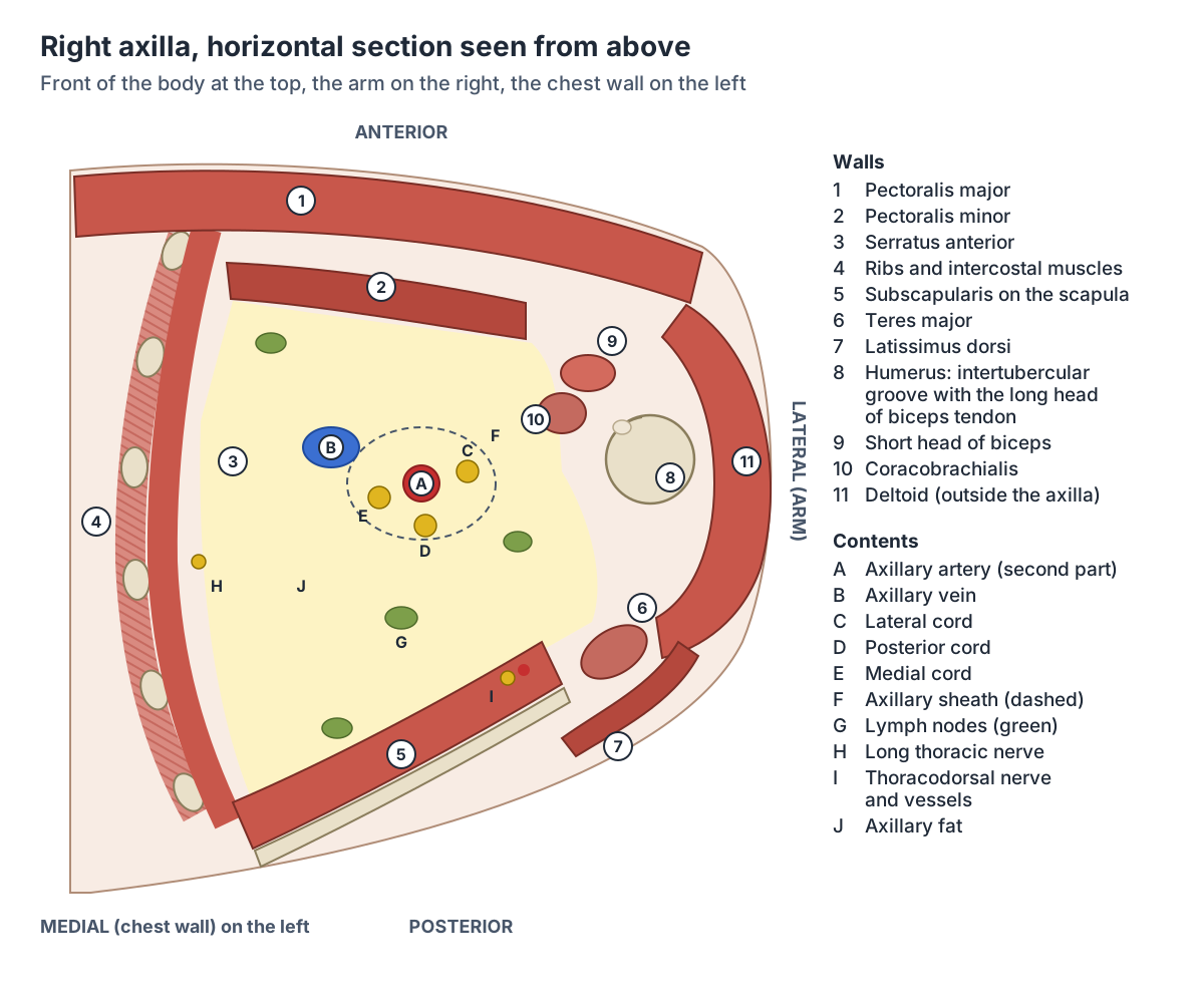
Boundaries of the axilla
| Boundary | Formed by | Notes |
|---|---|---|
| Apex (axillary inlet) | Clavicle (front), first rib (medial), superior border of the scapula (behind) | Opens into the root of the neck; artery and plexus enter here |
| Base (floor) | Axillary fascia and hair-bearing skin | The suspensory ligament of the axilla gives the armpit its hollow |
| Anterior wall | Pectoralis major, pectoralis minor, subclavius | Lower edge forms the anterior axillary fold |
| Posterior wall | Subscapularis, teres major, latissimus dorsi | Lower edge (latissimus dorsi and teres major) forms the posterior axillary fold |
| Medial wall | Serratus anterior over the upper ribs and intercostal muscles | Long thoracic nerve runs on serratus anterior |
| Lateral wall | Intertubercular groove of the humerus (with coracobrachialis and the short head of biceps) | The narrowest wall; some sources list the groove alone |
From TeachMeAnatomy, Kenhub (axillary region and shoulder fascias) and Lecturio.
Contents of the axilla
Embedded in axillary fat and enclosed within the axillary sheath, the contents of the axilla are the axillary artery and its branches, the axillary vein and its tributaries, the branches of the brachial plexus and the axillary lymph nodes. (Lecturio: Axilla and Brachial Plexus.)
The axillary sheath is a fibrous layer that wraps the axillary artery and the three cords of the brachial plexus. (TeachMeAnatomy: Arterial Supply to the Upper Limb.)
Radiopaedia explains where the sheath comes from: the prevertebral fascia of the neck extends down to ensheath the axillary artery and the cords of the plexus. (Radiopaedia: Brachial plexus.)
Kenhub describes the axilla as filled with adipose tissue that allows the passage of vessels, the nerve plexus, lymphatics and muscles, and points out that the axillary vein runs medial to the artery along its whole length. (Kenhub: Axillary region.)
The axillary artery and its three parts
At the lateral border of the first rib the subclavian artery enters the axilla and becomes the axillary artery. At the lower border of teres major it leaves the axilla and is renamed the brachial artery. (TeachMeAnatomy: Arterial Supply to the Upper Limb.)
Pectoralis minor crosses in front of the artery and divides it into three parts: the first part above (proximal to) pectoralis minor, the second part behind it and the third part below (distal to) it. Radiopaedia notes that the artery descends posterior to the axillary vein and that its branches are highly variable. (Radiopaedia: Axillary artery.)
The number of branches matches the number of the part: one from the first part (superior thoracic artery), two from the second (thoraco-acromial and lateral thoracic arteries) and three from the third (subscapular, anterior circumflex humeral and posterior circumflex humeral arteries). (Lecturio: Axilla and Brachial Plexus.)
The superior thoracic artery supplies the first and second intercostal spaces. (Kenhub: Axillary region.)
The full course, branches and variants of the main artery of the axilla. Axillary artery.
Parts of the axillary artery and their branches
| Part | Position | Branches |
|---|---|---|
| First | Above (proximal to) pectoralis minor | Superior thoracic artery |
| Second | Behind pectoralis minor; the cords surround it | Thoraco-acromial artery, lateral thoracic artery |
| Third | Below (distal to) pectoralis minor, to the lower border of teres major | Subscapular artery (gives thoracodorsal and circumflex scapular), anterior and posterior circumflex humeral arteries |
From Radiopaedia, TeachMeAnatomy, Lecturio and Kenhub.
The axillary vein
The axillary vein forms at the lower border of teres major from the union of the paired brachial veins and the basilic vein. It runs up through the axilla on the medial side of the artery and becomes the subclavian vein at the lateral border of the first rib. (Radiopaedia: Axillary vein.)
The cephalic vein joins the first part of the axillary vein above pectoralis minor after piercing the clavipectoral fascia, and five other tributaries correspond to the branches of the artery. About 14% of people have a double axillary vein; the lower of the two can be at risk during axillary surgery for breast cancer. (Radiopaedia: Axillary vein.)
Kenhub agrees that the vein is formed by the basilic and brachial veins. Some summaries, such as Lecturio's, describe it as formed by the cephalic and basilic veins; the cephalic vein actually joins higher up, so the Radiopaedia and Kenhub description is the one to learn. (Kenhub: Axillary region.)
The subclavian, axillary and brachial vessels and the cephalic and basilic veins. Arteries and veins of the arm.
Brachial plexus cords in the axilla
The brachial plexus is formed by the anterior rami of spinal nerves C5 to T1. Its roots form three trunks at the base of the neck; each trunk splits into an anterior and a posterior division, and in the axilla the divisions join to form three cords. (TeachMeAnatomy: The Brachial Plexus.)
The divisions form behind the middle of the clavicle, and the cords are named for their relationship to the second part of the axillary artery, the part behind pectoralis minor: lateral, posterior and medial. (Radiopaedia: Brachial plexus.)
The lateral cord comes from the anterior divisions of the superior and middle trunks and gives the lateral pectoral nerve, the musculocutaneous nerve and the lateral root of the median nerve. The medial cord comes from the anterior division of the inferior trunk and gives the medial pectoral nerve, the medial cutaneous nerves of the arm and forearm, the ulnar nerve and the medial root of the median nerve. The posterior cord collects all three posterior divisions and gives the upper and lower subscapular nerves, the thoracodorsal nerve, and the axillary and radial nerves. (TeachMeAnatomy: The Brachial Plexus.)
The thoracodorsal nerve (C6 to C8) from the posterior cord supplies latissimus dorsi. The long thoracic nerve (C5 to C7) arises directly from the roots and supplies serratus anterior on the medial wall. (Kenhub: Axillary region.)
The long thoracic nerve runs behind the axillary vessels and the brachial plexus and then lies on the surface of serratus anterior for its whole course, sending a branch to every slip of the muscle. It has a mean length of 27 cm. (Gooding BWT et al. Scapular winging. Shoulder Elbow. 2014.)
The intercostobrachial nerve crosses the axilla to supply the skin of the axilla, the lateral chest and the medial arm. A meta-analysis of 16 studies found that it arose from the second intercostal nerve in 90.6% of cases and communicated with the brachial plexus in about 41%. (Henry BM et al. Origin, branching, and communications of the intercostobrachial nerve: a meta-analysis. Cureus. 2017.)
The brachial plexus from roots to terminal branches and the course of each nerve. Nerves of the arm.
Cords of the brachial plexus in the axilla
| Cord | Formed from | Position (second part of the axillary artery) | Branches |
|---|---|---|---|
| Lateral | Anterior divisions of the superior and middle trunks (C5 to C7) | Lateral | Lateral pectoral nerve; musculocutaneous nerve; lateral root of the median nerve |
| Posterior | Posterior divisions of all three trunks (C5 to T1) | Posterior | Upper and lower subscapular nerves; thoracodorsal nerve; axillary nerve; radial nerve |
| Medial | Anterior division of the inferior trunk (C8, T1) | Medial | Medial pectoral nerve; medial cutaneous nerves of arm and forearm; ulnar nerve; medial root of the median nerve |
From TeachMeAnatomy and Radiopaedia (brachial plexus). Root values follow from the trunks each cord receives.
Axillary lymph nodes: groups and levels
There are about 20 to 30 axillary lymph nodes, and up to 40 have been recorded. They drain the whole upper limb, the breast and the trunk above the umbilicus. (Kenhub: Axillary lymph nodes.)
Anatomists divide them into five groups. The lateral (humeral) group of 4 to 6 nodes lies posteromedial to the axillary vein and receives most of the lymph from the upper limb. The anterior (pectoral) group lies along the lower border of pectoralis minor and drains the anterolateral abdominal wall above the umbilicus and the lateral breast. The posterior (subscapular) group of 6 to 7 nodes drains the back of the neck and upper back. The central group, at the base and centre of the axilla, receives lymph from these three. The apical group of 4 to 5 nodes, at the lateral border of the first rib, receives lymph from all the others. (Kenhub: Axillary lymph nodes.)
From the apical nodes lymph drains into the subclavian lymph trunk. The two sides differ: on the left the lymph reaches the thoracic duct, on the right the right lymphatic trunk. (Kenhub: Axillary lymph nodes.)
Radiopaedia calls the apical nodes the final common pathway for all the axillary nodes and notes lymphatic connections between the axillary and cervical nodes. (Radiopaedia: Axillary lymph nodes.)
Surgeons and radiologists use three levels instead of five groups, defined by pectoralis minor: level I nodes lie below the lower edge of pectoralis minor, level II nodes lie behind it and level III nodes lie above and medial to it. (Radiopaedia: Axillary lymph nodes.)
Lecturio adds a sixth, infraclavicular (deltopectoral) group, which lies outside the axilla and drains the superficial layers of the upper limb. (Lecturio: Axilla and Brachial Plexus.)
Lymph nodes throughout the body, how lymph flows and where the axillary nodes fit in. Lymphatic system.
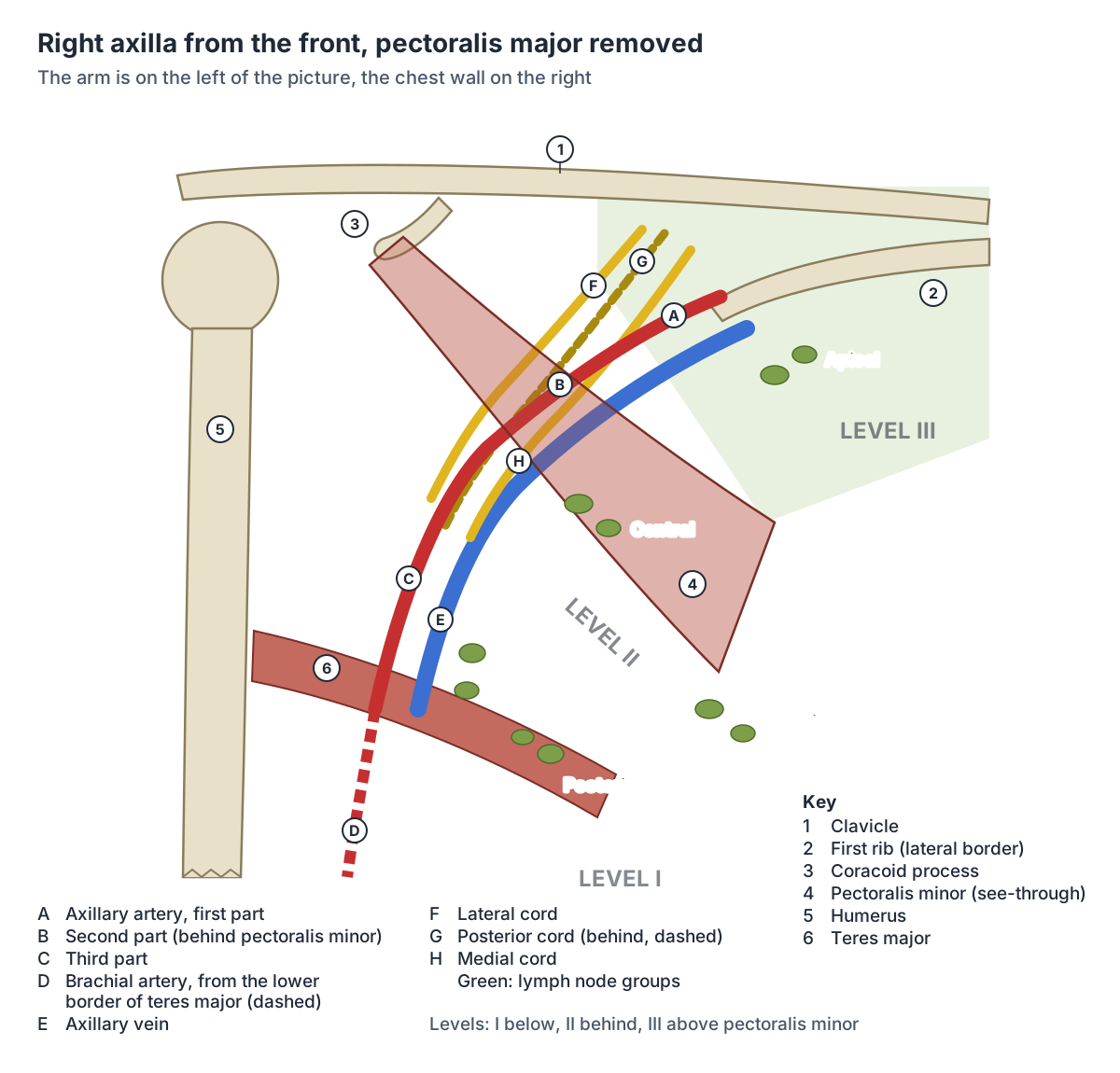
Axillary lymph node groups
| Group | Position | Number (Kenhub) | Main drainage |
|---|---|---|---|
| Lateral (humeral) | Along the axillary vein | 4 to 6 | Most of the upper limb |
| Anterior (pectoral) | Along the lower border of pectoralis minor | Not given | Anterolateral abdominal wall above the umbilicus; lateral breast |
| Posterior (subscapular) | Along the posterior wall | 6 to 7 | Back of the neck and upper back |
| Central | Base and centre of the axilla | Not given | Receives the lateral, anterior and posterior groups |
| Apical (subclavicular) | Lateral border of the first rib, at the apex | 4 to 5 | Receives all other groups; drains to the subclavian lymph trunk |
From Kenhub (axillary lymph nodes) and Radiopaedia. Surgical levels: I below, II behind and III above pectoralis minor.
Quadrangular space, triangular space and triangular interval
Three gaps in the posterior wall let structures pass from the axilla to the back of the shoulder and arm. The quadrangular space is bounded by subscapularis (teres minor from behind) above, teres major below, the long head of triceps medially and the surgical neck of the humerus laterally. The axillary nerve and the posterior circumflex humeral artery pass through it. (Kenhub: Anatomical spaces of the pectoral region.)
The triangular space carries the circumflex scapular artery to the scapular anastomoses. The triangular interval lies outside the axilla, below the lower border of teres major, and carries the radial nerve and the profunda brachii artery from the front to the back of the arm. (Kenhub: Anatomical spaces of the pectoral region.)
Their borders: the triangular space lies between teres minor above, teres major below and the long head of triceps; the triangular interval lies between teres major above and the long head of triceps medially and the lateral head laterally. (Kenhub: Fascias and spaces of the shoulder girdle region.)
Compression of the axillary nerve and posterior circumflex humeral artery in the quadrangular space causes quadrilateral space syndrome, an uncommon condition that mostly affects athletes. (Kenhub: Anatomical spaces of the pectoral region.)
The shoulder muscle supplied by the axillary nerve after it leaves the quadrangular space. Deltoid.
Clinical anatomy of the armpit
About 75% of the lymph from the breast drains into the axillary nodes, which is why they are examined and sampled in breast cancer. During removal of the axillary nodes (axillary clearance) the long thoracic nerve may be damaged, leading to a winged scapula. (TeachMeAnatomy: The Axilla.)
In breast cancer surgery the sentinel node, the first node that drains the breast, is identified and checked; if it contains cancer, the surrounding nodes may be removed. Removing axillary nodes carries a risk of lymphoedema, swelling of the arm and breast. (Kenhub: Axillary lymph nodes.)
A 2022 meta-analysis of 67 studies found arm lymphoedema in about 24% of patients more than two years after axillary lymph node dissection, compared with about 6% after sentinel node biopsy alone. (Che Bakri NA et al. Impact of axillary lymph node dissection and sentinel lymph node biopsy on upper limb morbidity in breast cancer patients. Ann Surg. 2023.)
Injury to the intercostobrachial nerve during axillary surgery is linked with numbness and nerve pain in the armpit and inner arm, and an estimated 10 to 60% of breast cancer survivors who had surgery report persistent pain. (Henry BM et al. Origin, branching, and communications of the intercostobrachial nerve: a meta-analysis. Cureus. 2017.)
Prolonged pressure on the axilla can compress the radial nerve and cause wrist drop. Injury to the long thoracic nerve leaves serratus anterior unable to hold the scapula against the chest wall, so the scapula wings. (Kenhub: Axillary region.)
At the apex, the vessels and nerves can be compressed between the bones that bound it, a cause of thoracic outlet syndrome. (TeachMeAnatomy: The Axilla.)
How lymph drains from the breast to the axillary nodes, and the axillary tail of the breast. Breast anatomy.
The axilla in the 3D model
The model opens from the front and highlights the axillary artery and vein with the thoraco-acromial trunk, the lateral thoracic, subscapular, thoracodorsal, circumflex scapular and both circumflex humeral arteries, and the matching veins, together with the muscles of the walls: pectoralis minor, subscapularis, teres major, serratus anterior, coracobrachialis and the short head of biceps. Pectoralis major is left unhighlighted so that you can see in. Rotate the model to look up into the armpit from below and the pyramid shape becomes clear. The model, BodyParts3D from one adult male, has no brachial plexus, lymph nodes or latissimus dorsi, so use the diagrams for those.
Follow the artery from the first rib to teres major and find pectoralis minor crossing in front of its middle: that crossing defines the three parts of the artery. (Radiopaedia: Axillary artery.)
Axilla (armpit) in the 3D model
- Anterior circumflex humeral artery
- The anterior circumflex humeral artery is a small branch of the axillary artery that wraps around the front of the surgical neck of the humerus.
- Axillary artery
- The axillary artery is the main artery of the armpit, continuing the subclavian artery.
- Axillary vein
- The axillary vein is the main vein of the armpit and carries blood from the upper limb.
- Circumflex scapular artery
- The circumflex scapular artery is the large branch of the subscapular artery that passes round the lateral border of the scapula.
- Coracobrachialis
- A slender muscle on the medial side of the upper arm.
- Lateral thoracic artery
- A branch of the axillary artery that runs along the lateral chest wall.
- Lateral thoracic vein
- A vein draining the lateral chest wall into the axillary vein.
- Pectoralis minor
- A thin triangular muscle lying deep to pectoralis major on the upper chest wall.
- Posterior circumflex humeral artery
- A branch of the axillary artery that wraps around the surgical neck of the humerus.
- Serratus anterior
- A broad muscle on the side of the thorax that holds the scapula against the chest wall.
- Short head of biceps brachii
- The medial head of biceps brachii at the front of the arm.
- Subscapular artery
- The largest branch of the axillary artery.
- Subscapular vein
- A vein that drains the scapular region into the axillary vein.
- Subscapularis
- The largest rotator cuff muscle, filling the subscapular fossa.
- Teres major
- A thick muscle of the posterior shoulder, sometimes called the lat's little helper.
- Thoracodorsal artery
- A branch of the subscapular artery to latissimus dorsi.
- Thoracodorsal vein
- A vein accompanying the thoracodorsal artery.
- Trunk of thoraco-acromial artery
- The main stem of the thoraco-acromial artery.
Frequently asked questions
What are the boundaries of the axilla?
The axilla has an apex, a base and four walls. The apex lies between the clavicle, the first rib and the upper border of the scapula. The base is the armpit skin and axillary fascia. The anterior wall is pectoralis major and minor, the posterior wall subscapularis, teres major and latissimus dorsi, the medial wall serratus anterior over the ribs and the lateral wall the intertubercular groove of the humerus.
What are the contents of the axilla?
The axilla contains the axillary artery and its branches, the axillary vein and its tributaries, the cords and branches of the brachial plexus and the axillary lymph nodes, all embedded in fat. The artery and the three cords are wrapped together in the axillary sheath, which is continuous with the prevertebral fascia of the neck. The vein lies on the medial side of the artery.
How many lymph nodes are in the armpit?
Each armpit usually holds about 20 to 30 lymph nodes, and up to 40 have been recorded. They are grouped as lateral, anterior (pectoral), posterior (subscapular), central and apical nodes, and surgeons also group them into three levels by their position below, behind or above pectoralis minor. Together they drain the arm, most of the breast and the trunk above the navel.
Why are the brachial plexus cords called lateral, medial and posterior?
The cords are named for where they lie around the second part of the axillary artery, the part behind pectoralis minor. The lateral cord lies on its lateral side, the medial cord on its medial side and the posterior cord behind it. All three are wrapped with the artery in the axillary sheath before they divide into the nerves of the arm.
Which nerve can be damaged during armpit surgery?
The long thoracic nerve, which runs down the medial wall on serratus anterior, can be injured during removal of the axillary lymph nodes; serratus anterior then cannot hold the scapula against the chest and the scapula wings. The intercostobrachial nerve, which crosses the axilla to the skin of the inner arm, is also at risk and its injury can cause numbness or pain.
Why do armpits smell?
The armpit skin contains apocrine sweat glands linked to its hair follicles. Their sweat is thicker than ordinary (eccrine) sweat because it contains organic compounds, and bacteria on the skin break these down, producing the odour. Eccrine glands, which are more numerous on the palms, soles and forehead, release a watery, odourless sweat used for cooling.
References
- OpenStax. Anatomy and Physiology 2e, §5.2 Accessory Structures of the Skin. Rice University, 2022.
- TeachMeAnatomy. The Axilla Region: borders and contents.
- TeachMeAnatomy. Arterial Supply to the Upper Limb.
- TeachMeAnatomy. The Brachial Plexus.
- Kenhub. Axillary region: anatomy, contents and borders.
- Kenhub. Axillary lymph nodes.
- Kenhub. Fascias and spaces of the shoulder girdle region.
- Kenhub. Anatomical spaces of the pectoral region.
- Lecturio. Axilla and Brachial Plexus: Anatomy.
- Cleveland Clinic. Axilla (Armpit) Anatomy.
- Radiopaedia. Axillary artery.
- Radiopaedia. Axillary vein.
- Radiopaedia. Brachial plexus.
- Radiopaedia. Axillary lymph nodes.
- Gooding BWT, Geoghegan JM, Wallace WA, Manning PA. Scapular winging. Shoulder Elbow. 2014;6(1):4-11.
- Henry BM, Graves MJ, Pękala JR, et al. Origin, branching, and communications of the intercostobrachial nerve: a meta-analysis with implications for mastectomy and axillary lymph node dissection in breast cancer. Cureus. 2017;9(3):e1101.
- Che Bakri NA, Kwasnicki RM, Khan N, et al. Impact of axillary lymph node dissection and sentinel lymph node biopsy on upper limb morbidity in breast cancer patients: a systematic review and meta-analysis. Ann Surg. 2023;277(4):572-580.
- Mitsuhashi N. et al. (2009). BodyParts3D: 3D structure database for anatomical concepts. Nucleic Acids Research 37, D782–D785. Data © The Database Center for Life Science, CC BY 4.0.
Related anatomy
- Axillary arteryAxillary artery anatomy: where it starts and ends, its three parts around pectoralis minor, all six branches…
- Upper limb nervesBrachial plexus diagram with roots, trunks, cords and branches; axillary, musculocutaneous, median, ulnar and radial…
- Upper limb blood vesselsArteries and veins of the arm and hand: subclavian, axillary branches, brachial, radial and ulnar arteries, palmar…
- Lymphatic systemLymph node anatomy with labelled diagrams: node structure, main node groups, lymph vessels, thoracic duct, spleen…
- Breast anatomyBreast anatomy with labelled diagrams and a 3D model: lobes, milk ducts, Cooper's ligaments, nipple, areola, blood…
- Chest musclesChest muscle anatomy with a 3D model and diagrams: pectoralis major and its three parts, pectoralis minor, serratus…
Explore the whole body in the interactive 3D human anatomy model, or browse the A–Z list of human structures.