

Printable anatomy worksheets: free labeling diagrams with answer keys
By Dr Shaista Andleeb · Last reviewed
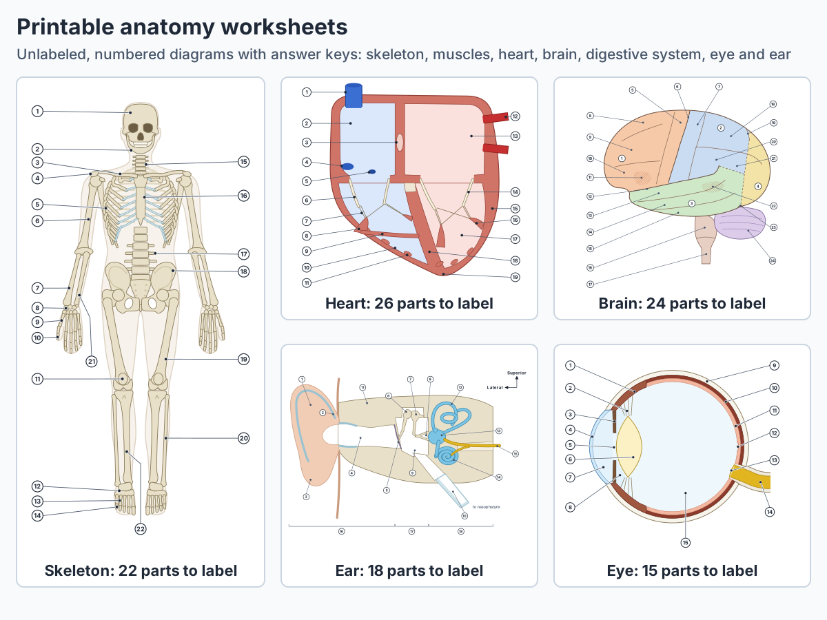
Free printable anatomy worksheets for seven parts of the human body: the skeleton, front muscles, heart, brain, digestive system, eye and ear. Each one is an unlabeled diagram with numbered structures. Write the name of each numbered part, then check the answer key under the sheet, or print it with the key on page 2.
Each worksheet goes with one of our human anatomy guides, which names and explains every numbered part, so you can learn a body system on its guide (and, for most sheets, in the 3D model) first, then test yourself here. The 3D model shows only the outer ear, so learn the middle and inner ear from the ear guide's diagrams. There is nothing to sign up for or install.
 Full size ⤢
What is an anatomy labeling worksheet?
An anatomy labeling worksheet is a diagram with the names taken off. Each structure keeps its pointer line, a number replaces its name, and you write the name on the matching numbered line. Covering the labels on a diagram and testing yourself, and labelling blank diagrams again and again, are both on LSU Health's list of ways to study anatomy actively. (LSU Health New Orleans School of Medicine: How to Study Anatomy in Your First Year of Medical School.)
Naming a structure from memory is a form of practice testing. A review of ten common study techniques rated practice testing and distributed practice (spreading study out over time) as the two with high utility, because they help learners of different ages and abilities. Rereading and highlighting were rated low. (Dunlosky J et al. Improving Students' Learning With Effective Learning Techniques. Psychol Sci Public Interest. 2013.)
The effect has been measured in anatomy. When 21 future medical and dental students answered nervous system questions three times, in a slightly different form each time, they scored almost 9% higher on the second quiz, with nothing in between but seeing the correct answers. On the third quiz, a week later, their scores were 29% higher than on the first. More of this evidence is on the anatomy quiz page. (Logan JM, Thompson AJ, Marshak DW. Testing to enhance retention in human anatomy. Anat Sci Educ. 2011.)
How to use an anatomy worksheet to study
Treat each sheet as a test. Learn the body system first, label the sheet from memory, check your answers, and come back to the parts you missed after a gap of a few days. The steps below follow the study advice of LSU Health's medical school and the University of North Carolina's Learning Center. (LSU Health New Orleans School of Medicine: How to Study Anatomy in Your First Year of Medical School.)
- Study first: Read the guide for the body system and turn the 3D human model to see where the main organs, bones and muscles lie. Each worksheet below links to its guide. (LSU Health New Orleans School of Medicine: How to Study Anatomy in Your First Year of Medical School.)
- Label from memory: Close your notes and write a name on every numbered line you can. Leave a blank rather than looking it up. (The Learning Center, University of North Carolina at Chapel Hill: Studying 101: Study Smarter Not Harder.)
- Check and mark: Open the answer key, mark each answer right or wrong, and read about the parts you missed on the guide.
- Relabel the misses: Print a fresh copy and label it again after about 1 day, 3 days and 1 week, the growing gaps LSU Health suggests, instead of cramming in one sitting. (LSU Health New Orleans School of Medicine: How to Study Anatomy in Your First Year of Medical School.)
- Say it or draw it: Explain each structure out loud as you label it, or sketch the diagram from memory on a blank page. Drawing from memory and explaining while drawing are both on LSU Health's list. (LSU Health New Orleans School of Medicine: How to Study Anatomy in Your First Year of Medical School.)
Why come back after a few days?
Spacing and self-testing both help. A 2024 systematic review in health professions education included 56 studies with 63 experiments on spaced practice and retrieval practice; 43 of them showed significant benefits of one or both methods over control or comparison groups. (Trumble E et al. Systematic review of distributed practice and retrieval practice in health professions education. Adv Health Sci Educ. 2024.)
For quick self-tests on bones, muscles, organs and the heart, try the anatomy quiz.
Printable human anatomy worksheets
Seven worksheets, with 15 to 26 numbered structures each. Print with answer key gives two pages: the diagram with blank numbered lines, then the key. Download diagram saves the sheet as an SVG file, which stays sharp at any print size. The answer key under each sheet stays closed until you open it.
Human skeleton worksheet: label the bones
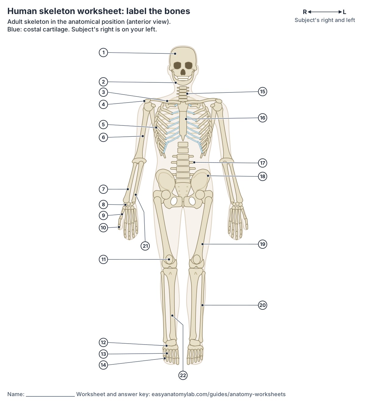
Answer key (22 labels)
- Skull (cranium)
- Mandible (lower jaw)
- Clavicle (collarbone)
- Scapula (acromion)
- Ribs
- Humerus
- Radius
- Carpals (wrist)
- Metacarpals (palm)
- Phalanges (fingers)
- Patella (kneecap)
- Tarsals
- Metatarsals
- Phalanges (toes)
- Cervical vertebrae
- Sternum (breastbone)
- Lumbar vertebrae
- Hip bone (coxal bone)
- Femur (thigh bone)
- Fibula
- Ulna
- Tibia (shin bone)
Human muscles worksheet: label the front muscles
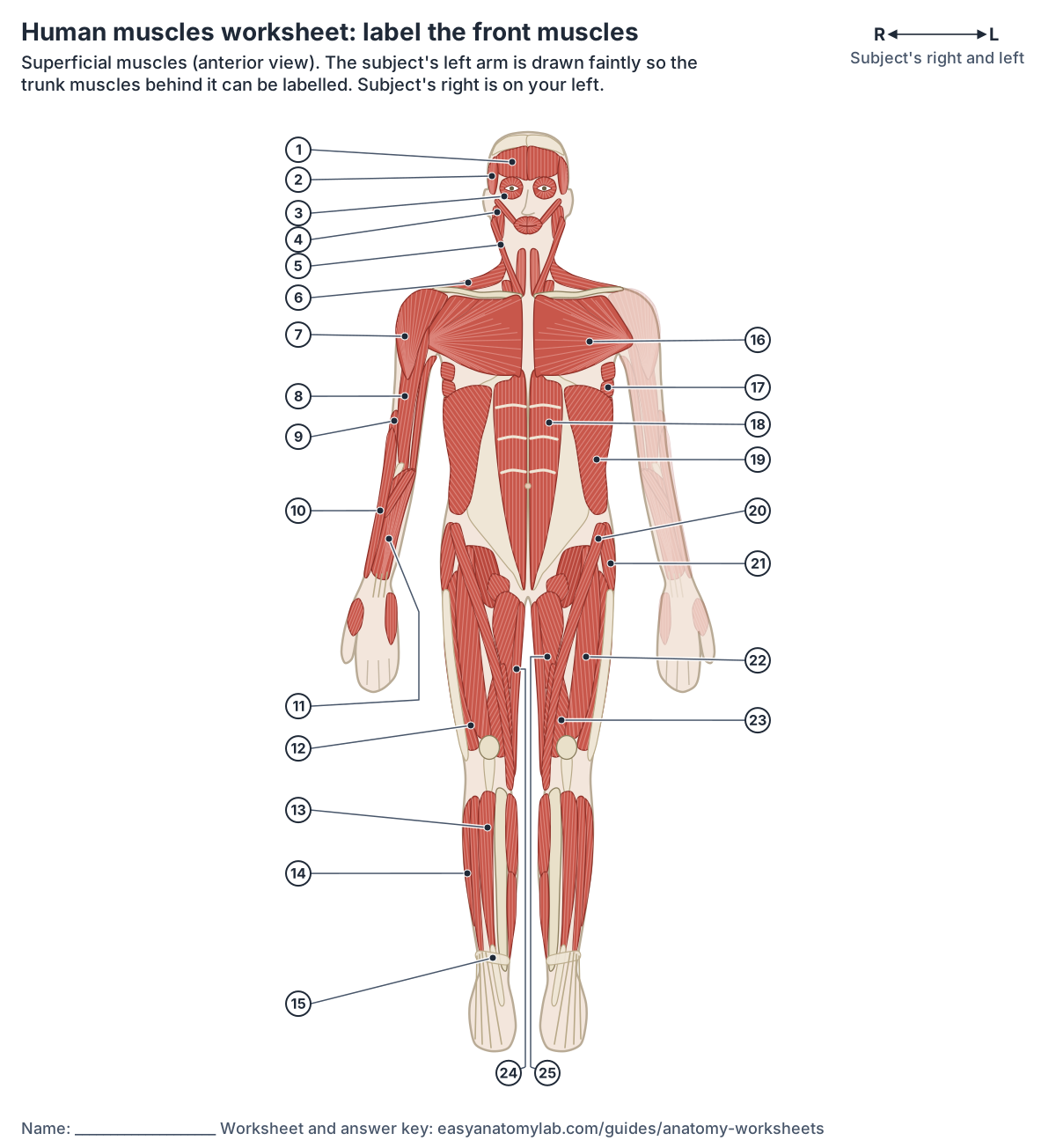
Answer key (25 labels)
- Frontalis (occipitofrontalis)
- Temporalis
- Orbicularis oculi
- Masseter
- Sternocleidomastoid
- Trapezius
- Deltoid
- Biceps brachii
- Brachialis
- Brachioradialis
- Forearm flexors
- Vastus lateralis
- Tibialis anterior
- Fibularis (peroneus) longus
- Extensor retinaculum
- Pectoralis major
- Serratus anterior
- Rectus abdominis
- External oblique
- Sartorius
- Tensor fasciae latae
- Rectus femoris
- Vastus medialis
- Gracilis
- Adductor longus
Heart worksheet: label the chambers and valves
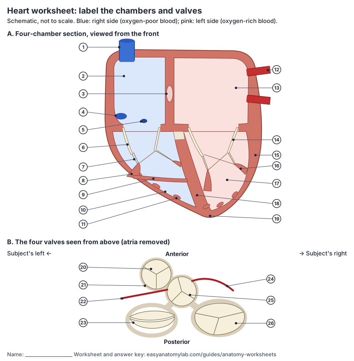
Answer key (26 labels)
- Superior vena cava
- Right atrium
- Fossa ovalis (on the interatrial septum)
- Opening of the inferior vena cava
- Coronary sinus opening
- Tricuspid valve
- Chordae tendineae
- Anterior papillary muscle
- Moderator band
- Right ventricle
- Trabeculae carneae
- Left pulmonary veins
- Left atrium
- Mitral (bicuspid) valve
- Thick left ventricle wall
- Papillary muscle
- Left ventricle
- Interventricular septum
- Apex
- Pulmonary valve
- Fibrous ring of the cardiac skeleton
- Left coronary artery
- Mitral valve
- Right coronary artery
- Aortic valve
- Tricuspid valve
Brain worksheet: label the lobes and gyri
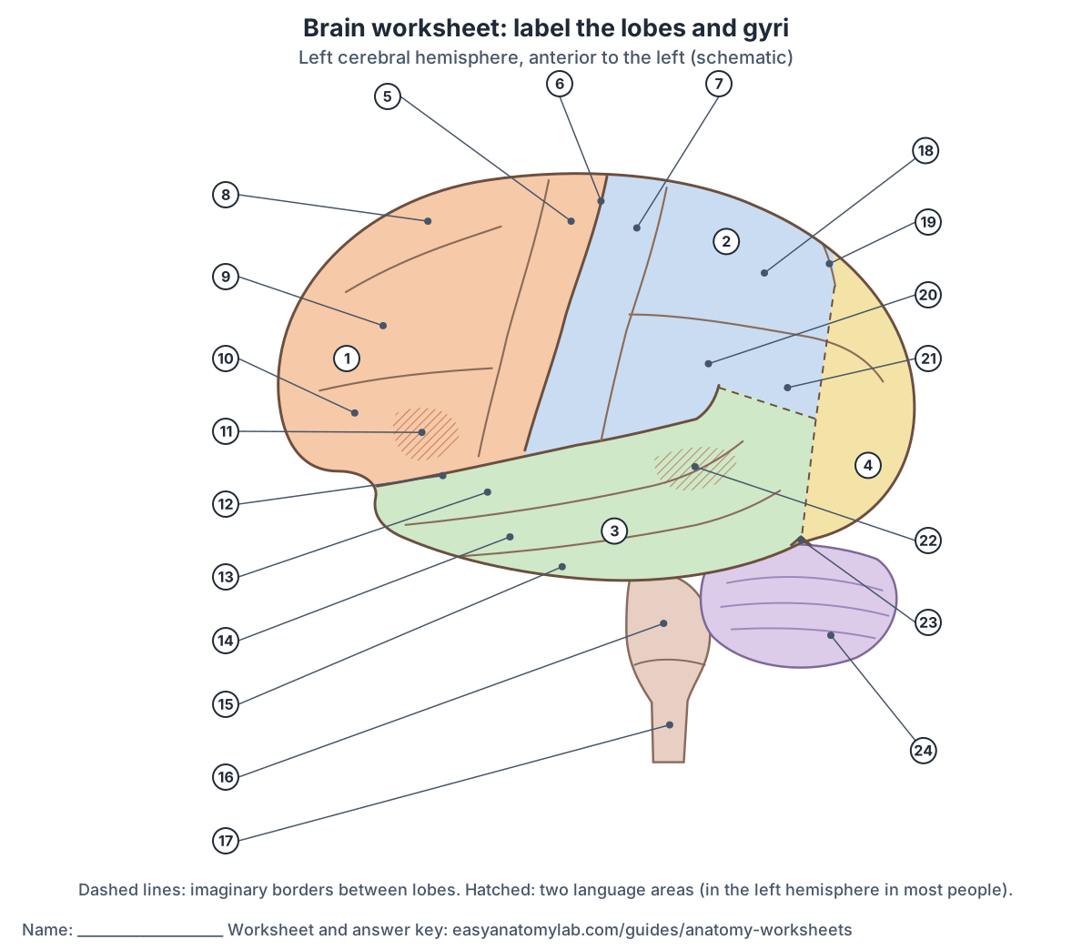
Answer key (24 labels)
- Frontal lobe
- Parietal lobe
- Temporal lobe
- Occipital lobe
- Precentral gyrus
- Central sulcus
- Postcentral gyrus
- Superior frontal gyrus
- Middle frontal gyrus
- Inferior frontal gyrus
- Broca's area
- Lateral sulcus
- Superior temporal gyrus
- Middle temporal gyrus
- Inferior temporal gyrus
- Pons
- Medulla oblongata
- Superior parietal lobule
- Parieto-occipital sulcus
- Supramarginal gyrus
- Angular gyrus
- Wernicke's area
- Preoccipital notch
- Cerebellum
Digestive system worksheet: label the organs
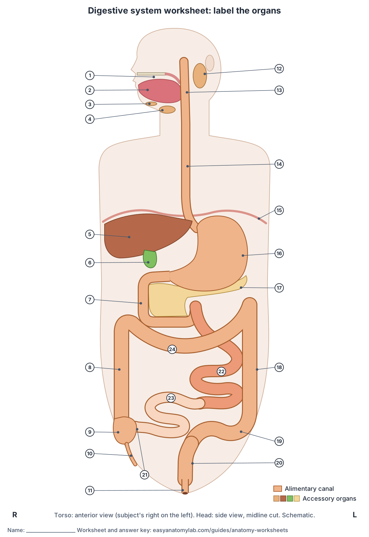
Answer key (24 labels)
- Mouth (oral cavity)
- Tongue
- Sublingual gland
- Submandibular gland
- Liver
- Gallbladder
- Duodenum
- Ascending colon
- Caecum (cecum)
- Appendix
- Anal canal and anus
- Parotid gland
- Pharynx
- Oesophagus (esophagus)
- Diaphragm
- Stomach
- Pancreas
- Descending colon
- Sigmoid colon
- Rectum
- Ileocaecal junction
- Jejunum
- Ileum
- Transverse colon
Eye worksheet: label the parts of the eyeball
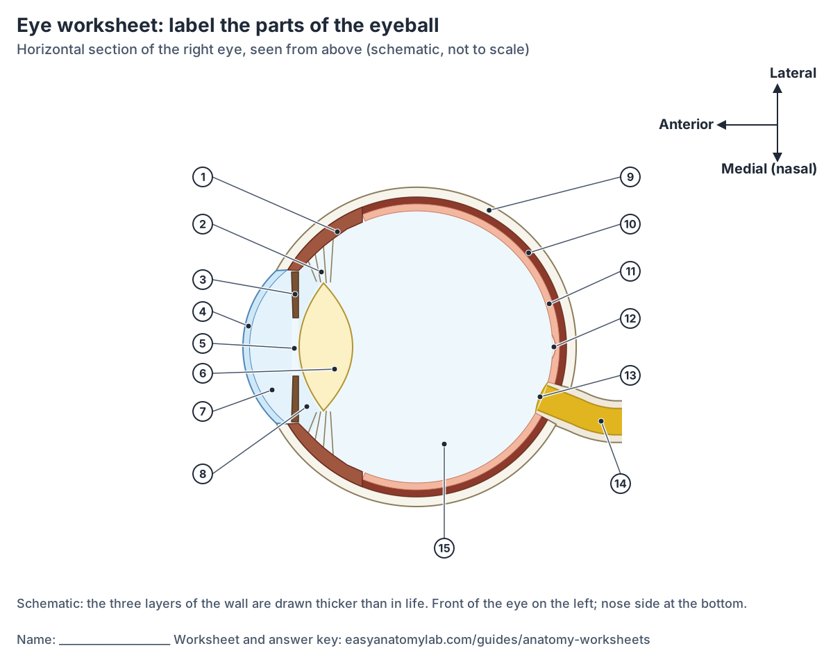
Answer key (15 labels)
- Ciliary body
- Suspensory ligament (zonular fibres)
- Iris
- Cornea
- Pupil
- Lens
- Anterior chamber (aqueous humour)
- Posterior chamber
- Sclera
- Choroid
- Retina
- Fovea (centre of the macula)
- Optic disc (blind spot)
- Optic nerve
- Vitreous body
Ear worksheet: label the outer, middle and inner ear
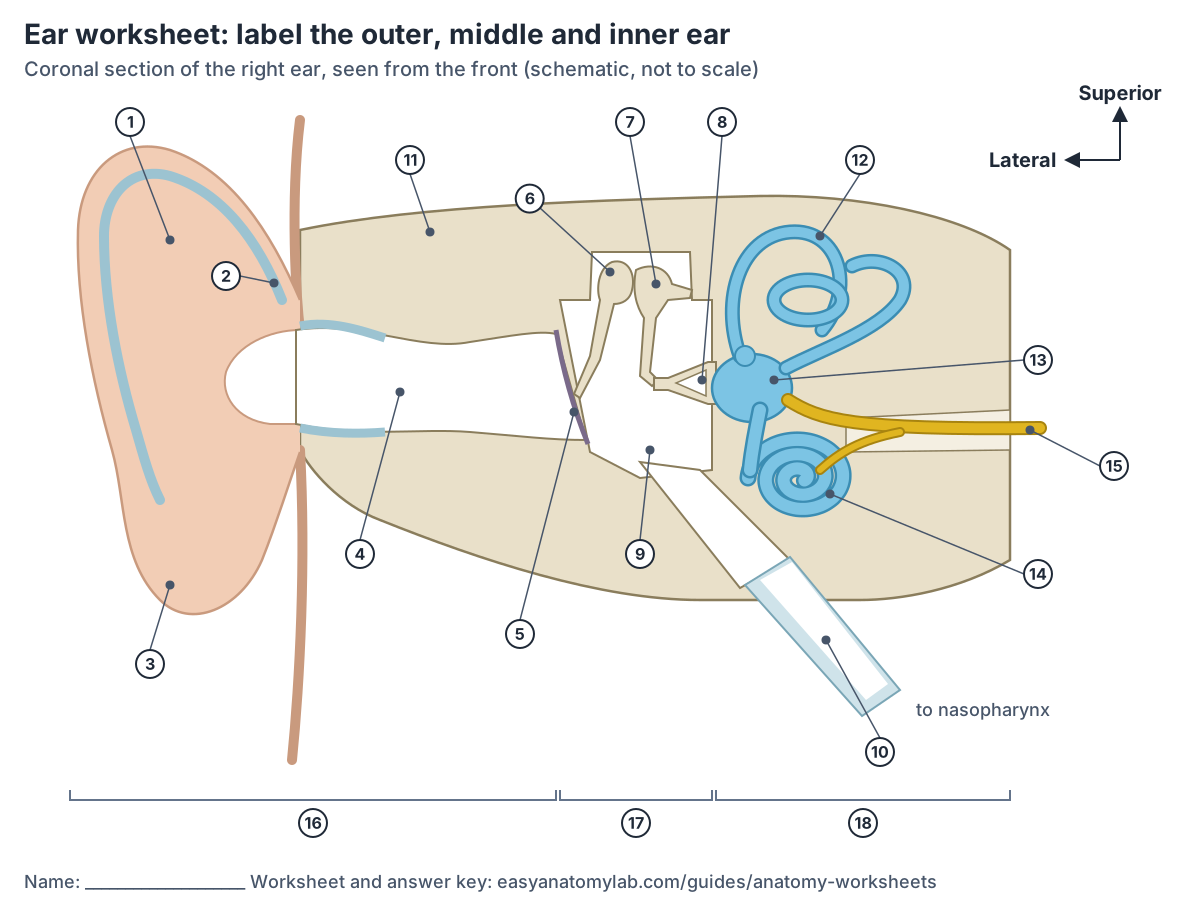
Answer key (18 labels)
- Auricle (pinna)
- Cartilage of the auricle
- Lobule
- External acoustic meatus (ear canal)
- Tympanic membrane (eardrum)
- Malleus
- Incus
- Stapes
- Tympanic cavity (the middle ear cavity)
- Eustachian (auditory) tube
- Temporal bone
- Semicircular canals
- Vestibule
- Cochlea
- Vestibulocochlear nerve (CN VIII)
- Outer (external) ear
- Middle ear
- Inner (internal) ear
Which guide goes with each worksheet?
Six of the worksheets are unlabeled copies of a diagram on the guide listed in the table, so that guide names and explains every numbered structure. The eye sheet was drawn for this page; the eye anatomy guide explains each of its parts, and the answer key under the sheet names them.
The views match the guides. Right and left always mean the subject's own right and left, so in a front (anterior) view the subject's right is on your left, as if the person were facing you. The eye sheet is a horizontal (transverse) section, a cut that divides a body part into upper and lower portions. (OpenStax Anatomy and Physiology 2e, 1.6 Anatomical Terminology.)
Worksheets and the guides that explain them
| Worksheet | View | Parts to label | Study it on |
|---|---|---|---|
| Human skeleton | Front (anterior) view; subject's right on your left | 22 bones and bone groups | Skeletal system |
| Human muscles | Front view of the superficial muscles | 25 | Muscular system |
| Heart | Four-chamber section from the front, and the four valves from above | 26 | Heart |
| Brain | Left cerebral hemisphere from the side, front to the left | 24 | Brain anatomy |
| Digestive system | Torso from the front, head from the side | 24 | Digestive system |
| Eye | Horizontal section of the right eyeball, seen from above | 15 | Eye anatomy |
| Ear | Coronal section of the right ear, seen from the front | 18 | Ear anatomy |
Each guide explains every numbered structure on its worksheet. Six guides show the same diagram with its labels; the eye sheet was drawn for this page.
Can you use the worksheets as anatomy coloring pages?
Yes, with one difference. In a University of Ottawa trial, students were asked to colour and label an anatomy colouring page. These sheets are shaded diagrams rather than outline drawings, so print one in black and white: the shapes come out in shades of grey, and you colour over each numbered structure and write its name on the matching line in the same colour. Name each part before you colour it, so the colouring does not replace the recall. (Fotsing S et al. A pilot randomized controlled trial of anatomy coloring books for first-year medical students. University of Ottawa Journal of Medicine. 2025.)
The evidence for colouring is small. In that pilot randomised trial, 17 first-year medical students spent 20 minutes on the anatomy of the mediastinum, either colouring and labelling a colouring page or reading an annotated atlas diagram. Retention test scores did not differ significantly between the groups, but the colouring group reported significantly lower anxiety afterwards, and 78% of them agreed that colouring helped them consolidate what they learned. The authors warn that the sample was small. (Fotsing S et al. A pilot randomized controlled trial of anatomy coloring books for first-year medical students. University of Ottawa Journal of Medicine. 2025.)
A second study followed 70 second-term medical students during a circulatory and respiratory system course. The 40% who preferred colouring-based methods scored significantly higher on the exams, but the students were grouped by the method they preferred, not at random, so the study cannot show that colouring caused the difference. (Uğuz Gençer C, Tetiker H. Contribution of coloring activity to learning in anatomy education. Medical Records. 2026 (Türk Medline record).)
How to turn a worksheet into anatomy flashcards
Use the answer key as the back of your cards. On the front, write the worksheet name and the number (or sketch the structure); on the back, write its name and one fact from the guide, such as what it does. LSU Health lists flashcards, including apps such as Anki, alongside practice questions and daily quizzing as ways to study actively. (LSU Health New Orleans School of Medicine: How to Study Anatomy in Your First Year of Medical School.)
Review the cards little and often. The University of North Carolina's Learning Center suggests making flashcards for material you have to memorise and reviewing them periodically through the day, and working a short time on each subject every day. (The Learning Center, University of North Carolina at Chapel Hill: Studying 101: Study Smarter Not Harder.)
Flashcard apps such as Anki use spaced repetition to schedule the reviews for you. At one US medical school, the 78 first-year students who used Anki scored significantly higher on three course exams and the Comprehensive Basic Science Exam than the 52 who did not. A cohort study like this shows an association, not proof that the app caused the higher scores. (Gilbert MM et al. A cohort study assessing the impact of Anki as a spaced repetition tool on academic performance in medical school. Med Sci Educ. 2023.)
For how often to review and more on self-testing, see the anatomy quiz page.
Can I print and share these worksheets?
Yes, for learning. You can print the worksheets to study from, and link to this page as often as you like. The terms of use let you use the site as a learning resource but do not give a blanket licence to republish its diagrams, so ask through the contact page before you post the sheets on another website or put them in something you sell.
The diagrams are EasyAnatomyLab's own schematic drawings, not scans from a textbook, and they are not to scale. Please keep the line at the bottom of each sheet, which points back to this page and its answer keys.
How were the worksheet diagrams checked?
Every label on the sheets was checked against cited textbook and anatomy sources before the labelled diagram went on its guide, and again for this page. Some of the details that are easy to get wrong, and how the sheets show them:
- Skeleton, forearm: the radius is the forearm bone on the thumb (lateral) side, and the ulna is the medial bone of the forearm. (OpenStax Anatomy and Physiology 2e, 8.2 Bones of the Upper Limb.)
- Skeleton, leg: the tibia (shin bone) is the larger, medial bone of the leg, and the thinner fibula lies on the outside. (OpenStax Anatomy and Physiology 2e, 8.4 Bones of the Lower Limb.)
- Heart: the superior and inferior venae cavae and the coronary sinus all open into the right atrium, and the moderator band crosses the right ventricle. (OpenStax Anatomy and Physiology 2e, 19.1 Heart Anatomy.)
- Brain: the central sulcus separates the frontal and parietal lobes, and the lateral sulcus separates the temporal lobe from the lobes above it. (TeachMeAnatomy: The Cerebrum.)
- Digestive system: the ascending colon runs up the right side of the abdomen and the descending colon down the left, so on a front view they appear on your left and right. (OpenStax Anatomy and Physiology 2e, 23.5 The Small and Large Intestines.)
- Eye: the sheet is a horizontal section of the right eye seen from above, so the optic disc (the blind spot) lies on the nasal side of the fovea, at the bottom of the drawing. (UTHealth Houston, Neuroscience Online: Lab 8, The Visual System.)
- Ear: the malleus is attached to the eardrum (tympanic membrane), and the stapes fits into the oval window of the inner ear. (TeachMeAnatomy: The Middle Ear.)
Frequently asked questions
Are these anatomy worksheets free?
Yes. All seven worksheets are free to view, print and download, with no account or sign-up. Each sheet has a print button that adds blank numbered lines and puts the answer key on a second page, and a download button that saves the diagram as an SVG file, which stays sharp at any print size.
Do the anatomy worksheets come with answer keys?
Yes. Every worksheet has a numbered answer key directly under it, closed until you open it, so you can test yourself first. The printed version puts the key on page 2, which you can take off before you start. The key uses the same names as the guide that explains each diagram.
What is the best way to study with an anatomy labeling worksheet?
Learn the body system first, then label the sheet from memory without notes, check the key and mark your misses. Label the missed parts again after about 1 day, 3 days and 1 week, as LSU Health's medical school suggests. Testing yourself like this, spread over days, has better research support than rereading or highlighting.
Does coloring help you learn anatomy?
It may help you stay engaged, but the evidence is small. In a pilot randomised trial with 17 medical students, colouring and labelling a page did not change retention test scores compared with studying an annotated diagram, though it lowered anxiety. A study of 70 students found higher exam scores with colouring, but the students were grouped by preference, not at random.
Can I make anatomy flashcards from these worksheets?
Yes. Use each answer key as the back of a set of cards: write the worksheet name and number on the front, and the structure's name and one fact on the back. Review a few cards every day, or enter them in a flashcard app such as Anki, which uses spaced repetition to schedule your reviews.
Can teachers use these worksheets in class?
Yes, as a learning resource: students can label the sheets in class or at home and check their answers with the keys on this page. The site's terms do not give a blanket licence to republish the diagrams, so ask through the contact page before posting them on another website or including them in material you sell.
Which parts of the body do the worksheets cover?
There are seven human worksheets: the skeleton from the front, the front muscles, the heart (chambers and valves), the brain (lobes and gyri of the left hemisphere), the digestive system, the eye (a horizontal section of the eyeball) and the ear (outer, middle and inner ear). Each one links to the guide that explains it.
References
- LSU Health New Orleans School of Medicine, Office of Student Affairs. How to Study Anatomy in Your First Year of Medical School.
- The Learning Center, University of North Carolina at Chapel Hill. Studying 101: Study Smarter Not Harder.
- Dunlosky J, Rawson KA, Marsh EJ, Nathan MJ, Willingham DT. Improving students' learning with effective learning techniques: promising directions from cognitive and educational psychology. Psychol Sci Public Interest. 2013.
- Logan JM, Thompson AJ, Marshak DW. Testing to enhance retention in human anatomy. Anat Sci Educ. 2011.
- Trumble E, Lodge J, Mandrusiak A, Forbes R. Systematic review of distributed practice and retrieval practice in health professions education. Adv Health Sci Educ Theory Pract. 2024.
- Gilbert MM, Frommeyer TC, Brittain GV, et al. A cohort study assessing the impact of Anki as a spaced repetition tool on academic performance in medical school. Med Sci Educ. 2023.
- Fotsing S, Xuan L, Khoury M, et al. A pilot randomized controlled trial of anatomy coloring books for first-year medical students: impact on anxiety and anatomical knowledge retention. University of Ottawa Journal of Medicine. 2025;15(2).
- Uğuz Gençer C, Tetiker H. Contribution of coloring activity to learning in anatomy education. Medical Records. 2026;8(1):1-6.
- Betts JG, Young KA, Wise JA, et al. Anatomy and Physiology 2e, 1.6 Anatomical Terminology. OpenStax; 2022.
- Betts JG, Young KA, Wise JA, et al. Anatomy and Physiology 2e, 8.2 Bones of the Upper Limb. OpenStax; 2022.
- Betts JG, Young KA, Wise JA, et al. Anatomy and Physiology 2e, 8.4 Bones of the Lower Limb. OpenStax; 2022.
- Betts JG, Young KA, Wise JA, et al. Anatomy and Physiology 2e, 13.2 The Central Nervous System. OpenStax; 2022.
- Betts JG, Young KA, Wise JA, et al. Anatomy and Physiology 2e, 14.1 Sensory Perception. OpenStax; 2022.
- Betts JG, Young KA, Wise JA, et al. Anatomy and Physiology 2e, 19.1 Heart Anatomy. OpenStax; 2022.
- Betts JG, Young KA, Wise JA, et al. Anatomy and Physiology 2e, 23.5 The Small and Large Intestines. OpenStax; 2022.
- TeachMeAnatomy. The Chambers of the Heart.
- TeachMeAnatomy. The Cerebrum.
- TeachMeAnatomy. The Middle Ear.
- UTHealth Houston McGovern Medical School. Neuroscience Online, Lab 8: The Visual System.
Related pages
- Human anatomy1,000+ bones, muscles, organs, vessels and nerves from real anatomical data.
- Anatomy quizFree anatomy quizzes and games: find-the-structure quizzes on 3D models and labelling practice.
- Skeletal systemHow many bones are in the human body? An adult has 206. See the skeletal system labeled in front and back diagrams, bones named by region, and a 3D skeleton.
- HeartHeart anatomy with labelled diagrams and a 3D model: location, pericardium, wall layers, four chambers, valves, great vessels and the conduction system.
- How to study anatomyHow to study anatomy: test yourself, space reviews over days, label and draw from memory, and use 3D models. The evidence, a weekly schedule and free tools.
About this guide
Published by Dr Shaista Andleeb. Drafted with AI assistance from the sources listed above and checked against them; last reviewed on 28 October 2026. The editorial policy explains how sources are chosen, how AI tools are used and how errors are corrected. To report a mistake, use the contact page.
More guides: How to study anatomy · Types of bone fractures · Best anatomy apps. Browse all anatomy guides, or explore the interactive 3D human anatomy model.