

Types of bone fractures: names, patterns and diagrams
By Dr Shaista Andleeb · Last reviewed
Bone fractures are named by several features at once. The main types are open or closed (is the skin broken?), complete or incomplete, and the shape of the break line: transverse, oblique, spiral or comminuted. Others are named by cause, such as stress, avulsion and compression fractures, and some, like greenstick and buckle fractures, happen mainly in children.
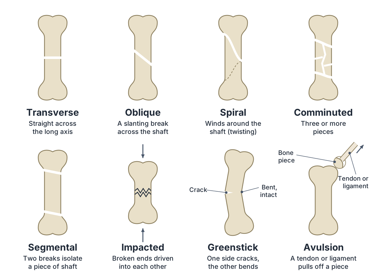
Most types below have a labelled line diagram, and each has a plain definition from cited textbook and clinical sources; the table compares the main types side by side, with an example of each. The names make more sense once you know the parts of a long bone, explained in our bone structure guide, and you can see every bone in 3D in the skeletal system guide.
This guide is for learning anatomy and is not medical advice. For an injury or symptoms, see a clinician. Read the educational disclaimer.
 Full size ⤢
What is a bone fracture?
A bone fracture is a broken bone. Fracture, break and crack all mean the same thing: doctors use "break" and "fracture" interchangeably. A bone can be fractured completely or only partly, and it can break crosswise, lengthwise or into several pieces. (AAOS OrthoInfo: Fractures (Broken Bones).)
Most fracture names describe a long bone, so its parts are worth knowing. The shaft is the diaphysis, a tube of hard compact bone around the medullary (marrow) cavity. Each end is an epiphysis. A membrane called the periosteum covers the outside, and tendons and ligaments attach to bone there. (OpenStax Anatomy and Physiology 2e, 6.3 Bone Structure.)
Between the shaft and each end is the metaphysis, the flared part of the bone where it widens; in a growing bone a layer of cartilage, the growth plate (epiphyseal plate, physis), separates the metaphysis from the epiphysis. (StatPearls: Greenstick Fracture.)
Fracture names combine. Doctors describe the type of fracture line, where it is in the bone, how far the pieces have moved and whether the skin is broken, so one fracture can be, for example, an open transverse fracture of the midshaft. (Merck Manual Professional Version: Overview of Fractures.)
For the parts of a long bone in more detail, with diagrams, see the bone structure guide.
How many types of bone fractures are there?
There is no single official number. OpenStax's anatomy textbook lists 8 common types, the American Academy of Orthopaedic Surgeons (AAOS) lists 5 common types, and the MSD Manual's table explains 21 fracture terms. The lists differ because each name describes a different feature of the same break. (OpenStax Anatomy and Physiology 2e, 6.5 Fractures: Bone Repair.)
It is easier to sort fractures by the question each name answers. Cleveland Clinic groups them by pattern, by how they happened, as open or closed, and as displaced or non-displaced, and Merck's manual describes the fracture line, its location and the displacement of the pieces. (Cleveland Clinic: Bone Fractures (Broken Bones).)
- Is the skin broken?: Open (compound) or closed (simple). (OpenStax Anatomy and Physiology 2e, 6.5 Fractures: Bone Repair.)
- Is the bone broken all the way through?: Complete, or incomplete like a greenstick or buckle fracture. (Radiopaedia: Complete fracture.)
- What shape is the break line?: Transverse, oblique, spiral, comminuted, segmental, impacted or longitudinal. (Merck Manual Professional Version: Overview of Fractures.)
- Have the pieces moved?: Displaced or non-displaced. (Cleveland Clinic: Bone Fractures (Broken Bones).)
- What caused it?: Stress, avulsion, compression or pathological fractures. (Cleveland Clinic: Bone Fractures (Broken Bones).)
- Is it a child's bone?: Greenstick, buckle and growth plate fractures happen in children. (Merck Manual Professional Version: Overview of Fractures.)
Types of bone fractures at a glance
| Type | What it looks like | Typical cause | Example |
|---|---|---|---|
| Transverse | A break straight across the bone, at a right angle to its long axis | Trauma, such as a fall or a crash | Midshaft of the tibia (shin bone) |
| Oblique | A straight break at a slant across the shaft | Trauma, such as a fall or a crash | Shaft of a long bone |
| Spiral | A break that winds around the shaft like a corkscrew | A twisting (rotational) force | Femur or tibia |
| Comminuted | The bone is broken into three or more pieces | A great force, such as a road crash | Femur shaft after a high-energy crash |
| Segmental | Two separate breaks that isolate a piece of the shaft | Trauma | Shaft of a long bone |
| Impacted | One broken end is driven into the other, so the bone looks shorter | Compression of the bone ends | Neck of the femur in a hip fracture |
| Greenstick | Cracked on one side and bent on the other; not broken through | Bending of a child's flexible bone | Forearm of a young child |
| Buckle (torus) | The outer bone wall bulges; there is no break line through it | An end-on (axial) load, often from a fall | Wrist end of the radius in a child |
| Stress (hairline) | A small crack | Repeated loading, for example in runners | Bones of the foot or lower leg |
| Avulsion | A piece of bone pulled off where a tendon or ligament attaches | A strong muscle pull or a ligament under force | Sports injuries in adolescents |
| Compression | The bone collapses into itself | Pressure on bone, often weakened by osteoporosis | Vertebrae of older adults |
| Pathological | A break through bone weakened by disease | Little force on bone weakened by disease | Bone weakened by a tumour |
| Open (compound) | A skin wound reaches the fracture; a bone end may stick out | Trauma | A bone end pushing out through the skin |
| Closed (simple) | The skin over the fracture is intact | Any cause | Any fracture with intact skin over it |
One fracture can have several of these names at once, for example an open transverse fracture. Where the sources tie no particular force to a pattern, the cause column gives the general one, trauma. Sources: OpenStax Anatomy and Physiology 2e, 6.5 Fractures: Bone Repair; AAOS OrthoInfo: Fractures (Broken Bones); Cleveland Clinic: Bone Fractures (Broken Bones); MSD Manual Consumer Version: Overview of Fractures; Merck Manual Professional Version: Overview of Fractures; Radiopaedia: Spiral fracture; Radiopaedia: Comminuted fracture; Radiopaedia: Greenstick fracture; Radiopaedia: Torus fracture; Radiopaedia: Avulsion fracture; Radiopaedia: Neck of femur fracture.
Open vs closed fractures (compound and simple)
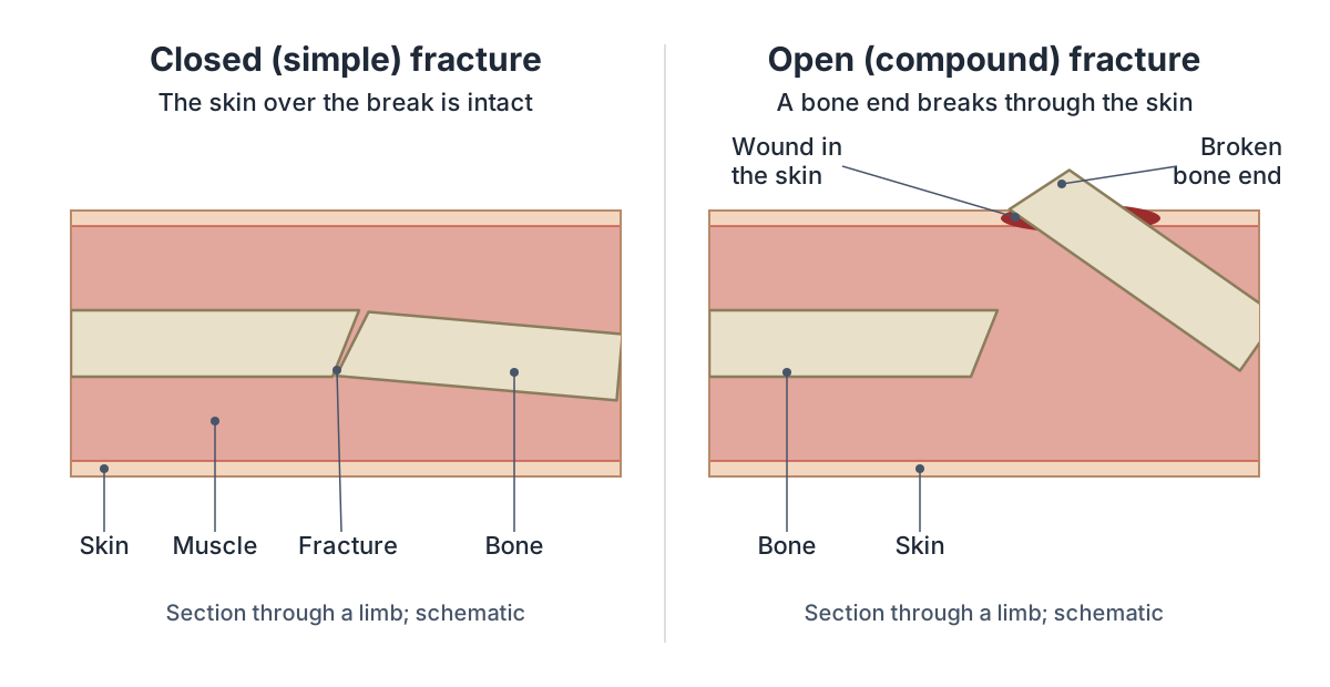
An open fracture is one where the skin is broken so that the fracture connects with the outside. Either a bone fragment sticks out through the skin, or a wound reaches down to the broken bone; the bone may or may not be visible. In a closed fracture the skin over the break is intact. (AAOS OrthoInfo: Fractures (Broken Bones).)
The skin wound does not have to sit directly over the break: StatPearls notes it may lie some distance away, so any fracture that comes with a wound is treated as open until shown otherwise. (StatPearls: Open Fracture Management.)
Compound is another name for an open fracture, and simple is another name for a closed one. Simple only means the skin is intact: closed fractures can still be serious. Open fractures carry a high risk of infection, in the wound and in the bone itself. (OpenStax Anatomy and Physiology 2e, 6.5 Fractures: Bone Repair.)
Complete and incomplete fractures
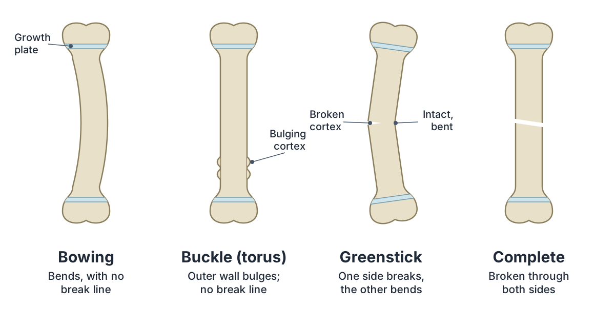
In a complete fracture the bone is broken all the way round its outer wall (cortex), so the pieces are fully separated. Transverse, oblique, spiral, longitudinal and comminuted fractures are all complete fractures. (Radiopaedia: Complete fracture.)
In an incomplete fracture the bone is not broken across its whole width, so the pieces do not come apart. Incomplete fractures happen mainly in children's long bones and include greenstick, buckle (torus) and bowing fractures. When the force only slightly exceeds a bone's breaking point, the bone may crack rather than break all the way through. (Radiopaedia: Incomplete fracture.)
Fracture patterns: transverse, oblique, spiral and comminuted
Pattern names describe the shape of the break line, what the break looks like. Transverse and oblique fractures break in a straight line; spiral, comminuted, segmental and greenstick fractures have an uneven break line. (Cleveland Clinic: Bone Fractures (Broken Bones).)
Transverse fracture
A transverse fracture runs straight across the bone, at a right angle to its long axis. In an upright limb the break line is horizontal, which is how AAOS describes it. Merck's manual shows one through the midshaft of the tibia. (Merck Manual Professional Version: Overview of Fractures.)
Oblique fracture
An oblique fracture is a straight break at a slant, at an angle that is not 90 degrees to the long axis of the bone. It usually crosses the shaft diagonally. (OpenStax Anatomy and Physiology 2e, 6.5 Fractures: Bone Repair.)
Spiral fracture
A spiral (torsion) fracture is caused by a twisting force that pulls the bone apart. The break line winds around the shaft like a corkscrew, so the broken ends can be sharp and slanted. On an X-ray, Merck's manual notes, part of a spiral fracture line runs parallel to the bone's long axis, which separates it from an oblique fracture. (MSD Manual Consumer Version: Overview of Fractures.)
Spiral fractures happen in long bones such as the tibia, femur and humerus. Radiopaedia describes them as usually caused by high-energy trauma and often displaced. (Radiopaedia: Spiral fracture.)
But the twist can also be fairly low-energy: studies of tibial shaft fractures put most spiral breaks down to low-energy injuries. (Suzuki T et al. Better discrimination of the concomitant peri-ankle fractures in the spiral tibial shaft fractures by thin-slice axial and three-dimensional CT. Medicine (Baltimore). 2021.)
In toddlers, a trip or jump with the foot fixed can cause an undisplaced spiral fracture of the lower tibia, called a toddler's fracture. The femur and fibula guides describe where spiral fractures occur in the leg. (Radiopaedia: Toddler fracture.)
Comminuted fracture
A comminuted fracture is a bone broken into three or more pieces, often many small fragments between two larger ends. It usually takes a great force, such as a car crash, though bone weakened by osteoporosis can also break this way. (MSD Manual Consumer Version: Overview of Fractures.)
Segmental fracture
A segmental fracture has two separate break lines that together isolate a segment of the shaft, which floats free between them. The Merck and MSD manuals count it as a kind of comminuted fracture; Radiopaedia and Cleveland Clinic list it as its own type. (Radiopaedia: Segmental fracture.)
Impacted fracture
In an impacted fracture one broken end is driven into the other, usually by compression, so the bone looks shorter. Merck's manual gives impacted fractures of the femoral neck (hip fractures) as an example. (Merck Manual Professional Version: Overview of Fractures.)
Longitudinal fracture
A longitudinal fracture runs along the length of the bone, parallel to its long axis, rather than across it. AAOS lists "lengthwise" alongside crosswise and multi-piece breaks. (Radiopaedia: Complete fracture.)
To see the long bones these patterns are drawn on, open the leg bones guide.
Displaced and non-displaced fractures
In a displaced fracture the broken pieces have moved so that they no longer line up, often leaving a gap. In a non-displaced fracture the pieces are still in their normal position with no space between them; AAOS also calls this a stable fracture. A non-displaced fracture is still a broken bone. (Cleveland Clinic: Bone Fractures (Broken Bones).)
Doctors describe displacement more exactly. An angulated fracture is one where the pieces lie at an angle to each other; Merck's manual measures angulation as the angle of the far (distal) piece from the near (proximal) one, and uses distraction for pieces pulled apart along the bone's length. (MSD Manual Consumer Version: Overview of Fractures.)
Fractures named by cause: stress, avulsion, compression and pathological
Some fractures are named for how they happened rather than for the shape of the break. Trauma, such as a fall, a road crash or a sports injury, causes most fractures; the four types below are named for the way the force acted or for the state of the bone that broke. (Cleveland Clinic: Bone Fractures (Broken Bones).)
Stress fracture (hairline fracture)
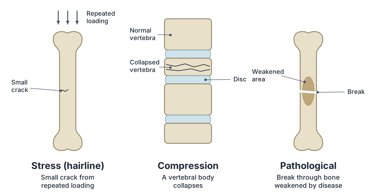
A stress fracture is a small crack caused by moderate force applied again and again, as in long-distance runners or soldiers marching with a heavy load. Bone normally repairs this small damage during rest; without rest the damage spreads into a crack. Stress fractures are also called hairline fractures. (Merck Manual Professional Version: Overview of Fractures.)
They usually affect the weight-bearing bones of the lower body, especially the foot and the lower leg (tibia and fibula). (Cleveland Clinic: Stress Fractures.)
Radiopaedia describes a stress fracture as a mismatch between bone strength and repeated mechanical stress, and separates two kinds: fatigue fractures (abnormal stress on normal bone) and insufficiency fractures (normal stress on weakened bone). (Radiopaedia: Stress fracture.)
Avulsion fracture
An avulsion fracture happens when a tendon or ligament pulls a piece of bone away from the main bone where it attaches. A ligament can pull off bone when an outside force is strong enough, as in a fall; a tendon can do it when its muscle contracts forcefully, as in young athletes. (MSD Manual Consumer Version: Overview of Fractures.)
Avulsion injuries are common in people who play sports, particularly adolescents. Examples include the hamstrings pulling on the ischial tuberosity of the pelvis (see the ischium guide) and the Achilles tendon pulling on the heel bone (calcaneus). (Radiopaedia: Avulsion fracture.)
Compression fracture
In a compression fracture the bone collapses into itself. It usually affects the vertebrae (vertebral crush fractures), most often in older adults whose bones are weakened by osteoporosis. The vertebral column guide shows the vertebral bodies that take this load. (MSD Manual Consumer Version: Overview of Fractures.)
Pathological fracture
A pathological fracture is a break through bone already weakened by disease, caused by little or no force. Causes include bone tumours (including cancer spread from elsewhere), infection and bone cysts. (Merck Manual Professional Version: Overview of Fractures.)
The sources differ on osteoporosis. The Merck and MSD manuals count fractures through osteoporotic bone as pathological; Merck adds that these are often called insufficiency or fragility fractures, while Radiopaedia mostly keeps the word pathological for tumours and describes breaks of osteoporotic bone as insufficiency or fragility fractures. (Radiopaedia: Pathological fracture.)
Types of fractures in children
Children's bones break differently from adults'. Growing bone contains more cartilage, is more flexible, has a thicker periosteum and has open growth plates, so it tends to bend under load instead of breaking right through. (StatPearls: Greenstick Fracture.)
StatPearls describes a spectrum. With no break through the cortex, the bone either bends (a bowing, or plastic deformation, fracture) or kinks (a buckle fracture). If one cortex breaks it is a greenstick fracture, and if both break it is a complete fracture. (StatPearls: Pediatric Torus Buckle Fracture.)
Greenstick fracture
A greenstick fracture is a partial fracture of a child's long bone: the cortex and periosteum break on one side while the opposite side stays intact and bends, like a fresh green twig. It is a childhood fracture and is common in the forearm and lower leg. (StatPearls: Greenstick Fracture.)
Buckle (torus) fracture
A buckle or torus fracture is an incomplete fracture where the outer wall of the bone bulges, or buckles, instead of breaking. It is caused by an end-on (axial) load, often a fall, and frequently affects the metaphysis of the radius near the wrist. (Radiopaedia: Torus fracture.)
Growth plate fracture (Salter-Harris types)
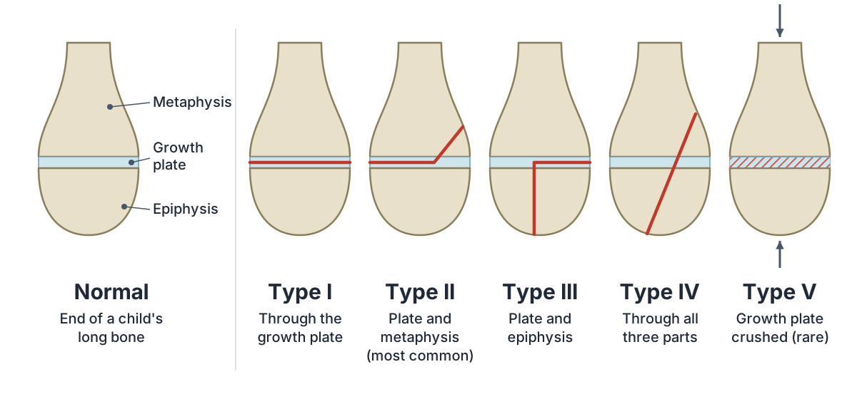
A growth plate fracture runs through the growth plate (physis), the cartilage between the widened end of the shaft (metaphysis) and the end of the bone (epiphysis). Growth plates are the last part of a child's bone to harden, so they are easily injured. These fractures only happen in children and adolescents, and an injured plate can stop growing or grow crookedly. (AAOS OrthoInfo: Growth Plate Fractures.)
Doctors most often grade them with the Salter-Harris system, by which parts the fracture crosses: (StatPearls: Salter-Harris Fracture.)
- Type I: straight through the growth plate, separating the bone end from the shaft. (AAOS OrthoInfo: Growth Plate Fractures.)
- Type II: across part of the growth plate and up through the metaphysis; the most common type. (Radiopaedia: Salter-Harris classification.)
- Type III: along part of the growth plate and down through the epiphysis, breaking off part of the bone end. (AAOS OrthoInfo: Growth Plate Fractures.)
- Type IV: through the metaphysis, the growth plate and the epiphysis. (Radiopaedia: Salter-Harris classification.)
- Type V: a crush of the growth plate by a compression force; rare. (AAOS OrthoInfo: Growth Plate Fractures.)
For the forearm bone most often involved in children's wrist fractures, see the radius guide.
Fractures named by bone and location
Fractures are also named for the bone and the part of it that broke. Merck's manual describes the location as the epiphysis (the bone end), the metaphysis (the neck between end and shaft) or the diaphysis, the shaft, which is divided into proximal, middle and distal thirds. (Merck Manual Professional Version: Overview of Fractures.)
A fracture that extends into a joint is an intra-articular (joint) fracture. It breaks the cartilage covering the bone ends, so the joint may move less well and osteoarthritis becomes more likely later. (MSD Manual Consumer Version: Overview of Fractures.)
When a joint is dislocated and one of its bones is also broken, the injury is called a fracture-dislocation. (Medical News Today: Fractures: Types, causes, symptoms, and treatment.)
Some bones have their own risks because of their anatomy. A scaphoid fracture in the wrist may not show on the first X-rays, and it can cut off part of the bone's blood supply so that the piece dies (osteonecrosis); displaced fractures of the femoral neck carry the same risk. (MSD Manual Consumer Version: Overview of Fractures.)
Some common fractures carry a person's name: the Colles fracture, a break across the distal radius often caused by a fall onto an outstretched hand, is the most frequent forearm fracture (see the radius guide for Colles and Smith fractures). (OpenStax Anatomy and Physiology 2e, 8.2 Bones of the Upper Limb.)
Our bone guides cover the fractures of each bone in context: the femur, tibia and fibula in the leg, the radius, ulna and humerus in the arm, the foot bones, the hand bones and the skull bones.
To find any bone of the body in 3D, use the skeletal system guide.
How does a broken bone heal?
A broken bone heals in four overlapping steps. Blood from torn vessels clots into a fracture haematoma. A soft callus of cartilage and new bone then forms inside and around the break and stabilises it. The cartilage is replaced by spongy (trabecular) bone, and finally the bone is remodelled back towards its original shape. (OpenStax Anatomy and Physiology 2e, 6.5 Fractures: Bone Repair.)
Clinical manuals group the same process into three stages: inflammation, repair and remodelling. Inflammation starts straight away and peaks within a couple of days. Repair begins within days and lasts weeks to months as the callus forms. Remodelling takes many months. (MSD Manual Consumer Version: Overview of Fractures.)
Children heal much faster than adults, and conditions that reduce blood flow, such as diabetes and peripheral vascular disease, slow healing. Setting a broken bone back in place is called reduction: closed reduction without surgery, open reduction with surgery to expose the break. (Merck Manual Professional Version: Overview of Fractures.)
Healing does not always go to plan. A fracture that heals more slowly than expected has delayed union; one that does not heal at all is a non-union; and one that heals with the pieces in the wrong position, leaving a deformity, is a malunion. Among the main reasons a fracture heals poorly are a damaged blood supply and a break that is not kept still enough. (Merck Manual Professional Version: Overview of Fractures.)
This guide explains names and anatomy only. If you think you have broken a bone, see a clinician: only an examination and imaging can show the type of fracture and the right treatment. (AAOS OrthoInfo: Fractures (Broken Bones).)
For bone and cartilage as tissues, see the tissue types guide.
Frequently asked questions
What are the main types of bone fractures?
The main types are open (compound) and closed (simple) fractures, complete and incomplete fractures, and the break-line patterns transverse, oblique, spiral and comminuted. Other fractures are named by cause, such as stress, avulsion, compression and pathological fractures, or are typical of children, such as greenstick, buckle and growth plate fractures. One fracture often has several of these names at once.
What is the difference between an open and a closed fracture?
In an open fracture the skin is broken: a bone end sticks out, or a wound reaches down to the break, even if no bone shows. In a closed fracture the skin over the break is intact. Open fractures carry a high risk of infection in the wound and the bone, so they are particularly serious, but closed fractures can be serious too.
Is a compound fracture the same as an open fracture?
Yes. Compound fracture is another name for an open fracture, one where the broken bone connects with the outside through a wound in the skin. In the same way, simple fracture is another name for a closed fracture. Simple describes the intact skin, not how minor the break is, so a simple fracture can still be severe.
Is a fracture the same as a broken bone?
Yes. A fracture is a broken bone, and doctors use fracture, break and crack interchangeably. A fracture can go all the way through the bone or only partly through it, and it can run across the bone, along it or into several pieces. Even a small crack, such as a stress fracture, counts as a broken bone.
What is a greenstick fracture?
A greenstick fracture is a partial break in a child's long bone. The bone cracks on one side while the other side stays intact and bends, like a fresh green twig. It happens because children's bones contain more cartilage and are more flexible than adults', so they tend to bend under load instead of breaking right through.
Is a hairline fracture the same as a stress fracture?
Usually, yes. Stress fractures are small cracks in bone, and both the MSD Manual and Cleveland Clinic say they are also called hairline fractures. They come from repeated force over time, such as running or marching with a heavy load, and mostly affect the weight-bearing bones of the foot and lower leg.
Which type of fracture is the most serious?
There is no single ranking. The risk of serious complications rises when the skin is broken (an open fracture, which can become infected) or when the injury damages blood vessels or nerves. Fractures of the femur or pelvis can also cause heavy internal bleeding. Any suspected broken bone needs to be seen by a clinician.
How long does a broken bone take to heal?
It varies from weeks to months. Healing time depends on the bone and the type of fracture, how severe the injury is, the person's age and their general health; children heal much faster than adults. The bone keeps remodelling for many months after it is strong enough to use, and a clinician will advise on each case.
References
- Betts JG, Young KA, Wise JA, et al. Anatomy and Physiology 2e, section 6.5 Fractures: Bone Repair. OpenStax; 2022.
- Betts JG, Young KA, Wise JA, et al. Anatomy and Physiology 2e, section 6.3 Bone Structure. OpenStax; 2022.
- American Academy of Orthopaedic Surgeons. Fractures (Broken Bones). OrthoInfo.
- American Academy of Orthopaedic Surgeons. Growth Plate Fractures. OrthoInfo.
- American Academy of Orthopaedic Surgeons. Stress Fractures. OrthoInfo.
- Cleveland Clinic. Bone Fractures (Broken Bones). Updated 15 January 2026.
- Cleveland Clinic. Stress Fractures.
- MSD Manual Consumer Version. Overview of Fractures. Merck & Co.
- Merck Manual Professional Version. Overview of Fractures. Merck & Co.
- Collins AC, Krishnaprasadh D. Greenstick Fracture. StatPearls. Treasure Island (FL): StatPearls Publishing.
- Asokan A, Kheir N. Pediatric Torus Buckle Fracture. StatPearls. Treasure Island (FL): StatPearls Publishing.
- Levine RH, Thomas A, Nezwek TA, Waseem M. Salter-Harris Fracture. StatPearls. Treasure Island (FL): StatPearls Publishing.
- Sop JL, Sop A. Open Fracture Management. StatPearls. Treasure Island (FL): StatPearls Publishing.
- Sheen JR, Mabrouk A. Fracture Healing Overview. StatPearls. Treasure Island (FL): StatPearls Publishing.
- Radiopaedia. Complete fracture.
- Radiopaedia. Incomplete fracture.
- Radiopaedia. Spiral fracture.
- Radiopaedia. Comminuted fracture.
- Radiopaedia. Segmental fracture.
- Radiopaedia. Greenstick fracture.
- Radiopaedia. Torus fracture.
- Radiopaedia. Avulsion fracture.
- Radiopaedia. Stress fracture.
- Radiopaedia. Pathological fracture.
- Radiopaedia. Salter-Harris classification.
- Radiopaedia. Neck of femur fracture.
- Radiopaedia. Toddler fracture.
- Suzuki T, Kurozumi T, Nakayama Y, et al. Better discrimination of the concomitant peri-ankle fractures in the spiral tibial shaft fractures by thin-slice axial and three-dimensional CT. Medicine (Baltimore). 2021;100(40):e27429.
- Betts JG, Young KA, Wise JA, et al. Anatomy and Physiology 2e, section 8.2 Bones of the Upper Limb. OpenStax; 2022.
- Brazier Y. Fractures: Types, causes, symptoms, and treatment. Medical News Today. Updated 10 November 2023.
Related pages
- Bone structureBone anatomy with a labeled long bone diagram and 3D femur: diaphysis, epiphyses, growth plate, periosteum, marrow, compact and spongy bone and bone cells.
- Skeletal systemHow many bones are in the human body? An adult has 206. See the skeletal system labeled in front and back diagrams, bones named by region, and a 3D skeleton.
- Leg bonesLeg bones labeled in a diagram and 3D model: the femur, patella, tibia and fibula, their landmarks and joints, which bone bears weight, and common fractures.
- Arm bonesArm bones labeled in a diagram and 3D model: the humerus, radius and ulna, their landmarks and joints, how the forearm bones differ, and common fractures.
- Anatomy quizFree anatomy quizzes and games: find-the-structure quizzes on 3D models and labelling practice.
About this guide
Published by Dr Shaista Andleeb. Drafted with AI assistance from the sources listed above and checked against them; last reviewed on 30 October 2026. The editorial policy explains how sources are chosen, how AI tools are used and how errors are corrected. To report a mistake, use the contact page.
More guides: How to study anatomy · Anatomy worksheets · Best anatomy apps. Browse all anatomy guides, or explore the interactive 3D human anatomy model.