

What is the vagus nerve?
The vagus nerve is the tenth cranial nerve (CN X). Its name comes from the Latin for "wandering", because it has the longest course of any cranial nerve, from the head to the abdomen. It is linked with the structures that develop from the fourth and sixth pharyngeal arches. (TeachMeAnatomy: The Vagus Nerve (CN X).)
It is a mixed nerve with parasympathetic, motor and sensory parts. It provides most of the parasympathetic supply to the heart and the gastrointestinal tract, motor fibres to the larynx and pharynx, and sensory fibres to the external acoustic meatus and the meninges. (Radiopaedia: Vagus nerve.)
The vagus is the largest single source of parasympathetic fibres, providing about 75% of the total parasympathetic outflow. It supplies the thoracic organs and most of the gastrointestinal tract through ganglia in plexuses close to the target organs. (TeachMeAnatomy: The Parasympathetic Nervous System.)
Most of its fibres carry information towards the brain: Bonaz and colleagues describe the nerve as about 80% afferent and 20% efferent fibres. (Bonaz B, Bazin T, Pellissier S. The Vagus Nerve at the Interface of the Microbiota-Gut-Brain Axis. Front Neurosci. 2018.)
Breit and colleagues give a range of 80% to 90% afferent and 10% to 20% efferent fibres, and call the nerve's afferent role its most important function. (Breit S et al. Vagus Nerve as Modulator of the Brain-Gut Axis in Psychiatric and Inflammatory Disorders. Front Psychiatry. 2018.)
There are two vagus nerves, a left and a right, which together contain about 75% of the parasympathetic nerve fibres. They exit the medulla and pass through the neck and chest to the abdomen, and the left and right nerves join to form the vagal trunks at the oesophageal hiatus. (Cleveland Clinic: Vagus Nerve.)
CN I to CN XII in order, with a diagram of where each leaves the brain and a table of skull openings. The 12 cranial nerves.
Course of the vagus nerve at a glance
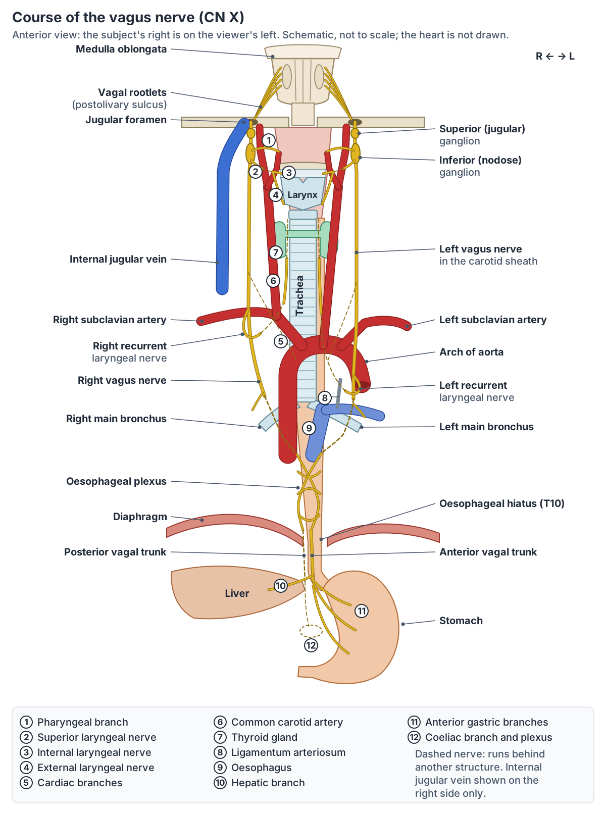
The vagus nerve arises from the medulla of the brainstem and leaves the cranium through the jugular foramen together with the glossopharyngeal (CN IX) and accessory (CN XI) nerves. In the neck it runs down inside the carotid sheath with the internal jugular vein and the common carotid artery. (TeachMeAnatomy: The Vagus Nerve (CN X).)
In the thorax the two nerves take different paths, pass behind the roots of the lungs and break up into the oesophageal plexus around the oesophagus. Near the diaphragm the plexus re-forms as the anterior and posterior vagal trunks. (Radiopaedia: Vagus nerve.)
The trunks pass through the oesophageal hiatus of the diaphragm at the level of T10, together with the oesophagus. TeachMeAnatomy lists the right and left vagus nerves among the contents of the hiatus. (TeachMeAnatomy: The Diaphragm.)
Kenhub's memory aid puts it simply: the oesophagus and vagus cross the diaphragm at the level of T10, and the hiatus transmits the oesophagus with the anterior and posterior vagal trunks. (Kenhub: Diaphragm.)
Vagus nerve nuclei in the medulla
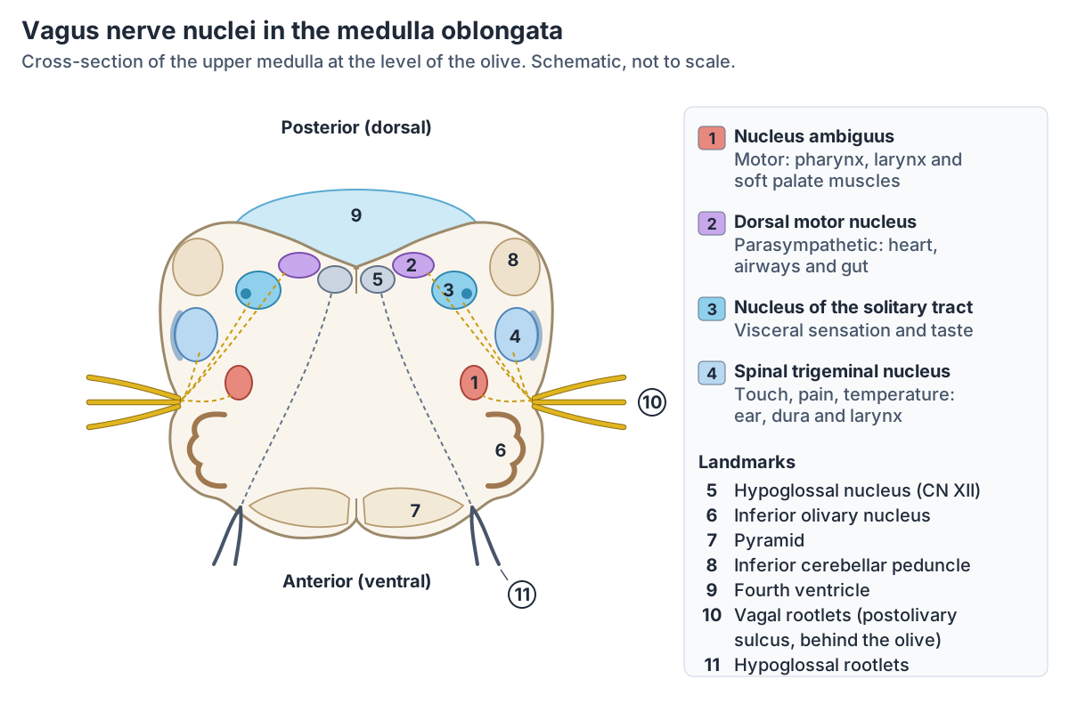
Four nuclei in the medulla oblongata contribute to the vagus nerve. The nucleus ambiguus sends motor fibres to the muscles of the soft palate, pharynx and larynx, plus preganglionic parasympathetic fibres to the heart. The dorsal motor nucleus sends parasympathetic fibres mainly to the gut and lungs. The nucleus of the solitary tract receives visceral sensation and taste, and the spinal trigeminal nucleus receives pain, temperature and touch from the outer ear, the dura of the posterior cranial fossa and the laryngeal mucosa. (Kenhub: Vagus nerve.)
Radiopaedia groups them the same way: two motor nuclei (the nucleus ambiguus, branchial motor, and the dorsal vagal nucleus, parasympathetic) and two sensory nuclei (the solitary tract nucleus for visceral sensation and taste, and the spinal trigeminal nucleus for general sensation from the skin). (Radiopaedia: Vagus nerve.)
The nucleus ambiguus is a long column in the lateral part of the upper medulla, dorsal to the inferior olivary nucleus and ventromedial to the spinal nucleus and tract of the trigeminal nerve. It also gives motor fibres to the glossopharyngeal and accessory nerves, and its fibres leave the medulla in the postolivary sulcus. (Radiopaedia: Nucleus ambiguus.)
The dorsal vagal nucleus lies on each side of the midline in the dorsal medulla, dorsal and lateral to the hypoglossal nucleus, and forms the vagal trigone in the floor of the fourth ventricle. Its preganglionic fibres reach the heart, airways, oesophagus, stomach, liver, pancreas, spleen, small intestine and proximal colon. (Radiopaedia: Dorsal vagal nucleus.)
The solitary nucleus surrounds the solitary tract in the dorsolateral medulla and lower pons. It is shared by the facial, glossopharyngeal and vagus nerves and receives taste and other sensory input. (Radiopaedia: Solitary nucleus.)
Vagus nerve nuclei and what they do
| Nucleus | Position in the medulla | Fibre type | What it supplies or receives |
|---|---|---|---|
| Nucleus ambiguus | Lateral upper medulla, dorsal to the inferior olive, ventromedial to the spinal trigeminal nucleus | Branchial motor (plus some parasympathetic) | Muscles of the soft palate, pharynx and larynx; preganglionic fibres that slow the heart |
| Dorsal motor nucleus of the vagus | Medial dorsal medulla, forming the vagal trigone in the floor of the fourth ventricle | Parasympathetic (visceral motor) | Heart, airways, oesophagus, stomach, liver, pancreas, spleen, small intestine and proximal colon |
| Nucleus of the solitary tract | Dorsolateral medulla, around the solitary tract | Visceral sensory and taste | Sensation from thoracic and abdominal organs; taste from the epiglottis region |
| Spinal trigeminal nucleus | Lateral medulla | General sensory | Pain, temperature and touch from the outer ear, the dura of the posterior cranial fossa and the laryngeal mucosa |
Positions from Radiopaedia; functions from Kenhub and Radiopaedia, which both list the same four nuclei.
Where the vagus nerve leaves the brainstem and skull
The vagus emerges as a row of rootlets in the retro-olivary (postolivary) groove of the medulla, between the olive and the inferior cerebellar peduncle. It leaves the skull through the jugular foramen, where it has two sensory swellings, the superior and inferior ganglia. The cranial root of the accessory nerve joins it just after the inferior ganglion. (Kenhub: Vagus nerve.)
Its fibres emerge between the glossopharyngeal nerve above and the cranial root of the accessory nerve below, cross the basal cistern and leave through the pars vascularis of the jugular foramen. The superior (jugular) ganglion lies in the jugular fossa and the inferior (nodose) ganglion about 1 cm below it. (Radiopaedia: Vagus nerve.)
The jugular foramen has two parts. The smaller anteromedial pars nervosa carries the inferior petrosal sinus and the glossopharyngeal nerve; the larger posterolateral pars vascularis carries the jugular bulb with the vagus and accessory nerves. In a three-part description all three nerves pass through the intrajugular part, between the petrosal and sigmoid parts, with CN IX anterolateral and CN X and XI posteromedial. (Radiopaedia: Jugular foramen.)
Breit and colleagues describe the same exit through the middle compartment of the jugular foramen and place the cell bodies of the vagal sensory fibres in the nodose ganglion. (Breit S et al. Vagus Nerve as Modulator of the Brain-Gut Axis in Psychiatric and Inflammatory Disorders. Front Psychiatry. 2018.)
Two small branches arise from the superior ganglion. The meningeal branch turns back through the jugular foramen to the dura of the posterior cranial fossa, and the auricular branch (Arnold's nerve) passes through the mastoid canaliculus to the outer surface of the eardrum and part of the outer ear. (Kenhub: Vagus nerve.)
The jugular foramen and the other openings of the skull base in 3D, with what passes through each. Skull foramina.
The vagus nerve in the neck: carotid sheath and cervical branches
In the neck the vagus descends within the carotid sheath, between the carotid artery and the internal jugular vein. (Kenhub: Vagus nerve.)
Radiopaedia places it at the back of the sheath, posterior to the space between the internal or common carotid artery and the internal jugular vein. From the inferior ganglion it also sends small branches to the carotid body. (Radiopaedia: Vagus nerve.)
The pharyngeal branches leave the inferior ganglion and join branches of the glossopharyngeal nerve, the external laryngeal nerve and sympathetic fibres to form the pharyngeal plexus. The plexus supplies the pharyngeal muscles, the pharyngeal mucosa and the soft palate, except stylopharyngeus and tensor veli palatini. The motor fibres in these branches come from the cranial root of the accessory nerve. (Kenhub: Vagus nerve.)
TeachMeAnatomy lists the muscles the vagus supplies here as the superior, middle and inferior constrictors, palatopharyngeus, salpingopharyngeus, most of the soft palate muscles and palatoglossus, the one tongue muscle not supplied by the hypoglossal nerve. Stylopharyngeus belongs to CN IX instead. (TeachMeAnatomy: The Vagus Nerve (CN X).)
The superior laryngeal nerve divides into two branches near the hyoid bone. The internal laryngeal nerve pierces the thyrohyoid membrane to supply the mucosa above the vocal folds (the glottis), and the external laryngeal nerve supplies the inferior pharyngeal constrictor and the cricothyroid muscle. Two superior cardiac branches on each side join the cardiac plexus. (Kenhub: Vagus nerve.)
The cartilages, vocal folds and intrinsic muscles supplied by the laryngeal branches of the vagus. Larynx anatomy.
Right vs left vagus nerve in the thorax
The right vagus passes in front of the right subclavian artery and behind the sternoclavicular joint to enter the thorax. The left vagus passes down between the left common carotid and left subclavian arteries, also behind the sternoclavicular joint. (TeachMeAnatomy: The Vagus Nerve (CN X).)
In the chest the left vagus crosses in front of the aortic arch and passes behind the left primary bronchus to reach the oesophagus, while the right vagus passes behind the oesophagus and the right primary bronchus. (Kenhub: Vagus nerve.)
Radiopaedia traces the right nerve from in front of the right subclavian artery into the fat around the brachiocephalic (innominate) vessels and the right paratracheal region, then behind the lung hilum. The left nerve crosses in front of the left subclavian artery, passes lateral to the aortic arch and descends behind the left lung hilum. (Radiopaedia: Vagus nerve.)
Thoracic branches differ slightly by side. The inferior cardiac nerve comes from the recurrent laryngeal nerve on the left but from the vagal trunk beside the trachea on the right, and both end in the deep cardiac plexus. Two or three anterior bronchial branches form the anterior pulmonary plexus, larger and more numerous posterior bronchial branches form the posterior pulmonary plexus, and oesophageal branches form the oesophageal plexus. (Kenhub: Vagus nerve.)
The common, internal and external carotid arteries that run with the vagus in the carotid sheath. Carotid arteries.
Right vs left vagus nerve
| Feature | Right vagus nerve | Left vagus nerve |
|---|---|---|
| Entry to the thorax | Crosses in front of the right subclavian artery, behind the sternoclavicular joint | Descends between the left common carotid and left subclavian arteries, behind the sternoclavicular joint |
| Recurrent laryngeal loop | Under the right subclavian artery, about T1 to T2 | Under the arch of the aorta beside the ligamentum arteriosum, about T4 to T5 |
| In the mediastinum | Beside the trachea, then behind the right lung root | Over the left side of the aortic arch, then behind the left lung root |
| Inferior cardiac branch | From the vagus beside the trachea | From the left recurrent laryngeal nerve |
| Below the oesophageal plexus | Mainly forms the posterior vagal trunk | Mainly forms the anterior vagal trunk |
| Abdominal branches | Most of the coeliac branches | Most of the hepatic branches |
From TeachMeAnatomy, Kenhub and Radiopaedia; vertebral levels of the loops from Kenhub.
Recurrent laryngeal nerves: why the left and right loops differ
Each vagus gives a recurrent laryngeal nerve that runs back up to the larynx, but the two hook around different arteries. The right nerve loops around the right subclavian artery at about the T1 to T2 vertebral level; the left loops around the aortic arch at about T4 to T5. Both then climb in the groove between the trachea and the oesophagus. (Kenhub: Recurrent laryngeal nerve.)
The right nerve branches from the vagus in front of the subclavian artery and passes under and behind it. The left branches at the level of the aortic arch, passes beneath the arch and loops through the aortopulmonary window posterolateral to the ligamentum arteriosum. Both ascend in the tracheo-oesophageal groove beside the medial surface of the thyroid lobes and enter the larynx below the lower border of the inferior constrictor, behind the cricothyroid joint. (Radiopaedia: Recurrent laryngeal nerve.)
A clinical review describes the same loops: the right recurrent laryngeal nerve passes behind the brachiocephalic artery before climbing the right tracheo-oesophageal groove, and the left passes below the arch behind the ligamentum arteriosum. It cites cadaver studies in which the left nerve is nearly 50% longer than the right. (Campbell BR et al. Unilateral Vocal Fold Immobility After Prolonged Endotracheal Intubation. JAMA Otolaryngol Head Neck Surg. 2020.)
The arteries the two nerves hook under come from the pharyngeal arches. The recurrent laryngeal nerve is the nerve of the sixth pharyngeal arch. On the left the sixth arch artery becomes the ductus arteriosus and the fourth arch artery forms the aortic arch; on the right the fourth arch artery becomes the proximal part of the subclavian artery. (TeachMeAnatomy: The Pharyngeal Arches.)
The ductus arteriosus connects the pulmonary trunk to the aorta before birth. After birth its muscle breaks down, leaving the connective tissue cord of the ligamentum arteriosum, the landmark the left nerve hooks beside. (OpenStax Anatomy and Physiology 2e, 20.6: Development of Blood Vessels and Fetal Circulation.)
Both recurrent nerves supply every intrinsic muscle of the larynx except cricothyroid, and carry sensory and secretomotor fibres to the laryngeal mucosa below the vocal folds and to the trachea. Galen was the first to describe the nerve as a branch of a cranial nerve, and "recurrent" comes from the Latin recurrere, to run back. (Radiopaedia: Recurrent laryngeal nerve.)
Kenhub adds that the interarytenoid muscle receives fibres from both sides, and that a branch links the superior and recurrent laryngeal nerves. (Kenhub: Vagus nerve.)
The lobes the recurrent laryngeal nerves pass behind, and why surgeons look for the nerve during thyroidectomy. Thyroid gland.
Non-recurrent laryngeal nerve (anatomical variant)
In a non-recurrent laryngeal nerve, the nerve leaves the vagus in the neck and enters the larynx directly instead of looping down into the thorax. Radiopaedia quotes one surgical series with an incidence of 0.57% on the right and 0.07% on the left. A right non-recurrent nerve usually follows partial regression of the fourth pharyngeal arch, which leaves an aberrant subclavian artery running behind the oesophagus. A left-sided case is very rare and is mostly reported with situs inversus. (Radiopaedia: Non-recurrent laryngeal nerve.)
A meta-analysis found a pooled prevalence of 0.7% for a right non-recurrent laryngeal nerve, and none in a sample of 20,006 left nerves. A right non-recurrent nerve was associated with an aberrant subclavian artery in 86.7% of cases according to the abstract (89.3% in the results section). (Henry BM et al. The Non-Recurrent Laryngeal Nerve: a meta-analysis and clinical considerations. PeerJ. 2017.)
The variant matters in neck surgery: if it is not recognised beforehand, the risk of injuring the nerve during an operation is about six times higher. (Radiopaedia: Non-recurrent laryngeal nerve.)
Oesophageal plexus and the anterior and posterior vagal trunks
Branches from both vagus nerves form the oesophageal plexus, which supplies the smooth muscle of the oesophagus. Below it, the right vagus forms the posterior vagal trunk and the left vagus the anterior vagal trunk, and the trunks pass into the abdomen through the oesophageal hiatus. (TeachMeAnatomy: The Vagus Nerve (CN X).)
Radiopaedia describes the left and right vagi joining near the hiatus to form the plexus, from which the anterior and posterior vagal trunks (also called gastric nerves) arise, the anterior trunk being mainly left-derived. (Radiopaedia: Vagus nerve.)
The food pipe the vagal plexus wraps around, its hiatus at T10 and its blood supply. Oesophagus (esophagus) anatomy.
The vagus nerve in the abdomen: stomach, liver and gut
The anterior vagal trunk gives anterior gastric branches; the posterior trunk gives posterior gastric branches and fibres to the coeliac (celiac), renal and superior mesenteric territories. The right vagus forms the posterior gastric plexus and the left the anterior gastric plexus, both running between the layers of the lesser omentum. Anterior gastric fibres reach the pylorus and upper duodenum. Coeliac branches come mainly from the right vagus and hepatic branches mainly from the left. (Kenhub: Vagus nerve.)
How far down the gut the vagus reaches depends on the source. TeachMeAnatomy says the terminal branches supply the oesophagus, stomach and small and large bowel up to the splenic flexure of the colon. (TeachMeAnatomy: The Vagus Nerve (CN X).)
Bonaz and colleagues note the disagreement directly: for some authors the vagus innervates the whole digestive tract, for others it stops at the left colonic flexure. (Bonaz B, Bazin T, Pellissier S. The Vagus Nerve at the Interface of the Microbiota-Gut-Brain Axis. Front Neurosci. 2018.)
Breit and colleagues describe the coeliac branch as supplying the intestine from the proximal duodenum to the distal part of the descending colon. (Breit S et al. Vagus Nerve as Modulator of the Brain-Gut Axis in Psychiatric and Inflammatory Disorders. Front Psychiatry. 2018.)
Radiopaedia's account of the dorsal vagal nucleus gives a shorter list ending at the proximal colon. (Radiopaedia: Dorsal vagal nucleus.)
The sacral parasympathetic outflow leaves through the pelvic splanchnic nerves, carrying preganglionic fibres from S2 to S4 to the pelvic organs. (TeachMeAnatomy: The Parasympathetic Nervous System.)
The curvatures, regions and nerve supply of the stomach that the vagal trunks reach. Stomach anatomy.
What does the vagus nerve do? Functions by fibre type
Kenhub lists five fibre types: general sensory fibres from parts of the ear and eardrum; taste fibres from the palate and epiglottis; visceral sensory fibres from the thoracic and abdominal organs; branchial motor fibres to fourth- and sixth-arch muscles (soft palate, pharynx and larynx); and parasympathetic fibres to smooth muscle and glands of the pharynx, larynx and the thoracic and abdominal organs. (Kenhub: Vagus nerve.)
On the heart, cardiac branches supply the sinoatrial and atrioventricular nodes and lower the resting heart rate. TeachMeAnatomy gives a resting rate of 60 to 80 beats per minute and about 100 without vagal input. In the gut the vagus stimulates smooth muscle contraction and gland secretion, and in the stomach it increases emptying and acid production. (TeachMeAnatomy: The Vagus Nerve (CN X).)
OpenStax agrees: without nervous input the sinoatrial node would set a rhythm of about 100 beats per minute, so parasympathetic stimulation normally slows the heart. The vagus nerve sends branches to both the sinoatrial and atrioventricular nodes and to parts of the atria and ventricles. (OpenStax Anatomy and Physiology 2e, 19.4: Cardiac Physiology.)
The vagus also carries blood pressure signals. Baroreceptors in the wall of the aorta send their axons in the vagus nerve, while those in the carotid sinus use the glossopharyngeal nerve. (OpenStax Anatomy and Physiology 2e, 15.2: Autonomic Reflexes and Homeostasis.)
In the cranial nerve exam the vagus is the motor limb of the gag reflex. The patient also says alternating sounds such as "lah-kah-pah" while the examiner watches the soft palate and the arches between the palate and tongue. (OpenStax Anatomy and Physiology 2e, 16.3: The Cranial Nerve Exam.)
Sympathetic vs parasympathetic outflow, and where the vagus fits in the craniosacral division. Autonomic nervous system.
Branches of the vagus nerve by region
| Region | Branch | Supplies |
|---|---|---|
| Jugular foramen (superior ganglion) | Meningeal branch | Dura of the posterior cranial fossa |
| Jugular foramen (superior ganglion) | Auricular branch (Arnold's nerve) | Skin of the back of the ear canal, outer surface of the eardrum and part of the outer ear |
| Neck (inferior ganglion) | Pharyngeal branches | Pharyngeal plexus: pharyngeal constrictors, palatopharyngeus, salpingopharyngeus, most soft palate muscles and palatoglossus |
| Neck | Superior laryngeal nerve: internal branch | Mucosa of the larynx above the vocal folds and the laryngopharynx |
| Neck | Superior laryngeal nerve: external branch | Cricothyroid and part of the inferior pharyngeal constrictor |
| Neck | Superior cardiac branches | Cardiac plexus |
| Root of neck (right) and thorax (left) | Recurrent laryngeal nerve | All intrinsic laryngeal muscles except cricothyroid; mucosa below the vocal folds; trachea and oesophagus |
| Thorax | Inferior cardiac branches | Deep cardiac plexus |
| Thorax | Anterior and posterior bronchial branches | Anterior and posterior pulmonary plexuses |
| Thorax | Oesophageal branches | Oesophageal plexus |
| Abdomen (anterior trunk) | Anterior gastric and hepatic branches | Front of the stomach to the pylorus and upper duodenum; liver |
| Abdomen (posterior trunk) | Posterior gastric and coeliac branches | Back of the stomach; coeliac plexus to the pancreas, spleen, kidneys and intestine |
Compiled from Kenhub and Radiopaedia; muscle lists cross-checked with TeachMeAnatomy.
Vagus nerve damage: clinical anatomy
A vagus lesion typically makes the uvula deviate away from the affected side, removes the pharyngeal (gag) reflex on that side and paralyses the vocal cord on that side. Because much of the nerve is parasympathetic, signs can be subtle or absent when the damage lies below the origins of the recurrent laryngeal nerves. (Radiopaedia: Vagus nerve.)
A one-sided upper motor neuron lesion usually causes little deficit, because the nucleus ambiguus receives corticobulbar fibres from both hemispheres. Lesions at the jugular foramen, including skull base fractures, tumours and Vernet syndrome, can also involve the nerve. (Radiopaedia: Vagus nerve.)
Damage to one recurrent laryngeal nerve causes a hoarse voice (dysphonia); damage to both causes loss of voice (aphonia) and noisy breathing (stridor). Causes include laryngeal or thyroid cancer and surgical complications. A patent ductus arteriosus can irritate the left recurrent laryngeal nerve. (TeachMeAnatomy: The Vagus Nerve (CN X).)
Kenhub describes a one-sided recurrent laryngeal lesion as a hoarse, breathy voice with a paramedian vocal cord, and bilateral lesions as a whispery voice with a risk of airway obstruction. A one-sided superior laryngeal nerve lesion generally causes no voice change, but bilateral lesions can limit control of pitch. (Kenhub: Vagus nerve.)
In vasovagal syncope the vagal supply to the heart overreacts to triggers such as heat, pain or stress, and blood pressure falls quickly, causing dizziness or fainting. In vagus nerve stimulation (VNS), a device under the skin of the chest sends mild electrical signals through the left vagus nerve; the FDA has approved it for epilepsy and for depression that does not respond to standard treatment. (Cleveland Clinic: Vagus Nerve.)
Kenhub adds that the stimulator's wire is wrapped around the left vagus in the neck and that its mechanism is not fully understood. Vagotomy, cutting part of the nerve to reduce stomach acid in peptic ulcer disease, was used in truncal, selective and highly selective forms. (Kenhub: Vagus nerve.)
Following the vagus nerve in the 3D model
The highlighted parts of the 3D model trace the route described above. Start at the medulla oblongata, follow the internal jugular vein and the common and internal carotid arteries down the neck (the vagus lies behind and between them), then find the right subclavian artery and the arch of the aorta, the two arteries the recurrent laryngeal nerves hook under. Below that, the trachea, main bronchi, pulmonary trunk, oesophagus and stomach show where the thoracic and abdominal branches go. The model contains no vagus nerve, so compare it with the course diagram.
In the model the internal jugular vein and common carotid artery mark the carotid sheath, where the vagus runs in the neck alongside them; the right nerve then passes in front of the subclavian artery and behind the sternoclavicular joint to enter the thorax. (TeachMeAnatomy: The Vagus Nerve (CN X).)
Vagus nerve in the 3D model
- Arch of aorta
- The aortic arch is the curved part of the aorta between the ascending and descending aorta, giving the great vessels to the head and arms.
- Brachiocephalic artery
- The brachiocephalic artery (trunk) is the first branch of the aortic arch and supplies the right head, neck and arm.
- Common carotid artery
- The common carotid artery is the main artery of the neck and splits into the external and internal carotid arteries.
- Cricoid cartilage
- The cricoid cartilage is a complete signet-ring-shaped cartilage forming the lower part of the larynx.
- Descending thoracic aorta
- The descending thoracic aorta is the part of the aorta in the posterior mediastinum.
- Esophagus
- The esophagus is a muscular tube about 22 to 25 cm long that carries swallowed food from the pharynx to the stomach.
- Internal carotid artery
- The internal carotid artery supplies the brain and the eye.
- Internal jugular vein
- The internal jugular vein is the main vein draining the head and neck.
- Main bronchus
- The left main (primary) bronchus, one of the two airways formed by the division of the trachea.
- Medulla oblongata
- The medulla oblongata is the lowest part of the brainstem, continuous with the spinal cord.
- Pulmonary trunk
- The large artery arising from the right ventricle that carries blood to the lungs.
- Stomach
- The stomach is a J-shaped, expandable part of the digestive tract that mixes food with acid and enzymes.
- Subclavian artery
- The major artery to the upper limb, neck and part of the brain.
- Thyroid cartilage
- The thyroid cartilage is the largest cartilage of the larynx, forming the laryngeal prominence.
- Trachea
- The trachea (windpipe) is a flexible air tube extending from the larynx to the bronchi.
Frequently asked questions
What does the vagus nerve do?
The vagus nerve moves the muscles of the pharynx, larynx and soft palate for swallowing and speech, carries about three-quarters of the body's parasympathetic outflow to slow the heart and drive digestion, and brings sensation back from the throat, heart, lungs and gut. Most of its fibres, about 80%, are sensory fibres carrying information to the brainstem.
Where is the vagus nerve located?
Each vagus nerve leaves the medulla oblongata, exits the skull through the jugular foramen and runs down the neck in the carotid sheath between the internal jugular vein and the common carotid artery. In the chest it passes behind the lung roots onto the oesophagus, and its trunks enter the abdomen through the oesophageal hiatus at T10 to reach the stomach and gut.
Is there one vagus nerve or two?
There are two, a left and a right vagus nerve, one on each side. They take slightly different routes in the chest and mix in the oesophageal plexus. Below that the left nerve mainly forms the anterior vagal trunk and the right nerve mainly forms the posterior vagal trunk.
Which foramen does the vagus nerve pass through?
The vagus nerve leaves the skull through the jugular foramen, together with the glossopharyngeal nerve (CN IX) and the accessory nerve (CN XI) and the internal jugular vein. It passes through the posterolateral part (pars vascularis) with the accessory nerve and the jugular bulb. Its superior and inferior sensory ganglia sit at and just below the foramen.
Why does the left recurrent laryngeal nerve loop under the aortic arch?
The left nerve hooks under the aortic arch beside the ligamentum arteriosum, the remnant of the ductus arteriosus, at about T4 to T5. The ductus is the left sixth arch artery, and the recurrent laryngeal nerve is the nerve of the sixth arch. The right nerve hooks under the subclavian artery at about T1 to T2, so the left nerve has the longer course.
What happens if the vagus nerve is damaged?
Damage to one vagus nerve can cause a hoarse voice, difficulty swallowing, loss of the gag reflex on that side and a uvula that points away from the damaged side. Injury to one recurrent laryngeal nerve, for example in thyroid surgery, causes hoarseness; injury to both can cause loss of voice and noisy breathing. Signs depend on where along the nerve the damage is.
How far down the gut does the vagus nerve go?
Sources differ. TeachMeAnatomy gives the splenic flexure of the colon as the limit, Radiopaedia's account of the dorsal vagal nucleus gives the proximal colon, and one review gives the distal descending colon; some authors include the whole digestive tract. The pelvic organs get their parasympathetic supply from the sacral outflow (S2 to S4) instead.
References
- TeachMeAnatomy. The Vagus Nerve (CN X).
- TeachMeAnatomy. The Parasympathetic Nervous System.
- TeachMeAnatomy. The Pharyngeal Arches.
- TeachMeAnatomy. The Diaphragm.
- Kenhub. Vagus nerve.
- Kenhub. Recurrent laryngeal nerve.
- Kenhub. Diaphragm.
- Radiopaedia. Vagus nerve.
- Radiopaedia. Recurrent laryngeal nerve.
- Radiopaedia. Non-recurrent laryngeal nerve.
- Radiopaedia. Jugular foramen.
- Radiopaedia. Nucleus ambiguus.
- Radiopaedia. Dorsal vagal nucleus.
- Radiopaedia. Solitary nucleus.
- OpenStax. Anatomy and Physiology 2e, §19.4 Cardiac Physiology. Rice University, 2022.
- OpenStax. Anatomy and Physiology 2e, §15.2 Autonomic Reflexes and Homeostasis. Rice University, 2022.
- OpenStax. Anatomy and Physiology 2e, §16.3 The Cranial Nerve Exam. Rice University, 2022.
- OpenStax. Anatomy and Physiology 2e, §20.6 Development of Blood Vessels and Fetal Circulation. Rice University, 2022.
- Cleveland Clinic. Vagus Nerve.
- Bonaz B, Bazin T, Pellissier S. The Vagus Nerve at the Interface of the Microbiota-Gut-Brain Axis. Front Neurosci. 2018;12:49.
- Breit S, Kupferberg A, Rogler G, Hasler G. Vagus Nerve as Modulator of the Brain-Gut Axis in Psychiatric and Inflammatory Disorders. Front Psychiatry. 2018;9:44.
- Henry BM, Sanna S, Graves MJ, et al. The Non-Recurrent Laryngeal Nerve: a meta-analysis and clinical considerations. PeerJ. 2017;5:e3012.
- Campbell BR, Shinn JR, Kimura KS, et al. Unilateral Vocal Fold Immobility After Prolonged Endotracheal Intubation. JAMA Otolaryngol Head Neck Surg. 2020;146(2).
- Mitsuhashi N. et al. (2009). BodyParts3D: 3D structure database for anatomical concepts. Nucleic Acids Research 37, D782–D785. Data © The Database Center for Life Science, CC BY 4.0.
Related anatomy
- Cranial nervesThe 12 cranial nerves in order with labelled diagrams, plus nerve anatomy of the face: facial nerve branches…
- Autonomic nervous systemThe autonomic nervous system explained: sympathetic vs parasympathetic outflow diagram, sympathetic trunk, effects on…
- LarynxLarynx anatomy with 3D model and diagrams: thyroid, cricoid, arytenoid and epiglottis cartilages, vocal folds…
- Esophagus (oesophagus)Esophagus (oesophagus) anatomy: its course and levels, four constrictions, upper and lower sphincters, muscle layers…
- Carotid arteriesCarotid arteries explained: common carotid, bifurcation, internal carotid, the 8 external carotid branches with a…
- Medulla oblongataMedulla oblongata: The medulla oblongata is the lowest part of the brainstem, continuous with the spinal cord. See…
Explore the whole body in the interactive 3D human anatomy model, or browse the A–Z list of human structures.