

How anatomical terms are built: roots, prefixes and suffixes
Anatomical terms come from ancient Greek and Latin. Because these languages are no longer used in everyday conversation, the meanings of their words do not change. OpenStax's example is hypertension: the prefix hyper- means high or over, and the root tension refers to pressure. (OpenStax Anatomy and Physiology 2e, 1.6: Anatomical Terminology.)
Most terms have up to three parts. A prefix is one or two syllables placed before a word to modify its meaning, and often describes the location or appearance of a part. The root, or stem, carries the basic meaning. A suffix is attached to the end of the root to modify it. Arthritis, for example, joins the Greek arthron, joint, and -itis, inflammation. (NCI SEER Training Modules: Prefixes, Word Roots and Suffixes.)
Parts combine in set patterns: a root plus a suffix (carcinoma), a prefix plus a root (neoplasm), a prefix, root and suffix (peri + card + itis, pericarditis), or two roots (erythroblast). A combining vowel, usually o, may be placed between parts to make the word easier to say. (NCI SEER Training Modules: Formation of Medical Terms.)
Directional prefixes appear all over anatomy. In OpenStax's list of Greek and Latin roots, ad means to or toward, ab means away from, sub means under, epi means on top of, and externus and internus mean outside and inside. So abduction moves a part away from the midline, and adduction brings it toward the midline. (OpenStax Anatomy and Physiology 2e, 11.2: Naming Skeletal Muscles.)
Superior, inferior, medial, lateral, proximal, distal and the anatomical position. Directional terms.
Common prefixes and suffixes in anatomical and medical terms
| Word part | Meaning | Example |
|---|---|---|
| ab- | Away from | Abduction: movement away from the midline |
| ad- | To, toward | Adduction: movement toward the midline |
| ante- | Before, forward | Antecubital: in front of the elbow |
| epi- | Upon, on top of | Epicondyle: a projection on or above a condyle |
| inter- | Between | Intertubercular sulcus: the groove between the humeral tubercles |
| infra- | Beneath, below | Infraspinous fossa: below the spine of the scapula |
| supra- | Above, upon | Supraspinous fossa: above the spine of the scapula |
| sub- | Under | Subscapular fossa: on the costal (front) surface of the scapula |
| peri- | Around | Periosteum: the membrane around a bone |
| endo- | Within, inner | Endosteum: the membrane lining the medullary cavity |
| hyper- | Above, beyond, excessive | Hyperextension: extension beyond the normal range |
| hypo- | Under, deficient | Hypodermis: the layer under the dermis |
| trans- | Across, through | Transverse: running across |
| contra- | Against (opposite) | Contralateral: on the opposite side |
| -itis | Inflammation | Arthritis: inflammation of a joint |
| -ectomy | Cut out, removal | Appendectomy: removal of the appendix |
| -otomy | Incision | Tracheotomy: incision into the trachea |
| -oma | Tumour | Osteoma: a tumour of bone |
| -megaly | Enlargement | Hepatomegaly: enlargement of the liver |
| -scopy | Examine | Arthroscopy: examining the inside of a joint |
Meanings from MedlinePlus (US National Library of Medicine) and OpenStax 11.2; examples built from those meanings.
Word roots for body parts
| Root | Meaning | Example |
|---|---|---|
| arthr/o | Joint | Arthritis |
| cardi/o | Heart | Pericardium |
| cephal/o | Head | Cephalic region |
| chondr/o | Cartilage | Chondrocyte |
| cost/o | Rib | Costal cartilage |
| derm/o | Skin | Dermis |
| gastr/o | Stomach | Gastric |
| gloss/o | Tongue | Hypoglossal |
| hem/o, hemat/o | Blood | Haematoma (hematoma) |
| hepat/o | Liver | Hepatic |
| my/o | Muscle | Endomysium |
| nephr/o, ren/o | Kidney | Nephron, renal |
| neur/o | Nerve | Neuron |
| oste/o | Bone | Osteon |
| ot/o | Ear | Otic region |
| rhin/o | Nose | Rhinitis |
| stomat/o | Mouth | Stomatitis |
| thorac/o | Chest | Thoracic cavity |
Root meanings from MedlinePlus Appendix A, Word Parts and What They Mean.
What are bone markings?
Bone markings are the named surface features of bones: bumps, ridges, smooth joint surfaces, grooves and holes. OpenStax sorts them into three classes. Articulations are where two bone surfaces meet to form a joint; projections are raised areas where tendons and ligaments attach; and holes are openings or grooves that let blood vessels and nerves enter the bone. (OpenStax Anatomy and Physiology 2e, 6.3: Bone Structure.)
OpenStax adds that the size and shape of a projection reflect the forces applied at that attachment, and the size of a hole reflects the size of the vessels and nerves that pass through it. (OpenStax Anatomy and Physiology 2e, 6.3: Bone Structure.)
Kenhub groups the markings differently, into projections that mainly serve as attachment sites (including the articular condyle and facet) and depressions and openings that hold nerves, vessels, tendons and muscles. Both schemes use the same names, so learn the names and their meanings rather than one system of groups. (Kenhub: Complete list of bone markings.)
What is a tubercle in anatomy? Tubercle vs tuberosity
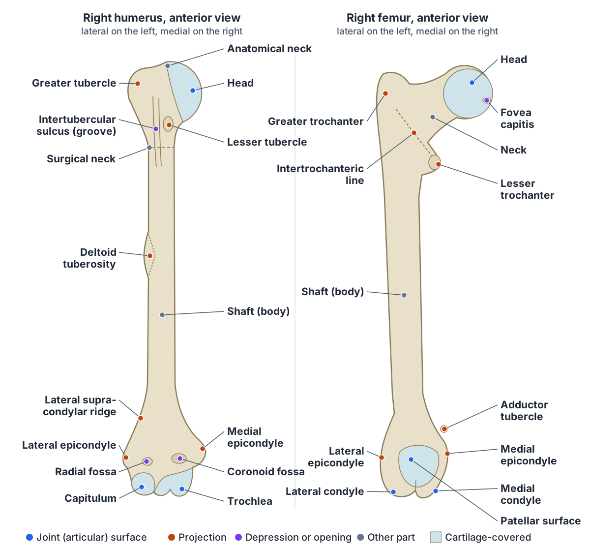
In anatomy, a tubercle is a small, rounded prominence on a bone, often where a tendon or ligament attaches. A tuberosity is a larger prominence, often with a rough surface. Examples are the adductor tubercle of the femur and the ulnar tuberosity. Kenhub notes that the two words are often confused. (Kenhub: Complete list of bone markings.)
OpenStax defines a tubercle as a small, rounded process and a tuberosity as a rough surface. On the humerus, the greater tubercle is an expanded area on the lateral side of the proximal end and the smaller lesser tubercle lies on its anterior aspect, with the narrow intertubercular groove (sulcus) between them. The deltoid tuberosity is a roughened, V-shaped area on the lateral shaft where the deltoid attaches. (OpenStax Anatomy and Physiology 2e, 8.2: Bones of the Upper Limb.)
Usage is not fully consistent. TeachMeAnatomy calls the same two humeral prominences the greater and lesser tuberosities, and gives their attachments: supraspinatus, infraspinatus and teres minor on the greater, and subscapularis on the lesser. (TeachMeAnatomy: The Humerus.)
On the lower limb, the tibial tuberosity is an elevated area on the front of the tibia near its proximal end, and the adductor tubercle is a small bump at the top of the medial epicondyle of the femur. (OpenStax Anatomy and Physiology 2e, 8.4: Bones of the Lower Limb.)
A trochanter is the largest of these projections: a large, blunt, irregular process found only on the femur, as the greater and lesser trochanters. (Kenhub: Complete list of bone markings.)
What is a condyle? Condyle vs epicondyle
A condyle is a rounded, knob-like surface at the end of a bone that forms a joint with another bone, such as the lateral condyle of the femur. An epicondyle is a projection on or just above a condyle; it does not form a joint but gives attachment to muscles and ligaments. (Kenhub: Complete list of bone markings.)
The distal femur shows the difference. Its medial and lateral condyles are smooth and articulate with the tibia below and the patella in front, while the epicondyles are non-articular elevations on the condyles, the medial being larger, from which the medial and lateral collateral ligaments of the knee arise. (TeachMeAnatomy: The Femur.)
OpenStax describes the same region: the epicondyles provide attachment for muscles and the supporting ligaments of the knee, a deep intercondylar fossa separates the condyles behind, and a wide groove at the front, the patellar surface, holds the patella. (OpenStax Anatomy and Physiology 2e, 8.4: Bones of the Lower Limb.)
At the elbow, the humerus has a much larger medial epicondyle and a smaller lateral one. The joint surfaces between them have their own names: the trochlea, a pulley-shaped surface on the medial side that articulates with the ulna, and the capitulum, a knob-like surface lateral to it that articulates with the radius. (OpenStax Anatomy and Physiology 2e, 8.2: Bones of the Upper Limb.)
The ulnar nerve passes in a groove on the back of the medial epicondyle, and both epicondyles lie outside the elbow joint capsule. (TeachMeAnatomy: The Humerus.)
The thigh bone's head, neck, trochanters, condyles and epicondyles in 3D. Femur.
What is a fossa in anatomy? Fossae, foramina and other openings
A fossa is a shallow depression or basin in a bone, often receiving another bone or holding a structure. Kenhub defines it as a broad, often shallow depression that may house brain structures or receive another bone, such as the temporal fossa of the skull. A fovea is similar but much smaller, such as the fovea capitis on the head of the femur. (Kenhub: Complete list of bone markings.)
Fossae on the humerus receive the forearm bones as the elbow moves. The coronoid fossa above the trochlea receives the coronoid process of the ulna when the elbow is flexed, the radial fossa above the capitulum receives the head of the radius, and the larger olecranon fossa on the back receives the olecranon when the elbow is fully extended. (OpenStax Anatomy and Physiology 2e, 8.2: Bones of the Upper Limb.)
The scapula has three named fossae: the shallow subscapular fossa on its costal (front) surface, and the supraspinous and infraspinous fossae on its back, separated by the scapular spine. Laterally, the glenoid fossa receives the head of the humerus. (Radiopaedia: Scapula.)
Openings have separate names. A foramen is a hole through which nerves and blood vessels pass, such as the jugular foramen; a fissure is an open slit, such as the inferior orbital fissure; a meatus is a short canal, such as the external acoustic meatus (ear canal); a notch is an indentation at the edge of a bone; a sulcus is a groove; and a sinus is a cavity, such as the paranasal sinuses. (Kenhub: Complete list of bone markings.)
OpenStax describes a fossa as an elongated basin, such as the mandibular fossa of the jaw joint, and gives the foramen magnum in the occipital bone as its example of a foramen. It defines a canal as a passage through bone and a sinus as an air-filled space in bone. (OpenStax Anatomy and Physiology 2e, 6.3: Bone Structure.)
Every major foramen of the skull and what passes through it. Skull foramina.
Processes, spines, crests, lines and heads
A process is any prominent projection, such as the transverse process of a vertebra. A spine is a sharp process, such as the ischial spine; a crest is a ridge, such as the iliac crest; a line is a slight, elongated ridge, such as the temporal lines of the parietal bones; and a protuberance is a part that protrudes outward, such as the chin. (OpenStax Anatomy and Physiology 2e, 6.3: Bone Structure.)
Kenhub's examples include the mastoid process behind the ear, the external occipital protuberance at the back of the skull, the linea aspera of the femur, the iliac crest and the spine of the sphenoid bone. It describes a facet as a flat, smooth articular area, such as the acromial facet of the clavicle. (Kenhub: Complete list of bone markings.)
The linea aspera is the roughened ridge down the back of the femoral shaft. Above, its lateral lip continues as the gluteal tuberosity, where gluteus maximus attaches, and its medial lip as the pectineal line. (TeachMeAnatomy: The Femur.)
A head is a prominent rounded end that forms a joint, and a neck is the narrower part that joins it to the rest of the bone. The rounded proximal end of the femur is its head, and the narrowed region below it is the neck; the greater trochanter projects upwards above the base of the neck and the lesser trochanter lies on the medial side. (OpenStax Anatomy and Physiology 2e, 8.4: Bones of the Lower Limb.)
The humerus has two necks: the anatomical neck, which separates the head from the tubercles, and the surgical neck below them, where the axillary nerve and circumflex humeral vessels lie against the bone. (TeachMeAnatomy: The Humerus.)
The upper arm bone with its tubercles, necks, epicondyles and fossae in 3D. Humerus.
Bone markings glossary
| Marking | Type | What it is | Example |
|---|---|---|---|
| Head | Joint surface | Prominent rounded end of a bone | Head of the femur |
| Condyle | Joint surface | Rounded knob that forms a joint | Occipital condyles; condyles of the femur |
| Facet | Joint surface | Flat, smooth articular area | Acromial facet of the clavicle; facets of vertebrae |
| Trochlea | Joint surface | Pulley-shaped articular surface | Trochlea of the humerus (with the ulna) |
| Capitulum | Joint surface | Small knob-like articular surface | Capitulum of the humerus (with the radius) |
| Process | Projection | Any prominent outgrowth | Transverse process of a vertebra; mastoid process |
| Spine | Projection | Sharp, slender process | Ischial spine; spine of the scapula |
| Tubercle | Projection | Small, rounded prominence | Greater tubercle of the humerus; adductor tubercle |
| Tuberosity | Projection | Larger, often rough prominence | Deltoid tuberosity; tibial tuberosity |
| Trochanter | Projection | Very large, blunt process (femur only) | Greater and lesser trochanters |
| Epicondyle | Projection | Non-articular prominence on or above a condyle | Medial epicondyle of the humerus |
| Crest | Projection | Prominent ridge | Iliac crest |
| Line (linea) | Projection | Slight, long ridge | Linea aspera; temporal lines |
| Protuberance | Projection | Swelling that protrudes outward | External occipital protuberance; chin |
| Fossa | Depression | Shallow basin | Olecranon fossa; mandibular fossa; supraspinous fossa |
| Fovea | Depression | Small pit | Fovea capitis of the femur |
| Sulcus (groove) | Depression | Groove, often for a vessel, nerve or tendon | Intertubercular sulcus of the humerus |
| Notch (incisure) | Depression | Indentation at the edge of a bone | Suprascapular notch |
| Foramen | Opening | Hole for nerves and blood vessels | Foramen magnum; jugular foramen |
| Fissure | Opening | Open slit or narrow cleft | Inferior orbital fissure |
| Meatus | Opening | Short canal into a bone | External acoustic meatus |
| Sinus | Cavity | Air-filled space in a bone | Paranasal sinuses |
Sources: OpenStax 6.3 (Table 6.2), 8.2 and 8.4; Kenhub, Complete list of bone markings; Radiopaedia, Scapula.
Parts of a long bone: diaphysis, epiphysis, periosteum
A long bone has a tubular shaft, the diaphysis, between its proximal and distal ends. Its hollow centre is the medullary cavity, filled with yellow marrow, and its walls are compact bone. Each wider end is an epiphysis, filled with spongy bone and red marrow; in adults the epiphysis meets the diaphysis at the metaphysis. (OpenStax Anatomy and Physiology 2e, 6.3: Bone Structure.)
The periosteum is the fibrous membrane covering the outer surface of the bone, except where joints form; it carries blood vessels, nerves and lymphatic vessels, and tendons and ligaments attach to it. The endosteum is a delicate membrane lining the medullary cavity, where bone growth, repair and remodelling take place. (OpenStax Anatomy and Physiology 2e, 6.3: Bone Structure.)
Compact bone is dense and strong and lies beneath the periosteum; its basic unit is the osteon. Spongy (cancellous) bone is a lighter lattice of struts called trabeculae, whose spaces in some bones hold red marrow, where blood cells form. (OpenStax Anatomy and Physiology 2e, 6.3: Bone Structure.)
Terms of movement: what does abduction mean in anatomy?
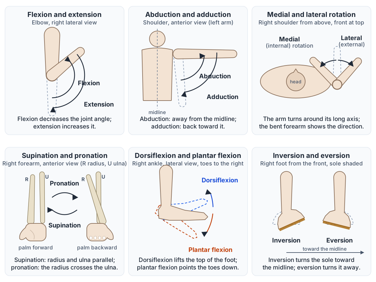
In anatomy, abduction moves a limb laterally, away from the midline of the body, as when you raise your arm out to the side or spread your fingers. Its opposite, adduction, brings the limb back toward the body or across the midline. Both are described from the anatomical position, and most movements come in such opposing pairs. (OpenStax Anatomy and Physiology 2e, 9.5: Types of Body Movements.)
Flexion decreases the angle between the bones of a joint and extension increases it, straightening the joint. Bending the elbow or knee is flexion. Extension beyond the normal range of motion is hyperextension, which can injure the knee or elbow, and bending the neck or trunk to one side is lateral flexion. (OpenStax Anatomy and Physiology 2e, 9.5: Types of Body Movements.)
TeachMeAnatomy notes two exceptions to the midline rule: for the fingers and toes, abduction and adduction are measured from the midline of the hand or foot, so spreading the fingers is abduction; and circumduction, often described as circular, is more accurately a conical movement of a limb driven from its joint of origin. (TeachMeAnatomy: Anatomical Terms of Movement.)
OpenStax describes circumduction as a sequence of flexion, abduction, extension and adduction in which one end of the limb stays relatively still and the other end traces a circle. It is possible at biaxial condyloid and saddle joints and multiaxial ball-and-socket joints. (OpenStax Anatomy and Physiology 2e, 9.5: Types of Body Movements.)
Which synovial joints allow which movements, from hinge to ball-and-socket. Types of joints.
Rotation, pronation and supination
Rotation turns a bone around its own long axis. At the shoulder and hip, medial (internal) rotation turns the anterior surface of the limb toward the midline and lateral (external) rotation turns it away. In the neck, the atlas (C1) rotates around the dens of the axis (C2) when you shake your head to say no. (OpenStax Anatomy and Physiology 2e, 9.5: Types of Body Movements.)
Pronation moves the forearm from the supinated position to the palm-backward position: the radius rotates at the proximal and distal radioulnar joints and crosses over the ulna, so the two bones form an X. Supination returns the palm forward, with the radius and ulna parallel again. (OpenStax Anatomy and Physiology 2e, 9.5: Types of Body Movements.)
TeachMeAnatomy describes the same movements with the elbow bent: supination turns the palm upward and pronation turns it downward, and it notes the subtle difference between these and medial and lateral rotation. (TeachMeAnatomy: Anatomical Terms of Movement.)
Kenhub defines pronation and supination as medial and lateral rotation of the radius, which is why they are named separately from medial and lateral rotation of the whole limb at the shoulder. (Kenhub: Anatomical terminology: planes, directions and regions.)
Movements of the foot, jaw, shoulder and thumb
Dorsiflexion lifts the front of the foot so its top moves toward the shin, and plantar flexion lifts the heel or points the toes down; these are the only movements of the ankle joint itself. Inversion turns the sole toward the midline and eversion turns it away; they happen at the intertarsal joints, not the ankle, and the foot inverts further than it everts. (OpenStax Anatomy and Physiology 2e, 9.5: Types of Body Movements.)
At the wrist, TeachMeAnatomy notes that the equivalent of dorsiflexion is simply called extension, and the hand's equivalent of plantarflexion is palmarflexion. It also notes that opposition of the thumb and little finger is limited to humans and some great apes. (TeachMeAnatomy: Anatomical Terms of Movement.)
Kenhub adds deviation: moving the hand toward the radial (thumb) side or the ulnar (little-finger) side at the wrist. (Kenhub: Anatomical terminology: planes, directions and regions.)
Elevation moves a part up and depression moves it down: shrugging elevates the scapulae, and elevating the mandible closes the mouth. Protraction moves the scapula or mandible forward and retraction pulls it back. Side-to-side movement of the mandible is excursion, lateral away from the midline and medial back to it. (OpenStax Anatomy and Physiology 2e, 9.5: Types of Body Movements.)
Opposition brings the tip of the thumb into contact with the tip of a finger and happens at the first carpometacarpal joint, a saddle joint, by combining flexion and abduction. Reposition returns the thumb to its place beside the index finger. (OpenStax Anatomy and Physiology 2e, 9.5: Types of Body Movements.)
Terms of movement and their opposites
| Movement | Opposite | What happens | Example |
|---|---|---|---|
| Flexion | Extension | Decreases the angle between bones | Bending the elbow or knee |
| Extension | Flexion | Increases the angle, straightening the joint | Straightening the knee |
| Lateral flexion | None (return to upright) | Bends the neck or trunk to one side | Tilting the trunk to the right |
| Abduction | Adduction | Moves a limb away from the midline | Raising the arm out to the side; spreading the fingers |
| Adduction | Abduction | Moves a limb toward or across the midline | Lowering the arm to the side |
| Circumduction | None | Conical movement combining flexion, abduction, extension and adduction | Circling the arm at the shoulder |
| Medial (internal) rotation | Lateral (external) rotation | Turns the front of the limb toward the midline | Shoulder and hip |
| Pronation | Supination | Turns the palm backward (or down); the radius crosses the ulna | Forearm |
| Supination | Pronation | Turns the palm forward (or up); radius and ulna parallel | Forearm |
| Dorsiflexion | Plantar flexion | Lifts the top of the foot toward the shin | Ankle |
| Plantar flexion | Dorsiflexion | Points the toes down or lifts the heel | Standing on tiptoe |
| Inversion | Eversion | Turns the sole toward the midline | Intertarsal joints of the foot |
| Elevation | Depression | Moves a part upward | Shrugging; closing the mouth |
| Protraction | Retraction | Moves a part forward | Reaching forward; jutting the jaw |
| Excursion | Medial excursion (back to the midline) | Side-to-side movement of the mandible | Moving the jaw from side to side |
| Opposition | Reposition | Brings the thumb tip to a fingertip | First carpometacarpal joint |
Sources: OpenStax 9.5, TeachMeAnatomy terms of movement and Kenhub anatomical terminology.
What is fascia? Fascia, aponeurosis, tendon and ligament
Fascia is connective tissue found below the skin. Superficial fascia is loose connective tissue beneath the dermis, with a fatty layer and a membranous layer; deep fascia is denser fibrous tissue that envelops the muscles, such as the transversalis fascia, the fascia lata of the thigh and the plantar fascia of the foot. (Radiopaedia: Fascia.)
There is no full agreement on fascia terminology. Terminologia Anatomica replaced superficial fascia with subcutaneous tissue or hypodermis, surgeons generally use fascia to mean deep fascia, and some authors call the connective tissue that lines body cavities and supports organs visceral fascia. (Radiopaedia: Fascia.)
Inside a skeletal muscle, connective tissue forms three wrappings: the epimysium around the whole muscle, the perimysium around bundles of fibres (fascicles) and the endomysium around each fibre. At the end of the muscle these continue as a tendon, which fuses with the periosteum of the bone, or as an aponeurosis, a broad, flat, tendon-like sheet. (OpenStax Anatomy and Physiology 2e, 10.2: Skeletal Muscle.)
A tendon is the dense connective tissue that attaches a muscle to bone, while ligaments are strong bands of fibrous connective tissue that hold bones together. Near some joints, a bursa, a thin connective tissue sac of lubricating fluid, and a tendon sheath around a tendon reduce friction. (OpenStax Anatomy and Physiology 2e, 9.4: Synovial Joints.)
Lumen, cortex, medulla and hilum
In anatomy, the lumen is the hollow passageway inside a tube-shaped structure, such as a blood vessel. Arteries have smaller lumens than veins, which helps maintain blood pressure, while veins are thin-walled vessels with large, irregular lumens. Around the lumen, a vessel wall has three layers: the tunica intima, tunica media and tunica externa. (OpenStax Anatomy and Physiology 2e, 20.1: Structure and Function of Blood Vessels.)
The cortex is the outer layer of an organ and the medulla is its inner region. A frontal section of a kidney shows an outer renal cortex, lighter in colour, and an inner medulla; the cortex contains the renal corpuscles and convoluted tubules. (OpenStax Anatomy and Physiology 2e, 25.3: Gross Anatomy of the Kidney.)
The adrenal gland has an outer cortex of glandular tissue, in three layers of lipid-storing cells, and an inner medulla of nervous tissue made of postganglionic sympathetic neurons. (OpenStax Anatomy and Physiology 2e, 17.6: The Adrenal Glands.)
Lymph nodes have the same plan: lymphoid follicles in the outer cortex and medullary cords of B cells and plasma cells in the inner medulla, from which lymph leaves through efferent lymphatic vessels. (OpenStax Anatomy and Physiology 2e, 21.1: Anatomy of the Lymphatic and Immune Systems.)
A hilum is the region where vessels, nerves and ducts enter and leave an organ. The renal hilum is the entry and exit site for the vessels, nerves, lymphatics and ureter of the kidney, and the renal pelvis emerges from it. (OpenStax Anatomy and Physiology 2e, 25.3: Gross Anatomy of the Kidney.)
Each lung also has a hilum, through which the pulmonary veins leave and the nerves of the pulmonary plexus enter; the visceral and parietal pleurae join each other there. (OpenStax Anatomy and Physiology 2e, 22.2: The Lungs.)
Renal cortex, medulla, pyramids and hilum in 3D and in section. Kidney.
Nervous system terms: grey matter, ganglion, nerve and plexus
In the peripheral nervous system, a ganglion is a group of neuron cell bodies, and a nerve is a bundle of axons. A plexus, in a general sense, is a network of fibres or vessels; the cervical, brachial, lumbar and sacral plexuses are networks of spinal nerves that give rise to the nerves of the neck, arm and leg. (OpenStax Anatomy and Physiology 2e, 13.4: The Peripheral Nervous System.)
In the brain, the cerebral cortex is a continuous layer of grey matter wrapping the forebrain, folded into ridges (gyri) separated by grooves (sulci). White matter carries signals between regions: the corpus callosum is the main pathway between the two hemispheres, and the white matter of the spinal cord is divided into columns. (OpenStax Anatomy and Physiology 2e, 13.2: The Central Nervous System.)
What does oblique (obliquely) mean in anatomy?
Oblique means at an angle: a structure that runs obliquely slants across the body, neither straight up and down nor straight across. OpenStax uses it this way when naming muscles by the direction of their fibres: rectus means straight, and the oblique muscles of the abdomen run at an angle. (OpenStax Anatomy and Physiology 2e, 11.2: Naming Skeletal Muscles.)
The two abdominal obliques show it well. The fibres of the external oblique run inferomedially, down and toward the midline, and the fibres of the internal oblique run superomedially, at right angles to them, while transversus abdominis runs transversely. (TeachMeAnatomy: The Abdominal Wall.)
The word also names a plane: an oblique plane cuts the body at an angle to the sagittal, frontal and transverse planes. (OpenStax Anatomy and Physiology 2e, 1.6: Anatomical Terminology.)
In radiology, an oblique projection is one in which the central X-ray beam passes through the body along a plane at an angle to the transverse or coronal plane, often used for angled views of the hand or foot. (Radiopaedia: Radiographic positioning terminology.)
External and internal obliques, transversus and rectus abdominis in 3D. Abdominal muscles.
How muscle names use anatomical terms
Muscle names put these terms together. They describe location (frontalis lies over the frontal bone), size (gluteus maximus, medius and minimus), length (longus and brevis), position relative to the midline (lateralis and medialis), fibre direction (rectus and oblique), number of origins (biceps, triceps), number of muscles in a group (quadriceps) and action (flexor, extensor, abductor, adductor). (OpenStax Anatomy and Physiology 2e, 11.2: Naming Skeletal Muscles.)
Where a name lists attachments, the origin comes first. The sternocleidomastoid arises from the sternum (sterno) and clavicle (cleido) and inserts on the mastoid process, a bone marking. (OpenStax Anatomy and Physiology 2e, 11.2: Naming Skeletal Muscles.)
Brachial, femoral, gluteal and the other region names used in muscle and nerve names. Regional terms.
Anatomical terms in the 3D model
- Femur
- The thigh bone, the longest, heaviest and strongest bone in the body.
- Hip bone
- The large irregular bone of the pelvis formed by the fusion of the ilium, ischium and pubis.
- Humerus
- The long bone of the upper arm.
- Mandible
- The lower jaw bone.
- Occipital bone
- The bone forming the back and base of the skull.
- Radius
- The lateral bone of the forearm.
- Scapula
- The shoulder blade, a flat triangular bone on the back of the thorax.
- Temporal bone
- A paired bone forming part of the side and base of the skull.
- Tibia
- The shinbone, the medial and main weight-bearing bone of the leg.
- Ulna
- The medial bone of the forearm.
Frequently asked questions
What is a tubercle in anatomy?
A tubercle is a small, rounded bump on a bone, usually where a tendon or ligament attaches. Examples are the greater and lesser tubercles of the humerus, where rotator cuff muscles attach, and the adductor tubercle of the femur, where adductor magnus attaches. A larger, rougher bump is called a tuberosity, such as the deltoid or tibial tuberosity.
What is the difference between a condyle and an epicondyle?
A condyle is a rounded joint surface at the end of a bone, covered in cartilage, such as the femoral condyles that rest on the tibia. An epicondyle is a projection on or just above a condyle that does not form a joint; muscles and ligaments attach to it, such as the medial epicondyle of the humerus.
What is a fossa in anatomy?
A fossa is a shallow depression or basin in a bone, often where another bone or a muscle fits. The olecranon fossa on the back of the humerus receives the olecranon of the ulna when the elbow straightens, the mandibular fossa holds the jaw joint, and the supraspinous and infraspinous fossae lie above and below the spine of the scapula.
What does abduction mean in anatomy?
Abduction is movement of a limb away from the midline of the body, such as raising the arm out to the side. Its opposite, adduction, brings the limb back toward the body. For the fingers and toes the reference line is the midline of the hand or foot, so spreading the fingers apart is abduction.
What is fascia?
Fascia is connective tissue beneath the skin. Superficial fascia, now usually called subcutaneous tissue, is a loose fatty layer under the dermis. Deep fascia is denser fibrous tissue that wraps muscles, such as the fascia lata of the thigh and the plantar fascia under the foot.
What does lumen mean in anatomy?
The lumen is the hollow space inside a tube-shaped structure. In a blood vessel it is the passageway through which blood flows, surrounded by the three layers of the vessel wall: tunica intima, tunica media and tunica externa. Veins have larger, more irregular lumens than arteries of similar size.
What is the cortex of an organ?
The cortex is the outer layer of an organ, and the medulla is its inner part. The kidney has an outer renal cortex containing the renal corpuscles and an inner medulla; the adrenal gland has a glandular cortex around a medulla of nervous tissue; and the cerebral cortex is the outer grey matter of the brain.
What does obliquely mean in anatomy?
Obliquely means at a slant, running at an angle rather than straight up and down or straight across. The external oblique muscle's fibres run obliquely down and toward the midline, and the internal oblique's run up and toward the midline. An oblique plane or X-ray view cuts the body at an angle to the standard planes.
References
- OpenStax. Anatomy and Physiology 2e, §1.6 Anatomical Terminology. Rice University, 2022.
- OpenStax. Anatomy and Physiology 2e, §6.3 Bone Structure. Rice University, 2022.
- OpenStax. Anatomy and Physiology 2e, §8.2 Bones of the Upper Limb. Rice University, 2022.
- OpenStax. Anatomy and Physiology 2e, §8.4 Bones of the Lower Limb. Rice University, 2022.
- OpenStax. Anatomy and Physiology 2e, §9.4 Synovial Joints. Rice University, 2022.
- OpenStax. Anatomy and Physiology 2e, §9.5 Types of Body Movements. Rice University, 2022.
- OpenStax. Anatomy and Physiology 2e, §10.2 Skeletal Muscle. Rice University, 2022.
- OpenStax. Anatomy and Physiology 2e, §11.2 Naming Skeletal Muscles. Rice University, 2022.
- OpenStax. Anatomy and Physiology 2e, §13.2 The Central Nervous System. Rice University, 2022.
- OpenStax. Anatomy and Physiology 2e, §13.4 The Peripheral Nervous System. Rice University, 2022.
- OpenStax. Anatomy and Physiology 2e, §17.6 The Adrenal Glands. Rice University, 2022.
- OpenStax. Anatomy and Physiology 2e, §20.1 Structure and Function of Blood Vessels. Rice University, 2022.
- OpenStax. Anatomy and Physiology 2e, §21.1 Anatomy of the Lymphatic and Immune Systems. Rice University, 2022.
- OpenStax. Anatomy and Physiology 2e, §22.2 The Lungs. Rice University, 2022.
- OpenStax. Anatomy and Physiology 2e, §25.3 Gross Anatomy of the Kidney. Rice University, 2022.
- Smith M. Complete list of bone markings. Kenhub, reviewed 2023.
- Rad A. Anatomical terminology: planes, directions and regions. Kenhub, reviewed 2023.
- TeachMeAnatomy. Anatomical Terms of Movement.
- TeachMeAnatomy. The Humerus.
- TeachMeAnatomy. The Femur.
- TeachMeAnatomy. The Scapula.
- TeachMeAnatomy. The Abdominal Wall.
- Deng F. Fascia. Radiopaedia.org (rID 68862).
- Luijkx T, et al. Scapula. Radiopaedia.org (rID 30653).
- Radiographic positioning terminology. Radiopaedia.org reference article.
- MedlinePlus, US National Library of Medicine. Appendix A: Word Parts and What They Mean.
- National Cancer Institute SEER Training Modules. Prefixes, Word Roots, and Suffixes.
- National Cancer Institute SEER Training Modules. Formation of Medical Terms.
- Mitsuhashi N. et al. (2009). BodyParts3D: 3D structure database for anatomical concepts. Nucleic Acids Research 37, D782–D785. Data © The Database Center for Life Science, CC BY 4.0.
Related anatomy
- Directional termsAnatomical position and directional terms with labelled diagrams: what proximal, distal, medial, lateral, anterior…
- Body regionsRegional terms in anatomy with front and back body-region diagrams: cephalic, cervical, brachial, femoral, crural and…
- Anatomical planesAnatomical planes explained: midsagittal vs parasagittal, coronal (frontal) and transverse (axial) planes, axes of…
- Types of jointsTypes of joints explained: fibrous, cartilaginous and synovial (diarthrosis) joints, a labelled synovial joint diagram…
- Skeletal systemExplore a realistic 3D human skeleton: the 206 bones, axial vs appendicular skeleton, bone types, bony landmarks and…
- Bone structureLong bone anatomy with a labeled section and 3D femur: diaphysis, epiphyses, growth plate, periosteum, marrow, compact…
Explore the whole body in the interactive 3D human anatomy model, or browse the A–Z list of human structures.