

What is the anatomical position?
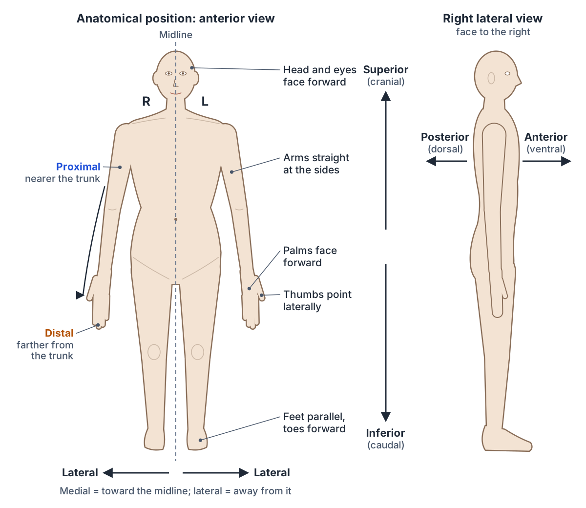
The anatomical position is the standard reference posture of the body: standing upright and facing forward, with the mouth closed, the arms straight and the hands by the hips with the palms facing forward, and the feet parallel with the toes pointing forward. Anatomists describe the location of every structure from this posture, so the description is clear and the same for everyone. (TeachMeAnatomy: Anatomical Position.)
Radiopaedia adds the details that matter for the bones and joints: the hips and knees are extended, the head faces forward with the eyes open and looking straight ahead, the shoulders are adducted so the arms hang at the sides, and the elbows are extended with the forearms supinated, so the radius and ulna lie parallel. (Radiopaedia: Anatomical position.)
Because the palms face forward, the thumbs point away from the body, that is, laterally. Lecturio describes the palms facing forward with the thumbs pointing laterally, and the head neutral and in line with the spine. (Lecturio: What is the anatomical position?.)
Sources describe the feet slightly differently. OpenStax places them at shoulder width and parallel, with the toes forward and the palms facing forward. Kenhub describes the feet as slightly apart, while Radiopaedia and TeachMeAnatomy put them together and parallel. All agree the toes point forward, and the difference does not change the meaning of any directional term. (OpenStax Anatomy and Physiology 2e, 1.6: Anatomical Terminology.)
Anatomical terms are always applied as if the body were in the anatomical position, whatever its real posture. OpenStax gives the example of a scar in the anterior carpal (wrist) region: it lies on the palm side of the wrist, and it is still called anterior when the hand rests palm down on a table. (OpenStax Anatomy and Physiology 2e, 1.6: Anatomical Terminology.)
Right and left always mean the subject's own sides, not the viewer's. When you look at a person or a picture from the front, their right side is on your left. The diagrams on this page mark the subject's right with an R. (OpenStax Anatomy and Physiology 2e, 1.6: Anatomical Terminology.)
Sagittal, frontal (coronal) and transverse planes, midsagittal vs parasagittal, and the axes of movement. Anatomical planes.
Anatomical position vs supine, prone and other positions
Supine describes a face-up orientation and prone a face-down orientation. Both terms are used in physical examinations and surgical procedures, and neither changes the directional terms: the front of the body is still anterior when a patient lies face down. (OpenStax Anatomy and Physiology 2e, 1.6: Anatomical Terminology.)
Radiographers name other positions too. Erect means standing or sitting, supine is lying on the back and prone is lying face down. In a lateral decubitus position the patient lies on one side, and in the Trendelenburg position the patient lies supine on a tilted table with the head lower than the feet. (Radiopaedia: Radiographic positioning terminology.)
Radiopaedia stresses that the anatomical position is the reference whether the patient is standing, supine, prone or sitting. Because gravity moves the internal organs, their positions are often described with the patient supine, as for the surface anatomy of the liver. (Radiopaedia: Anatomical position.)
The same words describe the forearm. Supination turns the palm upwards, so it lies supine, and pronation turns it downwards, so it lies prone. TeachMeAnatomy notes that the whole body is likewise supine when lying face up and prone when lying face down. (TeachMeAnatomy: Anatomical Terms of Movement.)
What are directional terms in anatomy?
Directional terms are anatomical words that describe where one body part lies in relation to another. They come in opposing pairs: anterior and posterior, superior and inferior, medial and lateral, proximal and distal, and superficial and deep, plus ventral and dorsal, cranial and caudal, and the hand and foot terms palmar, plantar and dorsal. (Kenhub: Anatomical terminology: planes, directions and regions.)
The terms are relative: a structure is only superior or medial compared with something else. OpenStax gives one example for each: the toes are anterior to the foot, the popliteus is posterior to the patella, the orbits (eye sockets) are superior to the oris (mouth), the pelvis is inferior to the abdomen, the brachium is proximal to the antebrachium, and the brain is deep to the skull. (OpenStax Anatomy and Physiology 2e, 1.6: Anatomical Terminology.)
Anatomical terms come from ancient Greek and Latin. Because these languages are no longer used in everyday conversation, the meanings of their words do not drift, which keeps the vocabulary stable. (OpenStax Anatomy and Physiology 2e, 1.6: Anatomical Terminology.)
The international standard for these names is Terminologia Anatomica. Radiopaedia groups the location words into relational terms (anterior, posterior, superior, inferior, medial, lateral, proximal, distal, superficial and deep), traditional terms (dorsal, ventral, caudal, cephalad, rostral, volar or palmar, and plantar) and terms of laterality (unilateral, bilateral, ipsilateral and contralateral). (Radiopaedia: Anatomical position.)
Bone markings, terms of movement, tissue words and the Greek and Latin roots behind them. Anatomical terms glossary.
Directional terms chart: each term, its opposite and an example
| Term | Meaning | Opposite | Example |
|---|---|---|---|
| Superior (cranial) | Above; toward the head | Inferior (caudal) | The head is superior to the neck |
| Inferior (caudal) | Below; toward the feet or tail | Superior (cranial) | The pelvis is inferior to the abdomen |
| Anterior (ventral) | Toward the front | Posterior (dorsal) | The toes are anterior to the foot |
| Posterior (dorsal) | Toward the back | Anterior (ventral) | The heart is posterior to the sternum |
| Medial | Toward the midline | Lateral | The nose is medial to the ears |
| Lateral | Away from the midline, toward the side | Medial | The eye is lateral to the nose |
| Proximal | Closer to a limb's attachment or a structure's origin | Distal | The knee joint is proximal to the ankle joint |
| Distal | Farther from the attachment or origin | Proximal | The wrist joint is distal to the elbow joint |
| Superficial (external) | Closer to the body surface | Deep (internal) | The skin is superficial to the bones |
| Deep (internal) | Farther from the body surface | Superficial (external) | The brain is deep to the skull |
| Ipsilateral | On the same side of the body | Contralateral | Left elbow and left shoulder |
| Contralateral | On the opposite side of the body | Ipsilateral | Right wrist and left wrist |
| Rostral (brain) | Toward the front of the cerebrum, or upward in the brainstem | Caudal | Above the midbrain, rostral means anterior |
| Palmar (volar) | Palm side of the hand | Dorsal (back of hand) | A scar on the palm side of the wrist |
| Plantar | Sole of the foot | Dorsal (top of foot) | The plantar fascia lies in the sole |
| Radial | Thumb side of the forearm and hand | Ulnar (little-finger side) | The radius is lateral to the ulna |
| Parietal | Lining the wall of a body cavity | Visceral (covering an organ) | Parietal and visceral pleura |
Sources: OpenStax 1.6, TeachMeAnatomy, Kenhub, Radiopaedia and OpenStax 8.2. All terms assume the anatomical position.
What does superior mean in anatomy? Superior vs inferior
In anatomy, superior means above, or closer to the head, and inferior means below, or closer to the feet. The head is superior to the neck and the umbilicus (navel) is inferior to the sternum. Other examples: the nose is superior to the mouth, the lungs are superior to the liver, and the appendix usually lies inferior to the transverse colon. (TeachMeAnatomy: Anatomical Terms of Location.)
OpenStax pairs each term with an older synonym: superior (cranial) means above or higher than another part of the body, and inferior (caudal) means below, toward the tail. (OpenStax Anatomy and Physiology 2e, 1.6: Anatomical Terminology.)
Kenhub defines superior as toward the top of the head and inferior as toward the feet, and lists cranial (toward the head) and caudal (toward the tail) as a separate pair. (Kenhub: Anatomical terminology: planes, directions and regions.)
Superior and inferior can mislead in the limbs, because the limbs move. Lift your arm above your head and your hand becomes higher than your elbow. TeachMeAnatomy points out that a change in limb position can reverse superior and inferior, which is why the limbs need a second pair of terms that does not depend on posture: proximal and distal. (TeachMeAnatomy: Anatomical Terms of Location.)
What does anterior mean in anatomy? Anterior vs posterior
Anterior means toward the front of the body and posterior means toward the back. The heart is posterior to the sternum, the triceps lie posterior to biceps brachii, pectoralis major is anterior to pectoralis minor, and the patella sits on the anterior side of the knee. (TeachMeAnatomy: Anatomical Terms of Location.)
OpenStax writes the pairs as anterior (ventral) and posterior (dorsal): in a standing human, the front of the body is also its ventral side and the back is its dorsal side. Its examples are the toes, which are anterior to the foot, and the popliteus muscle behind the knee, which is posterior to the patella. (OpenStax Anatomy and Physiology 2e, 1.6: Anatomical Terminology.)
Images are named the same way. An anterior view shows the body from the front and a posterior view from the back. In an AP (anteroposterior) radiograph the central X-ray beam passes from anterior to posterior, at right angles to the coronal plane; in a PA projection it passes from posterior to anterior. (Radiopaedia: Radiographic positioning terminology.)
Ventral vs dorsal: why the terms change in animals and the brain
Ventral means toward the belly and dorsal toward the back. On the human trunk they match anterior and posterior, but veterinary anatomy uses them differently: in a four-legged animal the belly faces the ground, so ventral points down and dorsal points up. The University of Minnesota's veterinary anatomy guide lists human anterior as conflicting with veterinary ventral, and human posterior with veterinary dorsal. (University of Minnesota Veterinary Anatomy: Veterinary vs human directional terms.)
The same guide explains that on the trunk, human superior corresponds to veterinary cranial and human inferior to veterinary caudal. In the head the terms do not conflict, because an animal's head points the same way as its body, while the human head sits at a right angle to the upright trunk. (University of Minnesota Veterinary Anatomy: Veterinary vs human directional terms.)
That right angle also bends the human brain's axis by roughly 90 degrees around the brainstem, so the terms rotate with it. Above the midbrain, rostral means anterior, caudal means posterior, dorsal means superior and ventral means inferior. Below the midbrain, in the brainstem and spinal cord, rostral means superior, caudal inferior, dorsal posterior and ventral anterior. (Kenhub: Brain orientation difficulties.)
So the dorsal horn of the spinal cord points backwards, while the dorsal surface of the cerebrum faces upwards. When a sentence about the brain uses rostral or dorsal, check which part of the nervous system it describes.
Lobes, brainstem and cerebellum, with the brain's own orientation terms in 3D. Brain anatomy.
What does medial mean in anatomy? Medial vs lateral
Medial means closer to the midline, the imaginary line that divides the body into equal right and left halves, and lateral means farther from it, toward the side. The eye is lateral to the nose, the nose is medial to the ears, and at the elbow the brachial artery lies medial to the biceps tendon. (TeachMeAnatomy: Anatomical Terms of Location.)
Kenhub separates three related words: median is the midline itself, medial is toward the median, and lateral is away from it. (Kenhub: Anatomical terminology: planes, directions and regions.)
Because the palms face forward in the anatomical position, the thumb (pollex) is lateral to the other digits, and the hallux, the big toe, is the medial toe. (OpenStax Anatomy and Physiology 2e, 1.6: Anatomical Terminology.)
Medial and lateral also put structures in order across a region. In the popliteal fossa behind the knee, the contents run from medial to lateral as the popliteal artery, popliteal vein, tibial nerve and common fibular nerve. (TeachMeAnatomy: The Popliteal Fossa.)
The most medial and deepest structure behind the knee, in 3D. Popliteal artery.
What does proximal mean in anatomy? Proximal vs distal
Proximal means closer to the origin of a structure, which for a limb is where it attaches to the trunk, and distal means farther from it. The wrist joint is distal to the elbow joint, the knee joint is proximal to the ankle joint, and the scaphoid lies in the proximal row of carpal bones, nearest the forearm. (TeachMeAnatomy: Anatomical Terms of Location.)
OpenStax defines proximal as closer to the trunk or the point of attachment and distal as farther from it. Its examples use regional names: the brachium (upper arm) is proximal to the antebrachium (forearm), and the crus (leg) is distal to the femur (thigh). (OpenStax Anatomy and Physiology 2e, 1.6: Anatomical Terminology.)
The pair is used for any structure with a clear beginning and end, such as limbs and blood vessels, not only for the arms and legs. Because it measures distance along the structure rather than height, it stays true in any posture: with the arm raised, the hand is higher than the shoulder but still distal to it. (TeachMeAnatomy: Anatomical Terms of Location.)
In the 3D model above, follow the highlighted upper limb bones from proximal to distal: humerus, radius and ulna, the eight carpal bones, the five metacarpals and the phalanges. The finger bones carry the terms in their names: each finger has a proximal, a middle and a distal phalanx, while the thumb has only a proximal and a distal one. In the lower limb the femur is proximal, then the patella, tibia and fibula.
What does superficial mean in anatomy? Superficial vs deep
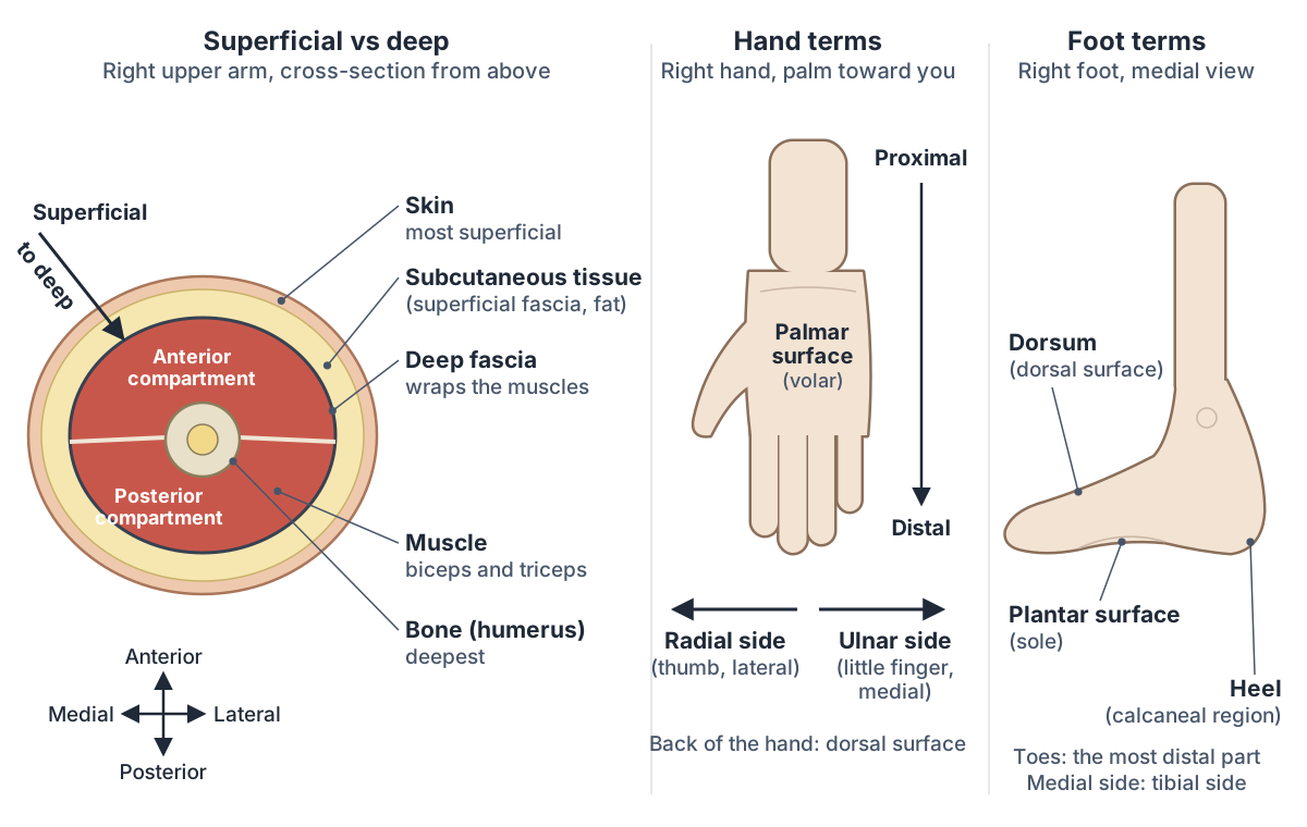
Superficial means closer to the surface of the body and deep means farther from it. The skin is superficial to the bones, and the brain is deep to the skull. Unlike the other pairs, superficial and deep describe depth beneath the skin rather than a direction across the body. (OpenStax Anatomy and Physiology 2e, 1.6: Anatomical Terminology.)
Kenhub lists a second pair with the same idea: external means toward the surface and internal means away from it. (Kenhub: Anatomical terminology: planes, directions and regions.)
The layers just under the skin have their own names. The superficial fascia is loose connective tissue beneath the dermis, with a fatty layer and a membranous layer; Terminologia Anatomica now calls it the subcutaneous tissue or hypodermis. The deep fascia is denser fibrous tissue that wraps the muscles, such as the fascia lata of the thigh and the plantar fascia of the foot. (Radiopaedia: Fascia.)
Depth also orders the contents of a space. In the popliteal fossa, the tibial and common fibular nerves are the most superficial contents and the popliteal artery is the deepest. (TeachMeAnatomy: The Popliteal Fossa.)
Two more depth words come from the serous membranes. The parietal layer lines the wall of a body cavity and the visceral layer covers the organ inside it, with a thin fluid-filled space between them. The pleura of the lungs, the pericardium of the heart and the peritoneum of the abdomen are built this way. (OpenStax Anatomy and Physiology 2e, 1.6: Anatomical Terminology.)
Ipsilateral vs contralateral, unilateral and bilateral
Ipsilateral means on the same side of the body and contralateral means on the opposite side. A fracture of the left elbow and an injury to the left shoulder are ipsilateral; a lesion in the right wrist is contralateral to one in the left wrist. (Radiopaedia: Radiographic positioning terminology.)
The terms matter most in the nervous system. Axons of the corticobulbar tract are ipsilateral: they run from the cortex to motor nuclei on the same side. Axons of the corticospinal tract are largely contralateral: most cross the midline at the pyramidal decussation, so each side of the brain controls muscles on the opposite side of the body. Axons that act on both sides are described as bilateral. (OpenStax Anatomy and Physiology 2e, 14.3: Motor Responses.)
Radiopaedia groups ipsilateral and contralateral with unilateral (one side) and bilateral (both sides) as the terms of laterality. (Radiopaedia: Anatomical position.)
The same-side and opposite-side terms drawn on the body, next to the anatomical planes. Ipsilateral vs contralateral diagram.
Directional terms for the hand and foot: palmar, plantar, dorsal, radial and ulnar
The hand and foot have their own surface terms. Palmar refers to the palm, the front of the hand, and dorsal to the back of the hand. Plantar refers to the sole of the foot, and dorsal to the top of the foot, its dorsum. (Kenhub: Anatomical terminology: planes, directions and regions.)
Volar is an older word for palmar. Radiopaedia lists volar or palmar among the traditional terms, alongside dorsal, ventral, caudal, cephalad, rostral and plantar. (Radiopaedia: Anatomical position.)
In the forearm, the ulna is the medial bone and the radius runs parallel to it on the lateral, thumb side. That is why the thumb side of the forearm and hand is called the radial side and the little-finger side the ulnar side. (OpenStax Anatomy and Physiology 2e, 8.2: Bones of the Upper Limb.)
The same idea works in both limbs. The preaxial border is the radial side of the upper limb and the tibial side of the lower limb, where the thumb and big toe lie; the postaxial border is the ulnar side of the upper limb and the fibular side of the lower limb. These borders come from the embryonic limb bud, and superficial veins mark them: the cephalic and great saphenous veins on the preaxial side, the basilic and small saphenous veins on the postaxial side. (Radiopaedia: Preaxial and postaxial borders.)
For the fingers and toes, abduction and adduction are measured from the midline of the hand or foot rather than the midline of the body, so spreading the fingers apart is abduction. (TeachMeAnatomy: Anatomical Terms of Movement.)
Bones, muscles, tendons and nerves of the hand, with the palmar and dorsal sides labelled. Hand anatomy.
Combined terms: superomedial, inferolateral, anterolateral and more
Two directional terms can be joined to describe a direction between them. The first part takes an -o ending: superomedial means above and toward the midline, inferolateral below and toward the side, anterolateral in front and to the side, and posteromedial behind and toward the midline.
The popliteal fossa shows four of them at once. Its diamond shape has a superomedial border (semimembranosus), a superolateral border (biceps femoris), an inferomedial border (the medial head of gastrocnemius) and an inferolateral border (the lateral head of gastrocnemius and plantaris). (TeachMeAnatomy: The Popliteal Fossa.)
Muscle fibre directions use the same words. In the abdominal wall, the fibres of the external oblique run inferomedially, those of the internal oblique run superomedially at right angles to them, and those of transversus abdominis run transversely. (TeachMeAnatomy: The Abdominal Wall.)
Bones are described this way too. On the femur, the lesser trochanter projects from the posteromedial side just below the neck, and the intertrochanteric line runs inferomedially between the two trochanters. (TeachMeAnatomy: The Femur.)
Movements can be described in the same way: protraction moves the scapula anterolaterally around the chest wall, as when reaching forward, and retraction moves it posteromedially, as when pulling something toward you. (TeachMeAnatomy: Anatomical Terms of Movement.)
External and internal obliques, transversus abdominis and rectus abdominis, with fibre directions. Abdominal muscles.
Directional terms vs regional terms
Directional terms say where something is relative to something else; regional terms name the part of the body. The two are combined in everyday anatomy: an anterior carpal scar is on the front (palm side) of the wrist. (OpenStax Anatomy and Physiology 2e, 1.6: Anatomical Terminology.)
Regional names are precise about the limbs. OpenStax reserves brachium (arm) for the upper arm and antebrachium for the forearm, and uses femur for the thigh and crus (leg) only for the part of the lower limb between the knee and the ankle. (OpenStax Anatomy and Physiology 2e, 1.6: Anatomical Terminology.)
Cephalic, cervical, brachial, femoral, crural and every other region, front and back. Regional terms and body regions.
Directional terms in the 3D model
- Capitate
- The largest carpal bone, with a rounded head.
- Distal phalanx of index finger
- The smallest, end bone of the index finger (second digit), supporting the fingertip and nail.
- Distal phalanx of little finger
- The smallest, end bone of the little finger (fifth digit), supporting the fingertip and nail.
- Distal phalanx of middle finger
- The smallest, end bone of the middle finger (third digit), supporting the fingertip and nail.
- Distal phalanx of ring finger
- The smallest, end bone of the ring finger (fourth digit), supporting the fingertip and nail.
- Distal phalanx of thumb
- The end bone of the thumb, supporting the nail and pulp.
- Femur
- The thigh bone, the longest, heaviest and strongest bone in the body.
- Fibula
- The slender lateral bone of the leg.
- Fifth metacarpal bone
- The little finger metacarpal. One of the five long bones of the palm.
- First metacarpal bone
- The thumb metacarpal, shorter and stouter than the others. One of the five long bones of the palm.
- Fourth metacarpal bone
- The ring finger metacarpal. One of the five long bones of the palm.
- Hamate
- A wedge-shaped carpal bone of the distal row with a hook.
- Humerus
- The long bone of the upper arm.
- Lunate
- A crescent-shaped carpal bone in the middle of the proximal row.
- Middle phalanx of index finger
- The middle of the three phalanges of the index finger (second digit).
- Middle phalanx of little finger
- The middle of the three phalanges of the little finger (fifth digit).
- Middle phalanx of middle finger
- The middle of the three phalanges of the middle finger (third digit).
- Middle phalanx of ring finger
- The middle of the three phalanges of the ring finger (fourth digit).
- Patella
- The kneecap, a triangular sesamoid bone in the tendon of quadriceps femoris.
- Pisiform
- A small, pea-shaped carpal bone on the palmar side of the triquetrum.
- Proximal phalanx of index finger
- The longest of the three phalanges of the index finger (second digit), nearest the palm.
- Proximal phalanx of little finger
- The longest of the three phalanges of the little finger (fifth digit), nearest the palm.
- Proximal phalanx of middle finger
- The longest of the three phalanges of the middle finger (third digit), nearest the palm.
- Proximal phalanx of ring finger
- The longest of the three phalanges of the ring finger (fourth digit), nearest the palm.
- Proximal phalanx of thumb
- The first of the thumb's two phalanges.
- Radius
- The lateral bone of the forearm.
- Scaphoid
- The largest bone of the proximal carpal row, shaped like a boat.
- Second metacarpal bone
- The index finger metacarpal, usually the longest. One of the five long bones of the palm.
- Third metacarpal bone
- The middle finger metacarpal. One of the five long bones of the palm.
- Tibia
- The shinbone, the medial and main weight-bearing bone of the leg.
- Trapezium
- A four-sided carpal bone at the thumb side of the distal row.
- Trapezoid
- A small wedge-shaped carpal bone of the distal row.
- Triquetral
- A pyramid-shaped carpal bone on the ulnar side of the proximal row.
- Ulna
- The medial bone of the forearm.
Frequently asked questions
What are the main directional terms in anatomy?
The main directional terms come in opposite pairs: superior and inferior (above and below), anterior and posterior (front and back), medial and lateral (toward and away from the midline), proximal and distal (nearer to and farther from a limb's attachment) and superficial and deep (nearer to and farther from the surface). Ventral, dorsal, cranial and caudal are older synonyms used on the trunk.
What is the anatomical position and why is it important?
The anatomical position is the body standing upright and facing forward, arms at the sides, palms facing forward and feet parallel with the toes forward. Every directional term, plane and movement is defined from this posture, so a description means the same thing whether the person is standing, lying face up (supine) or lying face down (prone).
Is the elbow proximal or distal to the wrist?
The elbow is proximal to the wrist, because it is closer to where the upper limb attaches to the trunk; the wrist is distal to the elbow. Proximal and distal describe position along the limb, not height, so the elbow stays proximal to the wrist even when the hand is raised above the head.
What is the difference between anterior and ventral?
In human anatomy, anterior and ventral both mean the front of the body, and posterior and dorsal both mean the back, because humans stand upright. In four-legged animals they differ: ventral means the belly side facing the ground, while the front end of the body is cranial. Inside the human brain, ventral means inferior above the midbrain.
What does superficial mean in anatomy?
Superficial means closer to the surface of the body, and its opposite, deep, means farther from the surface. The skin is superficial to the muscles and bones, and the brain is deep to the skull. In the popliteal fossa behind the knee, the tibial nerve is superficial and the popliteal artery is the deepest structure.
Is the thumb medial or lateral?
The thumb is lateral. In the anatomical position the palms face forward, so the thumbs point away from the body, and the radius, the forearm bone on the thumb side, lies lateral to the ulna. The big toe, by contrast, is the medial toe of the foot.
What is the difference between superior and proximal?
Superior means higher, toward the head, and is used mainly for the trunk and head. Proximal means closer to the point of attachment and is used for limbs, vessels and other structures with a beginning and an end. The two can disagree: with the arm raised, the hand is superior to the shoulder but still distal to it.
What does caudal mean in anatomy?
Caudal means toward the tail, which in a standing human is the same as inferior, toward the feet. Its opposite is cranial, toward the head. In the brain, rostral and caudal follow the bend of the nervous system: above the midbrain caudal means posterior, while in the brainstem and spinal cord it means inferior.
References
- OpenStax. Anatomy and Physiology 2e, §1.6 Anatomical Terminology. Rice University, 2022.
- OpenStax. Anatomy and Physiology 2e, §8.2 Bones of the Upper Limb. Rice University, 2022.
- OpenStax. Anatomy and Physiology 2e, §14.3 Motor Responses. Rice University, 2022.
- TeachMeAnatomy. Anatomical Position.
- TeachMeAnatomy. Anatomical Terms of Location.
- TeachMeAnatomy. Anatomical Terms of Movement.
- TeachMeAnatomy. The Popliteal Fossa.
- TeachMeAnatomy. The Abdominal Wall.
- TeachMeAnatomy. The Femur.
- Hacking C, Jones J, Bell D, et al. Anatomical position. Radiopaedia.org (rID 36890).
- Radiographic positioning terminology. Radiopaedia.org reference article.
- Burlak K. Preaxial and postaxial borders. Radiopaedia.org (rID 55389).
- Deng F. Fascia. Radiopaedia.org (rID 68862).
- Rad A. Anatomical terminology: planes, directions and regions. Kenhub, reviewed 2023.
- Solomon N. Brain orientation difficulties. Kenhub, reviewed 2022.
- Lecturio. What is the anatomical position?
- University of Minnesota College of Veterinary Medicine. Veterinary Anatomy: Veterinary vs human directional terms.
- Mitsuhashi N. et al. (2009). BodyParts3D: 3D structure database for anatomical concepts. Nucleic Acids Research 37, D782–D785. Data © The Database Center for Life Science, CC BY 4.0.
Related anatomy
- Anatomical planesAnatomical planes explained: midsagittal vs parasagittal, coronal (frontal) and transverse (axial) planes, axes of…
- Body regionsRegional terms in anatomy with front and back body-region diagrams: cephalic, cervical, brachial, femoral, crural and…
- Anatomical termsAnatomical terms explained with diagrams: bone markings like tubercle, fossa and condyle, movements like abduction…
- Types of jointsTypes of joints explained: fibrous, cartilaginous and synovial (diarthrosis) joints, a labelled synovial joint diagram…
- Skeletal systemExplore a realistic 3D human skeleton: the 206 bones, axial vs appendicular skeleton, bone types, bony landmarks and…
- Human body diagramHuman anatomy diagram set: eight labelled drawings of body parts, internal organs, skeleton and muscles front and…
Explore the whole body in the interactive 3D human anatomy model, or browse the A–Z list of human structures.