

What are regional terms in anatomy?
Regional terms are the anatomical names of the body's areas, each one fixed to a specific part. OpenStax uses them carefully for the limbs: brachium, or arm, is reserved for the upper arm, antebrachium, or forearm, is used rather than lower arm, femur, or thigh, is the thigh, and the leg, or crus, is only the part of the lower limb between the knee and the ankle. (OpenStax Anatomy and Physiology 2e, 1.6: Anatomical Terminology.)
Regional and directional terms work together. A regional term says which part of the body you mean; a directional term says where within or next to it. OpenStax's example is a scar in the anterior carpal region, which is on the palm side of the wrist. (OpenStax Anatomy and Physiology 2e, 1.6: Anatomical Terminology.)
Regional terms are also the basis of regional anatomy, the study of how all the structures in one body region, such as the abdomen, relate to one another. Systemic anatomy instead follows one body system, such as all the skeletal muscles of the muscular system. (OpenStax Anatomy and Physiology 2e, 1.1: Overview of Anatomy and Physiology.)
Superior, inferior, medial, lateral, proximal, distal and the anatomical position. Directional terms.
The main regions of the body
Kenhub divides the body into seven main regions: the head, neck, thorax, abdomen, pelvis, upper limb (upper extremity) and lower limb (lower extremity). Each is divided further: the upper limb into the shoulder and arm, elbow and forearm, wrist and hand, and the lower limb into the hip, gluteal region and thigh, the knee and leg, and the ankle and foot. (Kenhub: Anatomical terminology: planes, directions and regions.)
OpenStax groups the smaller regions into four combined regions: the cephalic region, which is all of the head; the trunk; the upper limb; and the lower limb. (OpenStax Anatomy and Physiology 2e, 1.6: Anatomical Terminology.)
The regions on the trunk have no visible borders, so anatomists define them with surface lines and landmarks. Kenhub names the axillary, deltoid, pectoral, scapular, vertebral and gluteal regions this way. (Kenhub: Anatomical terminology: planes, directions and regions.)
Head and neck regions: cephalic and cervical
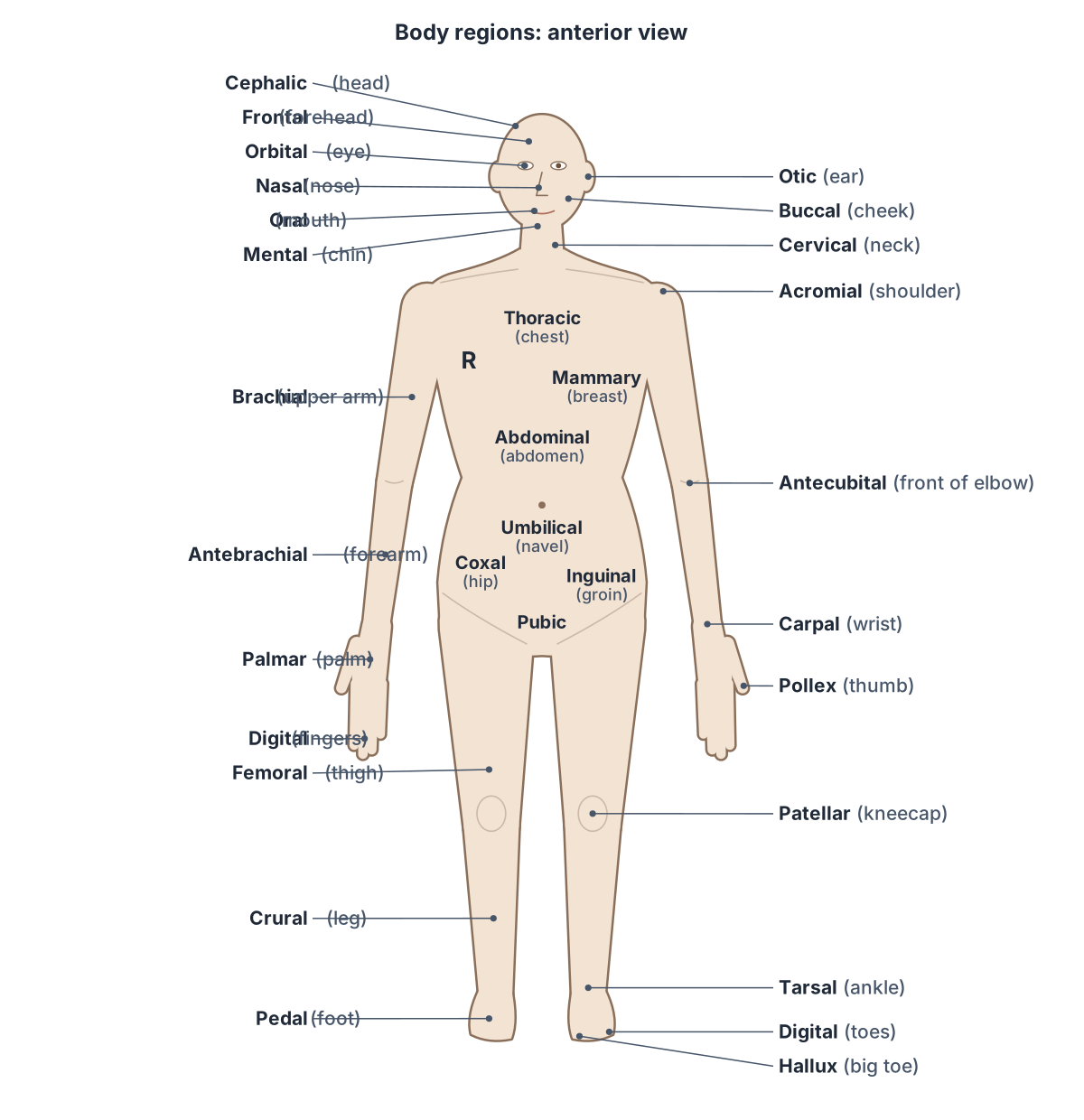
The cephalic region is the whole head. OpenStax divides it into the cranial region (the upper head) and the facial region (the lower head), and names smaller regions within the face: ocular (eyes), auricle or otic (ears), nasal (nose), buccal (cheeks) and mental (chin). The neck is the cervical region. (OpenStax Anatomy and Physiology 2e, 1.6: Anatomical Terminology.)
Other textbooks add more names. Lange and colleagues' open Human Anatomy text labels the frontal region (forehead), orbital or ocular region (eye) and oral region (mouth) as well as cephalic, cranial, facial, cervical, nasal, otic, buccal and mental. (Lange J et al. Human Anatomy, 1.06: Anatomical Atlas, Anatomical Terminology (LibreTexts).)
Kenhub notes that the head and neck regions are named after the structures within them, such as the orbital, buccal and occipital regions and the submandibular triangle of the neck. (Kenhub: Anatomical terminology: planes, directions and regions.)
Skull, face, brain and the main structures of the head and neck. Head anatomy.
Trunk regions: thoracic, abdominal, pelvic and inguinal
On the front of the trunk, OpenStax names the thoracic region (chest), the mammary region (breast), the abdominal region, the umbilicus or navel, the coxal region, the pubic region and the inguinal, or groin, region. The axillary region is the armpit. (OpenStax Anatomy and Physiology 2e, 1.6: Anatomical Terminology.)
Sources place the coxal region differently: OpenStax's figure labels it at the belt line, while Lange and colleagues label coxal as the hip. Their text also adds the pectoral region (upper chest) and the pelvic and genital regions. (Lange J et al. Human Anatomy, 1.06: Anatomical Atlas, Anatomical Terminology (LibreTexts).)
The umbilicus, or navel, lies in the midline of the anterior abdominal wall. Because its position is fairly constant between people, it is used to divide the abdomen into quadrants and regions. (Kenhub: Umbilicus.)
Abdominal regions and quadrants in brief
The abdomen is divided in two ways. Two lines crossing at the umbilicus make four quadrants: right upper (RUQ), left upper (LUQ), right lower (RLQ) and left lower (LLQ), the system most used in medicine. Two vertical lines dropped from the midpoints of the clavicles and two horizontal lines, one below the ribs and one above the pelvis, make nine regions. (OpenStax Anatomy and Physiology 2e, 1.6: Anatomical Terminology.)
The nine regions are the right and left hypochondriac, epigastric, right and left lumbar, umbilical, right and left inguinal (iliac) and hypogastric regions. The vertical lines are the midclavicular lines, each passing through the middle of the clavicle. (Texas Tech University Health Sciences Center El Paso, Medical Gross Anatomy: Topographical anatomy of the abdomen.)
The four quadrants and nine regions in detail, with the organs in each. Abdominal anatomy.
Upper limb regions: brachial, antebrachial, carpal and manual
From the shoulder to the fingers, OpenStax names the acromial region (the point of the shoulder), brachial (upper arm), antecubital (front of the elbow), olecranal (back of the elbow), antebrachial (forearm), carpal (wrist), palmar (palm), digital or phalangeal (fingers) and pollex (thumb). The manual region is the hand. (OpenStax Anatomy and Physiology 2e, 1.6: Anatomical Terminology.)
The antecubital region contains the cubital fossa, a triangular depression over the front of the elbow that marks the change from arm to forearm. The median cubital vein crosses it, joining the cephalic and basilic veins, and is a common site for taking blood (venepuncture). (TeachMeAnatomy: The Cubital Fossa.)
The cubital fossa is bounded by brachioradialis laterally, pronator teres medially and a line between the humeral epicondyles above. From lateral to medial it contains the radial nerve, the biceps tendon, the brachial artery and the median nerve. (TeachMeAnatomy: The Cubital Fossa.)
Bones, muscles, vessels and nerves of the upper limb, in 3D. Arm anatomy.
Lower limb regions: femoral, patellar, crural, sural and pedal
OpenStax names the lower limb regions as femoral (thigh), patellar (knee), crural (leg), tarsal (ankle), pedal (foot), digital or phalangeal (toes) and hallux (great toe) on the front, and gluteal (buttocks), popliteal (back of the knee), sural (back of the lower leg) and plantar (sole of the foot) on the back. (OpenStax Anatomy and Physiology 2e, 1.6: Anatomical Terminology.)
The thigh is the part of the lower limb between the hip joint and the knee joint, and the leg is the region between the knee joint and the ankle joint. So in anatomy the leg means only the crural region, not the whole lower limb. (OpenStax Anatomy and Physiology 2e, 8.4: Bones of the Lower Limb.)
Sources draw the crural and sural regions slightly differently. OpenStax labels crural as the lower leg and sural as the back of the lower leg; Lange and colleagues label sural as the calf and add the calcaneal region for the heel. (Lange J et al. Human Anatomy, 1.06: Anatomical Atlas, Anatomical Terminology (LibreTexts).)
The popliteal region contains the popliteal fossa, a diamond-shaped area on the back of the knee that is the main route for vessels and nerves between the thigh and the leg. It holds the popliteal artery and vein and the tibial and common fibular nerves. (TeachMeAnatomy: The Popliteal Fossa.)
The bones, muscles and vessels of the lower limb from hip to foot. Leg anatomy.
Regions on the back of the body
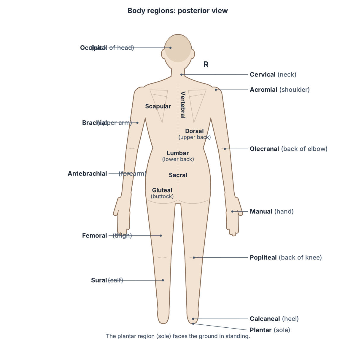
On the posterior view, OpenStax labels the cervical region (neck), dorsal region (upper back), lumbar region (lower back), acromial region (shoulder), brachial and antebrachial regions, olecranal region (back of the elbow), manual region (hand), gluteal region (buttocks), femoral region, popliteal region (back of the knee), sural region and plantar region (sole). (OpenStax Anatomy and Physiology 2e, 1.6: Anatomical Terminology.)
Lange and colleagues add the vertebral region over the spine and the sacral region over the sacrum, and Kenhub names the scapular region over the shoulder blade. (Lange J et al. Human Anatomy, 1.06: Anatomical Atlas, Anatomical Terminology (LibreTexts).)
Several back regions match the bones beneath them, which you can see in the 3D model: the scapular region overlies the scapula, the vertebral region the vertebral column, the lumbar region the five lumbar vertebrae, the sacral region the sacrum, and the calcaneal region the calcaneus, the heel bone.
Spine, back muscles and the regions of the back, in 3D. Back anatomy.
Everyday names and their anatomical terms
Most regional terms are Latin or Greek words for the body part, so learning the root unlocks the term. MedlinePlus, the US National Library of Medicine's word-part list, gives cephal- for head, thorac- for chest, cost- for rib, derm- for skin, oste- for bone, my- for muscle, ot- for ear, rhin- for nose and stomat- for mouth. (MedlinePlus (US National Library of Medicine): Appendix A, Word Parts and What They Mean.)
The table below pairs each everyday name with its regional term. Note the ones that catch students out: crural is the leg between the knee and ankle while sural is only its back (the calf), antebrachial is the forearm, and antecubital and olecranal are the front and back of the elbow.
Everyday names and anatomical regional terms
| Everyday name | Regional term | Front or back |
|---|---|---|
| Whole head | Cephalic | Both |
| Upper head (skull) | Cranial | Both |
| Face | Facial | Front |
| Forehead | Frontal | Front |
| Eye | Orbital or ocular | Front |
| Ear | Otic (auricle) | Both |
| Nose | Nasal | Front |
| Cheek | Buccal | Front |
| Mouth | Oral | Front |
| Chin | Mental | Front |
| Back of the head | Occipital | Back |
| Neck | Cervical | Both |
| Chest | Thoracic (upper chest: pectoral) | Front |
| Breast | Mammary | Front |
| Armpit | Axillary | Front |
| Belly | Abdominal | Front |
| Navel (belly button) | Umbilical | Front |
| Hip | Coxal | Front |
| Groin | Inguinal | Front |
| Area above the genitals | Pubic | Front |
| Upper back | Dorsal | Back |
| Shoulder blade | Scapular | Back |
| Spine | Vertebral | Back |
| Lower back (loin) | Lumbar | Back |
| Over the sacrum | Sacral | Back |
| Buttock | Gluteal | Back |
| Point of the shoulder | Acromial | Both |
| Upper arm | Brachial | Both |
| Front of the elbow | Antecubital | Front |
| Back of the elbow | Olecranal | Back |
| Forearm | Antebrachial | Both |
| Wrist | Carpal | Front |
| Hand | Manual | Both |
| Palm | Palmar | Front |
| Fingers | Digital (phalangeal) | Front |
| Thumb | Pollex | Front |
| Thigh | Femoral | Both |
| Kneecap | Patellar | Front |
| Back of the knee | Popliteal | Back |
| Leg (knee to ankle) | Crural | Front |
| Calf | Sural | Back |
| Ankle | Tarsal | Front |
| Foot | Pedal | Front |
| Heel | Calcaneal | Back |
| Sole | Plantar | Underside |
| Toes | Digital (phalangeal) | Front |
| Big toe | Hallux | Front |
Sources: OpenStax 1.6 (Figure 1.12), Lange et al. Human Anatomy (LibreTexts) and Kenhub. OpenStax places the coxal region at the belt line; Lange et al. label it the hip.
Body cavities and serous membranes
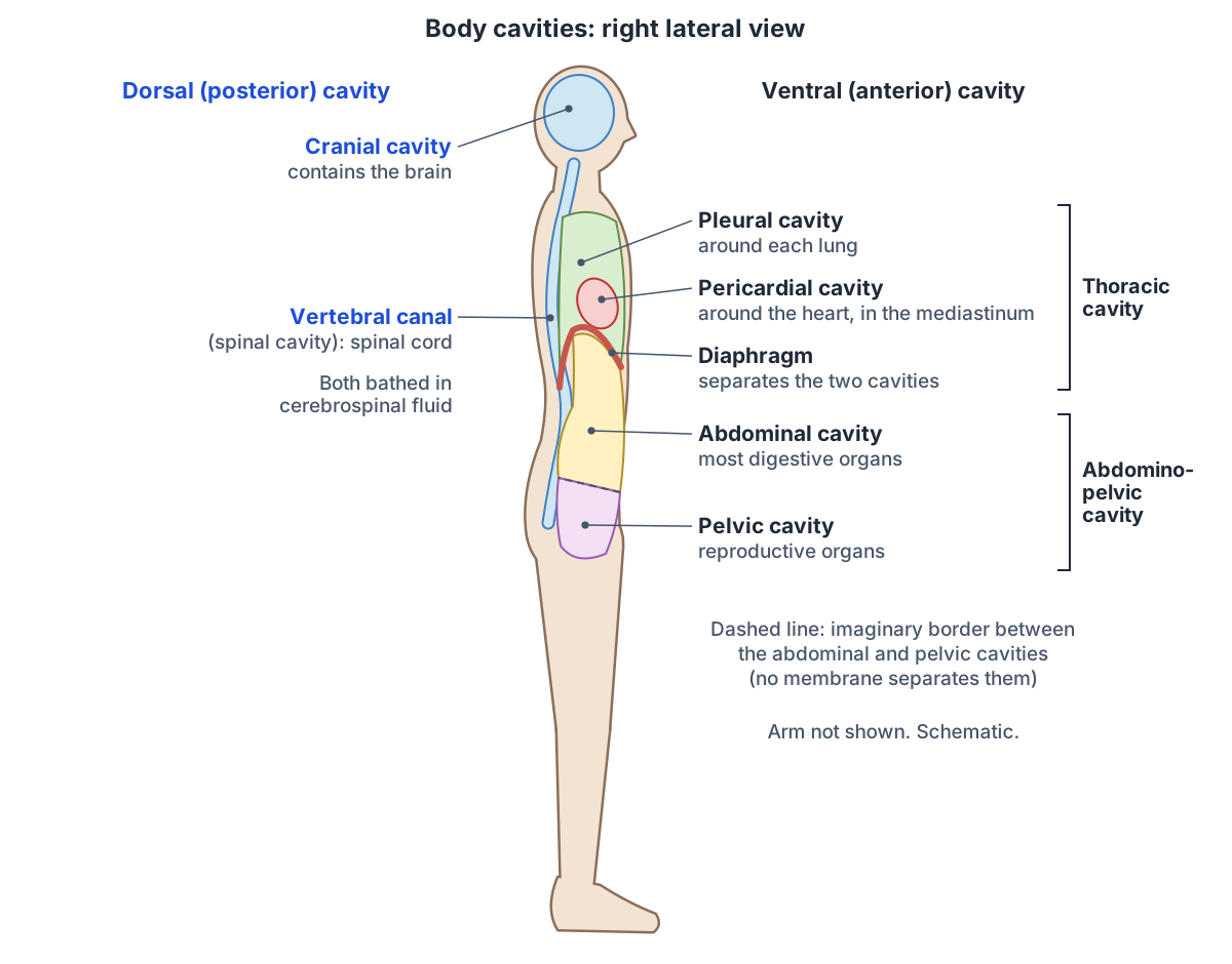
The body has two main cavities. The dorsal (posterior) cavity holds the cranial cavity, which contains the brain, and the spinal (vertebral) cavity, which contains the spinal cord; the two are continuous and both are protected by bone and cerebrospinal fluid. The ventral (anterior) cavity holds the thoracic and abdominopelvic cavities. (OpenStax Anatomy and Physiology 2e, 1.6: Anatomical Terminology.)
The thoracic cavity, enclosed by the rib cage, contains the lungs and the heart, which sits in the mediastinum. The diaphragm separates it from the abdominopelvic cavity. The abdominal part of that cavity holds most of the digestive organs and the pelvic part the reproductive organs, and no membrane divides the two. (OpenStax Anatomy and Physiology 2e, 1.6: Anatomical Terminology.)
Kenhub describes the same cavities with anterior and posterior names: the posterior cavity is the cranial cavity and vertebral canal, bathed in cerebrospinal fluid, and the anterior cavity is the thoracic cavity, with its mediastinal and pleural cavities, and the abdominopelvic cavity. The pericardial cavity around the heart lies within the mediastinum. (Kenhub: Anatomical terminology: planes, directions and regions.)
Organs in the ventral cavity are wrapped in serous membranes with two layers: a parietal layer lining the cavity wall and a visceral layer covering the organ, with a thin space of fluid between them that reduces friction. The pleura surrounds the lungs, the pericardium the heart and the peritoneum the abdominopelvic organs. (OpenStax Anatomy and Physiology 2e, 1.6: Anatomical Terminology.)
Where each internal organ sits in the thoracic and abdominopelvic cavities. Organs of the body.
Surface anatomy: landmarks you can see and feel
Surface anatomy studies the structures that can be seen or felt through the skin and uses them as landmarks for deeper parts. Clinicians use these landmarks to count ribs, find vertebral levels and place stethoscopes, needles and incisions.
At the top of the sternum, the jugular (suprasternal) notch is visible under the skin. Below it, the sternal angle can be felt as a horizontal ridge where the manubrium meets the body of the sternum; it marks the level of the second costal cartilage. (TeachMeAnatomy: The Sternum.)
A horizontal plane through the sternal angle passes through the intervertebral disc between the T4 and T5 vertebrae. The midclavicular line runs vertically through the middle of the clavicle and the midaxillary line through the middle of the armpit. (Texas Tech University Health Sciences Center El Paso, Medical Gross Anatomy: Topographical anatomy of the thorax.)
Radiopaedia gives the same level: the sternal angle lies at the second costal cartilage and the T4/T5 intervertebral disc, an important palpable landmark in clinical examination. (Radiopaedia: Manubriosternal joint.)
At the back of the neck, the seventh cervical vertebra is called the vertebra prominens because its long spinous process is easily felt, and it marks the change from the cervical to the thoracic spine. (Texas Tech University Health Sciences Center El Paso, Medical Gross Anatomy: Topographical anatomy of the back.)
Radiopaedia confirms that the name vertebra prominens comes from the long, easily palpable spinous process of C7, which ends in a rounded tubercle and is not bifid (split in two). (Radiopaedia: C7 vertebra.)
On the pelvis, you can feel the iliac crest, the curved upper margin of the ilium, when you put your hands on your waist, and its rounded front end, the anterior superior iliac spine, at the anterolateral hip. The hip bone itself is also called the coxal bone, which is where the coxal region gets its name. (OpenStax Anatomy and Physiology 2e, 8.3: The Pelvic Girdle and Pelvis.)
The transpyloric plane is an imaginary horizontal plane halfway between the jugular notch and the upper border of the pubic symphysis, at about the level of the L1 vertebral body. Traditionally the pylorus of the stomach, the hila of both kidneys and the origin of the superior mesenteric artery lie at this level, though positions vary between people. (Radiopaedia: Transpyloric plane.)
The sternum, sternal angle and how to count ribs, in 3D. Rib cage.
How reliable are the umbilicus and iliac crest landmarks?
Textbook tables put the umbilicus at about the level of the L3/L4 intervertebral disc in people who are not obese, and a line across the tops of the iliac crests (the intercristal or Tuffier's line) at the level of the L4 vertebra. Kenhub also gives the L3 to L4 disc for the umbilicus. (Texas Tech University Health Sciences Center El Paso, Medical Gross Anatomy: Topographical anatomy of the abdomen.)
Imaging shows more variation. In a CT study of 834 adults aged 18 to 35 without spinal disease, the umbilicus was most often level with the L4 vertebral body, and its position did not depend on height, weight, body mass index, sex or ethnicity. The iliac crests lay above the L4/L5 disc space in 26.5% of patients and at it in 29.8%, and sat higher (more cephalad) in women than in men. (Shin D et al. Are the umbilicus and iliac crests truly at the level of L4 to L5? A computed tomography-based study. Int J Spine Surg. 2024.)
In a review of 571 radiographs, the intercristal line crossed anywhere from the L3 to the L5 vertebral body, most often the L4 body. On lateral views it passed through L5 in 32.8% of women but only 5.7% of men, so this study found the line lower in women, the opposite direction to the CT study: even the sex difference is uncertain. (Chowdhury A, Sharma H. A review of 571 radiographs on Tuffier's inter-cristal line and its application in lumbar spinal surgery. J Spine Surg. 2018.)
Palpation adds error. When anaesthetists used Tuffier's line to choose a lumbar interspace, they picked the correct one in 55.8% of 52 patients, and the authors concluded the line is not a reliable landmark in every case. Surface landmarks are a guide, not a measurement. (Lin N et al. Abdominal circumference but not the degree of lumbar flexion affects the accuracy of lumbar interspace identification by Tuffier's line palpation method. BMC Anesthesiol. 2015.)
Surface landmarks and their vertebral levels
| Landmark | How to find it | Approximate level | Agreement between sources |
|---|---|---|---|
| Vertebra prominens | Most prominent spinous process at the base of the back of the neck | C7 | Radiopaedia and TTUHSC agree |
| Jugular (suprasternal) notch | Notch at the top of the manubrium, between the ends of the clavicles | Not given in our sources | Visible under the skin |
| Sternal angle | Horizontal ridge where the manubrium meets the body of the sternum; second costal cartilage | T4/T5 disc | Radiopaedia and TTUHSC agree |
| Transpyloric plane | Halfway between the jugular notch and the pubic symphysis | L1 vertebral body | Radiopaedia and TTUHSC agree; varies between people |
| Umbilicus | Midline of the anterior abdominal wall | L3/L4 disc (textbooks) or L4 body (CT study) | Kenhub and TTUHSC: L3/L4; Shin et al. 2024: most often L4 |
| Iliac crests (intercristal line) | Line across the highest points of the iliac crests | L4 traditionally; L3 to L5 in imaging | TTUHSC: L4; Chowdhury and Sharma 2018: L3 to L5 |
| Cubital fossa | Triangular hollow in front of the elbow | Not applicable | TeachMeAnatomy |
| Popliteal fossa | Diamond-shaped hollow behind the knee | Not applicable | TeachMeAnatomy |
Levels are approximate and vary with build, age and sex; landmarks guide but do not measure.
Body regions in the 3D model
- Body of sternum
- The long middle part of the sternum.
- Calcaneus
- The largest tarsal bone, forming the heel.
- Capitate
- The largest carpal bone, with a rounded head.
- Clavicle
- The collarbone, an S-shaped long bone running between the sternum and the shoulder.
- Cuboid bone
- A cube-shaped tarsal bone on the lateral side of the foot.
- Femur
- The thigh bone, the longest, heaviest and strongest bone in the body.
- Fibula
- The slender lateral bone of the leg.
- Fifth lumbar vertebra
- The fifth of the five lumbar vertebrae, labelled L5, in the lower back.
- First lumbar vertebra
- The first of the five lumbar vertebrae, labelled L1, in the lower back.
- Fourth lumbar vertebra
- The fourth of the five lumbar vertebrae, labelled L4, in the lower back.
- Hamate
- A wedge-shaped carpal bone of the distal row with a hook.
- Hip bone
- The large irregular bone of the pelvis formed by the fusion of the ilium, ischium and pubis.
- Humerus
- The long bone of the upper arm.
- Intermediate cuneiform bone
- The smallest of the three cuneiform bones.
- Lateral cuneiform bone
- A wedge-shaped bone of the midfoot.
- Lunate
- A crescent-shaped carpal bone in the middle of the proximal row.
- Manubrium
- The upper part of the sternum.
- Medial cuneiform bone
- The largest of the three cuneiform bones.
- Navicular bone of foot
- A boat-shaped tarsal bone on the medial side of the foot.
- Patella
- The kneecap, a triangular sesamoid bone in the tendon of quadriceps femoris.
- Pisiform
- A small, pea-shaped carpal bone on the palmar side of the triquetrum.
- Radius
- The lateral bone of the forearm.
- Sacrum
- A triangular bone of five fused sacral vertebrae.
- Scaphoid
- The largest bone of the proximal carpal row, shaped like a boat.
- Scapula
- The shoulder blade, a flat triangular bone on the back of the thorax.
- Second lumbar vertebra
- The second of the five lumbar vertebrae, labelled L2, in the lower back.
- Seventh cervical vertebra
- The seventh of the seven cervical (neck) vertebrae, labelled C7.
- Talus
- A tarsal bone that sits between the tibia and fibula above and the calcaneus below.
- Third lumbar vertebra
- The third of the five lumbar vertebrae, labelled L3, in the lower back.
- Tibia
- The shinbone, the medial and main weight-bearing bone of the leg.
- Trapezium
- A four-sided carpal bone at the thumb side of the distal row.
- Trapezoid
- A small wedge-shaped carpal bone of the distal row.
- Triquetral
- A pyramid-shaped carpal bone on the ulnar side of the proximal row.
- Ulna
- The medial bone of the forearm.
- Xiphoid process
- The small lowest part of the sternum.
Frequently asked questions
What are the main regions of the body?
The body is usually divided into the head, neck, thorax (chest), abdomen, pelvis, upper limbs and lower limbs. OpenStax groups these into four combined regions: the cephalic region (head), the trunk, the upper limb and the lower limb. Each is divided into smaller named regions, such as brachial (upper arm) and crural (leg between knee and ankle).
What is the difference between regional and directional terms?
Regional terms name a part of the body, such as carpal for the wrist or femoral for the thigh. Directional terms describe where one structure lies relative to another, such as anterior, medial or distal. They are often combined: a scar in the anterior carpal region is on the palm side of the wrist.
What is the crural region?
The crural region is the leg in the strict anatomical sense: the part of the lower limb between the knee and the ankle. Its back, the calf, is called the sural region. The thigh above the knee is the femoral region, so in anatomy the word leg does not include the thigh.
What is the difference between brachial and antebrachial?
Brachial refers to the brachium, the upper arm between the shoulder and the elbow, which contains the humerus. Antebrachial refers to the antebrachium, the forearm between the elbow and the wrist, which contains the radius and ulna. Between them, the antecubital region at the front of the elbow marks the change from arm to forearm.
What is the antecubital region?
The antecubital region is the front of the elbow. It contains the cubital fossa, a triangular hollow bounded by the brachioradialis and pronator teres muscles, which holds the brachial artery, the median nerve and the biceps tendon. The median cubital vein crosses it and is a common site for taking blood.
What is the popliteal region?
The popliteal region is the back of the knee. It contains the popliteal fossa, a diamond-shaped hollow bordered by two hamstring muscles above and the two heads of gastrocnemius below. The popliteal artery, popliteal vein, tibial nerve and common fibular nerve pass through it between the thigh and the leg.
What are the body cavities?
The dorsal (posterior) cavity contains the cranial cavity with the brain and the vertebral canal with the spinal cord. The ventral (anterior) cavity contains the thoracic cavity, with the lungs and the heart in the mediastinum, and below the diaphragm the abdominopelvic cavity, which holds the digestive organs in its abdominal part and the reproductive organs in its pelvic part.
What is surface anatomy?
Surface anatomy is the study of structures you can see or feel through the skin, such as the sternal angle, the vertebra prominens (C7), the iliac crests and the umbilicus, and how they relate to deeper organs and vertebral levels. Clinicians use these landmarks to count ribs, examine organs and guide needles, though their levels vary between people.
References
- OpenStax. Anatomy and Physiology 2e, §1.1 Overview of Anatomy and Physiology. Rice University, 2022.
- OpenStax. Anatomy and Physiology 2e, §1.6 Anatomical Terminology. Rice University, 2022.
- OpenStax. Anatomy and Physiology 2e, §8.3 The Pelvic Girdle and Pelvis. Rice University, 2022.
- OpenStax. Anatomy and Physiology 2e, §8.4 Bones of the Lower Limb. Rice University, 2022.
- Lange J, et al. Human Anatomy, §1.06 Anatomical Atlas: Anatomical Terminology. LibreTexts Medicine, CC BY-NC-SA 4.0.
- Rad A. Anatomical terminology: planes, directions and regions. Kenhub, reviewed 2023.
- Stewart S. Umbilicus. Kenhub, reviewed 2023.
- TeachMeAnatomy. The Cubital Fossa.
- TeachMeAnatomy. The Popliteal Fossa.
- TeachMeAnatomy. The Sternum.
- Texas Tech University Health Sciences Center El Paso. Medical Gross Anatomy: Topographical Anatomy of the Thorax.
- Texas Tech University Health Sciences Center El Paso. Medical Gross Anatomy: Topographical Anatomy of the Back.
- Texas Tech University Health Sciences Center El Paso. Medical Gross Anatomy: Topographical Anatomy of the Abdomen.
- Ling J, Knipe H, et al. Manubriosternal joint. Radiopaedia.org (rID 50776).
- Luijkx T, et al. C7 vertebra. Radiopaedia.org (rID 30851).
- Murthy S, Yan E, Gaillard F. Transpyloric plane. Radiopaedia.org (rID 52002).
- Shin D, Nguyen K, Small E, et al. Are the umbilicus and iliac crests truly at the level of L4 to L5? A computed tomography-based study of surface anatomy of the anterior lumbar spine. Int J Spine Surg. 2024;18(6):660-666. doi:10.14444/8651.
- Chowdhury A, Sharma H. A review of 571 radiographs on Tuffier's inter-cristal line and its application in lumbar spinal surgery. J Spine Surg. 2018;4(2):383-387.
- Lin N, Li Y, Bebawy JF, Dong J, Hua L. Abdominal circumference but not the degree of lumbar flexion affects the accuracy of lumbar interspace identification by Tuffier's line palpation method: an observational study. BMC Anesthesiol. 2015;15:9.
- MedlinePlus, US National Library of Medicine. Appendix A: Word Parts and What They Mean.
- Mitsuhashi N. et al. (2009). BodyParts3D: 3D structure database for anatomical concepts. Nucleic Acids Research 37, D782–D785. Data © The Database Center for Life Science, CC BY 4.0.
Related anatomy
- Directional termsAnatomical position and directional terms with labelled diagrams: what proximal, distal, medial, lateral, anterior…
- Anatomical planesAnatomical planes explained: midsagittal vs parasagittal, coronal (frontal) and transverse (axial) planes, axes of…
- Anatomical termsAnatomical terms explained with diagrams: bone markings like tubercle, fossa and condyle, movements like abduction…
- AbdomenAbdominal anatomy with labelled quadrant and nine-region diagrams and a 3D model: organs on each side, abdominal wall…
- Organs of the bodyHuman anatomy organs explained with a labelled internal organs diagram: organs by body system and location, left vs…
- Human body diagramHuman anatomy diagram set: eight labelled drawings of body parts, internal organs, skeleton and muscles front and…
Explore the whole body in the interactive 3D human anatomy model, or browse the A–Z list of human structures.