

How to read a human anatomy diagram
Anatomy diagrams show the body in the anatomical position: standing upright, feet at shoulder width and parallel, toes forward, with the upper limbs held out at each side and the palms facing forward. Describing every part from this one reference position keeps directions consistent. (OpenStax Anatomy and Physiology 2e, 1.6: Anatomical Terminology.)
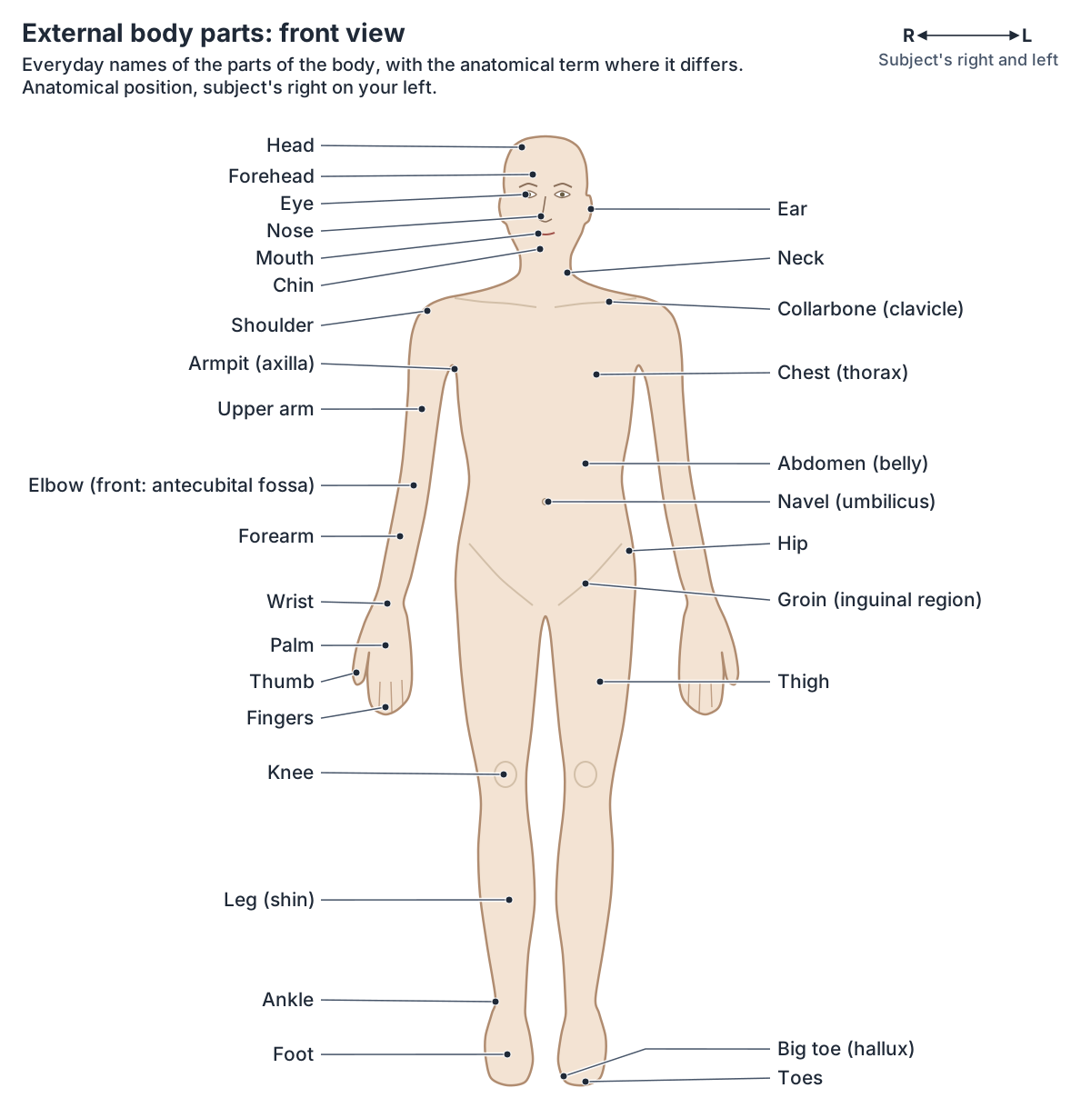
Right and left always mean the subject's right and left, not the viewer's. In a front (anterior) view the subject's right side is therefore on your left; in a back (posterior) view it is on your right. The whole-body diagrams on this page mark this with an R and L arrow. (OpenStax Anatomy and Physiology 2e, 1.6: Anatomical Terminology.)
Anatomists use a few limb names more strictly than everyday speech. The arm (brachium) is only the upper arm, and the forearm (antebrachium) is the part between the elbow and the wrist. The thigh (femoral region) runs from hip to knee, and the leg (crus) is only the part between the knee and the ankle. (OpenStax Anatomy and Physiology 2e, 1.6: Anatomical Terminology.)
Anterior, posterior, medial, lateral, proximal and distal are explained with diagrams in the guide to Directional terms.
Anatomy body parts: the external parts of the body
Every external body part has a regional name in anatomy. The neck is the cervical region; the trunk combines the chest (thoracic), mammary, abdominal, navel and hip (coxal) regions; the armpit is the axillary region and the front of the elbow the antecubital region. In the lower limb, the thigh is the femoral region and the lower leg the crural region, with the groin (inguinal region) where the thigh meets the trunk. (OpenStax Anatomy and Physiology 2e, 1.6: Anatomical Terminology.)
A few surface marks help to place the parts. The navel (umbilicus) marks roughly the level of the disc between the third and fourth lumbar vertebrae in people who are not obese, and the nipple lies over the fourth space between the ribs in males and in girls before puberty. (Texas Tech University Health Sciences Center El Paso: Topographical anatomy of the abdomen.)
In the chest, the bump where the manubrium meets the body of the sternum is the sternal angle, where the second rib's cartilage joins the breastbone. (Texas Tech University Health Sciences Center El Paso: Topographical anatomy of the thorax.)
Every regional term from cephalic to pedal is mapped front and back in the guide to Body regions.
Back of the body: parts seen from behind
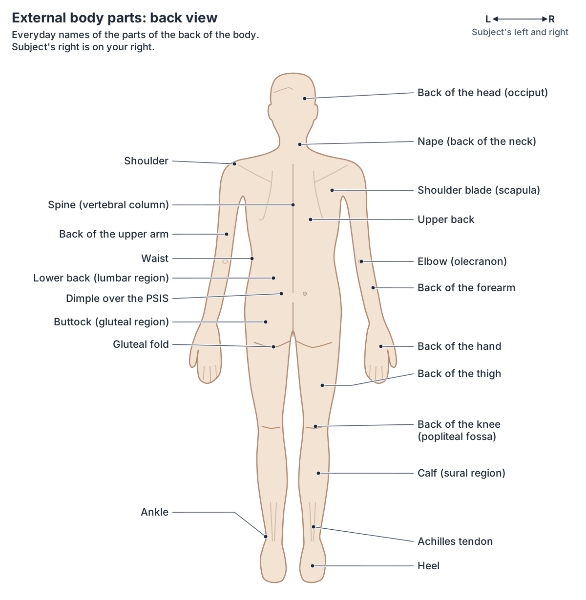
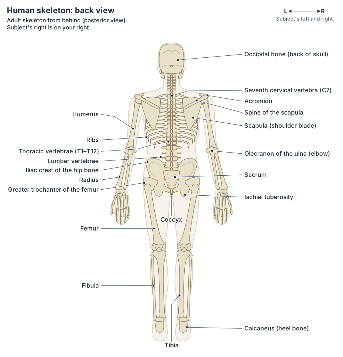
Down the middle of the back runs the vertebral furrow, a groove that marks the spines of the vertebrae, with the muscle masses of the erector spinae on each side. At the base of the neck the spine of the seventh cervical vertebra is easy to see and feel, and between the buttocks lies the natal cleft. (Texas Tech University Health Sciences Center El Paso: Topographical anatomy of the back.)
The shoulder blade (scapula) lies on the back of the shoulder; the long, prominent ridge across its upper part is the spine of the scapula. (OpenStax Anatomy and Physiology 2e, 8.1: The Pectoral Girdle.)
The dimples belong to the hip bones: the posterior superior iliac spine is surrounded but not covered by muscles and ligaments, which is why it shows as a depression in the skin. (OpenStax Anatomy and Physiology 2e, 8.3: The Pelvic Girdle and Pelvis.)
The spine, back muscles and lower back are covered together in Back anatomy.
Internal organs diagram: where the organs are
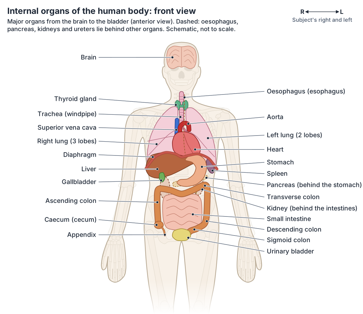
Most organs sit in the two largest body compartments. In the dorsal (posterior) cavity, the cranial cavity houses the brain and the spinal cavity encloses the spinal cord. The ventral (anterior) cavity is divided by the diaphragm into the thoracic cavity, which contains the lungs and the heart, and the abdominopelvic cavity, whose abdominal part houses the digestive organs and whose pelvic part houses the organs of reproduction. (OpenStax Anatomy and Physiology 2e, 1.6: Anatomical Terminology.)
The heart lies in the mediastinum between the lungs, behind the sternum and costal cartilages, with its apex just left of the sternum. It is about the size of a fist. (OpenStax Anatomy and Physiology 2e, 19.1: Heart Anatomy.)
The lungs are paired, pyramid-shaped organs that rest on the diaphragm. The right lung has three lobes and the left lung two, with a dent called the cardiac notch where the heart sits. (OpenStax Anatomy and Physiology 2e, 22.2: The Lungs.)
Below the diaphragm, the liver fills the right upper quadrant and is the largest gland in the body, weighing about 1.4 kg (3 lb) in an adult. The gallbladder sits on the back of its right lobe, and the pancreas lies across the back of the abdomen behind the stomach. (OpenStax Anatomy and Physiology 2e, 23.6: Accessory Organs in Digestion: The Liver, Pancreas, and Gallbladder.)
On the four-quadrant map of the abdomen, which crosses at the navel, the stomach lies in the left upper quadrant with the lower left ribs, and the right side of the liver in the right upper quadrant. (OpenStax Anatomy and Physiology 2e, 1.6: Anatomical Terminology.)
The large intestine frames the small intestine: the ascending colon runs up the right side, the transverse colon crosses from right to left and the descending colon runs down the left side to the S-shaped sigmoid colon, which enters the pelvis. (OpenStax Anatomy and Physiology 2e, 23.5: The Small and Large Intestines.)
The kidneys lie on either side of the spine behind the abdominal lining, the left at about the T12 to L3 vertebrae and the right a little lower because the liver pushes it down. (OpenStax Anatomy and Physiology 2e, 25.3: Gross Anatomy of the Kidney.)
The urinary bladder lies in the pelvis, behind the pubic bone and in front of the rectum; in females it lies in front of the uterus. (OpenStax Anatomy and Physiology 2e, 25.2: Gross Anatomy of Urine Transport.)
Each organ's location, size and left or right side is covered in the guide to the Organs of the body.
Skeleton diagram: the bones from the front
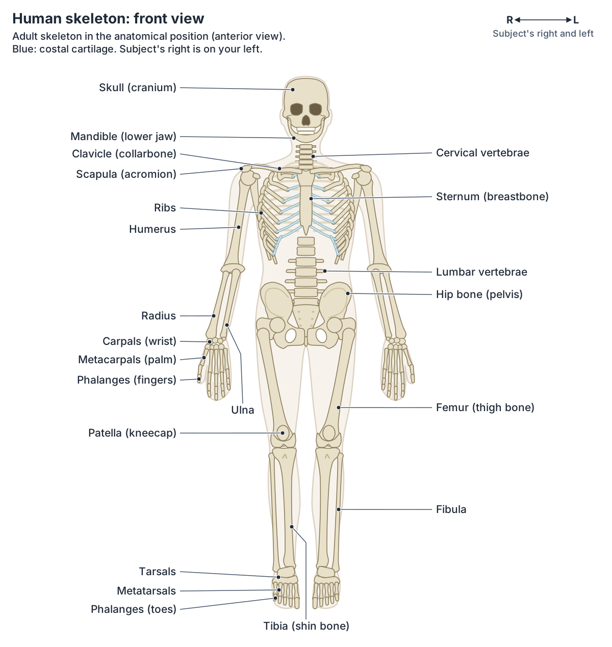
An adult skeleton has 206 bones. The axial skeleton has 80: the skull and its associated bones, the vertebral column and the thoracic cage. The appendicular skeleton has 126: the bones of the upper and lower limbs and the girdles that attach them to the axial skeleton. (OpenStax Anatomy and Physiology 2e, 7.1: Divisions of the Skeletal System.)
The National Cancer Institute's SEER anatomy course gives the same split, 80 axial and 126 appendicular bones. (SEER Training Modules (National Cancer Institute): Skeletal System.)
The thoracic cage is formed by 12 pairs of ribs with their costal cartilages and the sternum. Ribs 1 to 7 are true ribs that reach the sternum through their own cartilage; ribs 8 to 12 are false ribs, and the last two, 11 and 12, are floating ribs with no attachment to the sternum. (OpenStax Anatomy and Physiology 2e, 7.4: The Thoracic Cage.)
Each upper limb has 30 bones: the humerus, the radius and ulna, 8 carpals, 5 metacarpals and 14 phalanges. In the anatomical position the radius lies on the lateral (thumb) side of the forearm and the ulna on the medial side. (OpenStax Anatomy and Physiology 2e, 8.2: Bones of the Upper Limb.)
Each lower limb also has 30 bones: the femur, patella, tibia, fibula, 7 tarsals, 5 metatarsals and 14 phalanges. The femur is the longest and strongest bone of the body. (OpenStax Anatomy and Physiology 2e, 8.4: Bones of the Lower Limb.)
Every bone in a rotatable 3D skeleton, with bone types and functions, is in the guide to the Skeletal system.
Skeleton from the back
From behind, the vertebral column is the centre of the diagram. The adult column has 24 vertebrae, 7 cervical, 12 thoracic and 5 lumbar, plus the sacrum, formed from five fused vertebrae, and the coccyx, formed from four smaller fused ones. The prominent spine of C7 can be felt at the base of the neck. (OpenStax Anatomy and Physiology 2e, 7.3: The Vertebral Column.)
The scapula is a flat, triangular bone on the back of the shoulder. Its spine runs across its upper part and ends laterally in the acromion, which forms the bony tip of the shoulder. (OpenStax Anatomy and Physiology 2e, 8.1: The Pectoral Girdle.)
Each hip bone forms from three bones, the ilium, ischium and pubis, which fuse in the late teens. The iliac crest is the curved upper margin of the ilium that you can feel along your waistline. (OpenStax Anatomy and Physiology 2e, 8.3: The Pelvic Girdle and Pelvis.)
At the elbow, the olecranon of the ulna forms the bony tip of the elbow. (OpenStax Anatomy and Physiology 2e, 8.2: Bones of the Upper Limb.)
At the foot, the calcaneus, the largest bone of the foot, forms the heel, and the lateral malleolus of the fibula and the medial malleolus of the tibia form the bumps on either side of the ankle. (OpenStax Anatomy and Physiology 2e, 8.4: Bones of the Lower Limb.)
The vertebrae, discs and curves of the spine are covered in the guide to the Vertebral column.
Bones of the human skeleton by region
| Region | Bones | Number |
|---|---|---|
| Skull and associated bones | Cranial and facial bones, hyoid, 6 ear ossicles | 22 + 7 |
| Vertebral column | 7 cervical, 12 thoracic, 5 lumbar vertebrae, sacrum, coccyx | 26 |
| Thoracic cage | 12 pairs of ribs, sternum | 25 |
| Upper limbs (each) | Humerus, radius, ulna, 8 carpals, 5 metacarpals, 14 phalanges | 30 |
| Lower limbs (each) | Femur, patella, tibia, fibula, 7 tarsals, 5 metatarsals, 14 phalanges | 30 |
| Girdles | Clavicle and scapula on each side; the two hip bones | 4 + 2 |
| Total | Axial 80 + appendicular 126 | 206 |
Counts from OpenStax Anatomy and Physiology 2e, sections 7.1, 7.3, 7.4, 8.2 and 8.4. The sacrum and coccyx each count as one bone.
Muscle diagram: the major muscles from the front
There are more than 600 muscles in the body, which together make up about 40 percent of a person's weight. (SEER Training Modules (National Cancer Institute): Muscle Groups.)
Cleveland Clinic gives the same figure of more than 600 muscles. (Cleveland Clinic: Muscle.)
Muscle names describe the muscle: its shape, its size compared with its neighbours, its location, its attachments, the number of origins it has, or its action. (OpenStax Anatomy and Physiology 2e, 11.2: Naming Skeletal Muscles.)
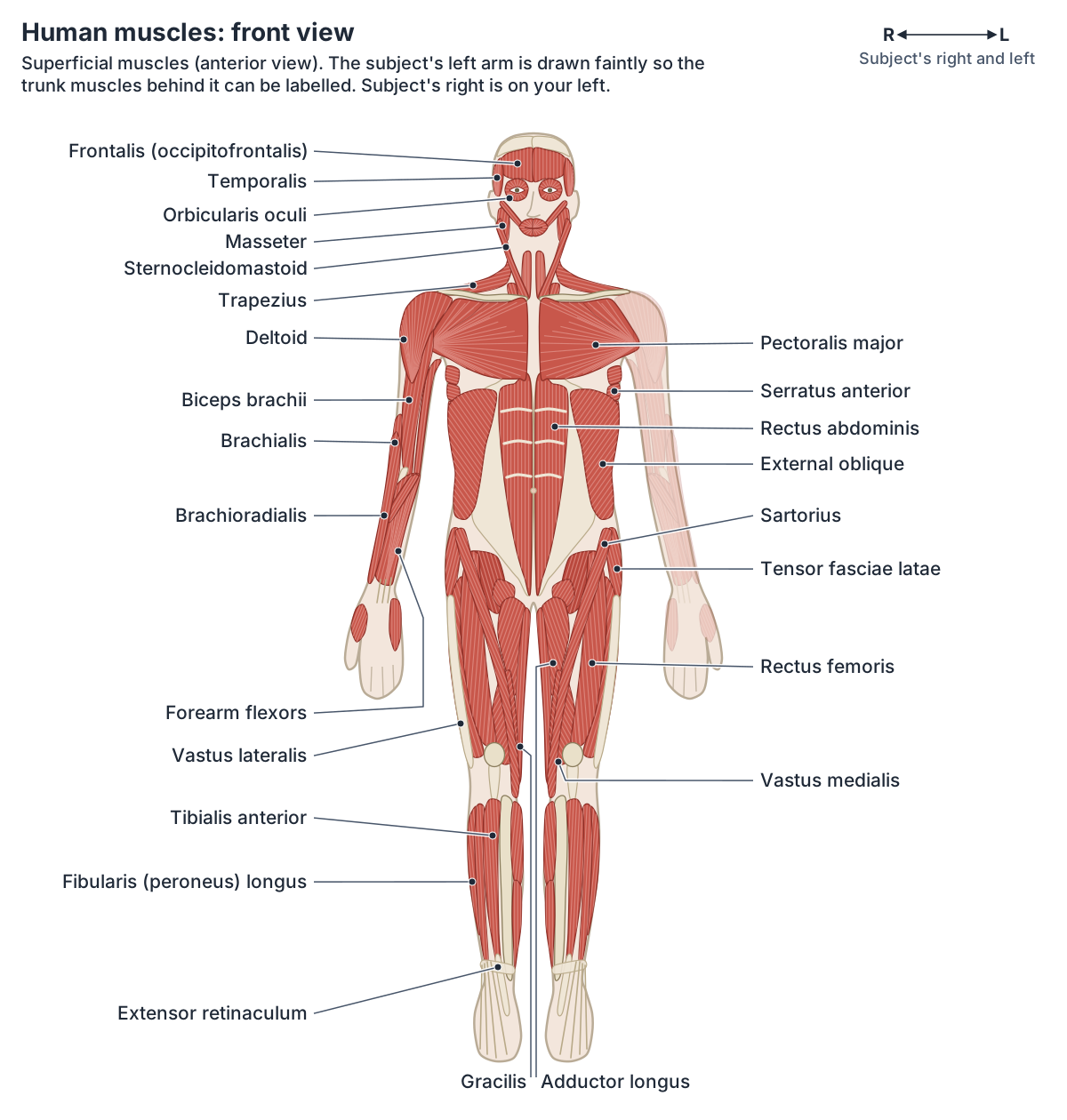
On the front of the chest, pectoralis major is thick and fan-shaped and covers much of the upper thorax. The deltoid creates the rounded line of the shoulder, and the two-headed biceps brachii crosses the shoulder and elbow to flex the forearm. (OpenStax Anatomy and Physiology 2e, 11.5: Muscles of the Pectoral Girdle and Upper Limbs.)
The rectus abdominis is crossed by three tendinous intersections, which give the trained abdomen its six-pack look, and the white linea alba runs down the midline between the two muscles. The external oblique, the outermost layer of the side of the abdomen, has fibres that run down and towards the midline. (OpenStax Anatomy and Physiology 2e, 11.4: Axial Muscles of the Abdominal Wall and Thorax.)
The front of the thigh holds the quadriceps femoris, four muscles that extend the knee: rectus femoris and the vastus lateralis, medialis and intermedius. The band-like sartorius starts at the anterior superior iliac spine and crosses the thigh, and tibialis anterior lies on the lateral side of the shin. (OpenStax Anatomy and Physiology 2e, 11.6: Appendicular Muscles of the Pelvic Girdle and Lower Limbs.)
Muscle groups, naming and a 3D model with every modelled muscle are in the guide to the Muscular system.
Back muscles diagram: the major muscles from behind
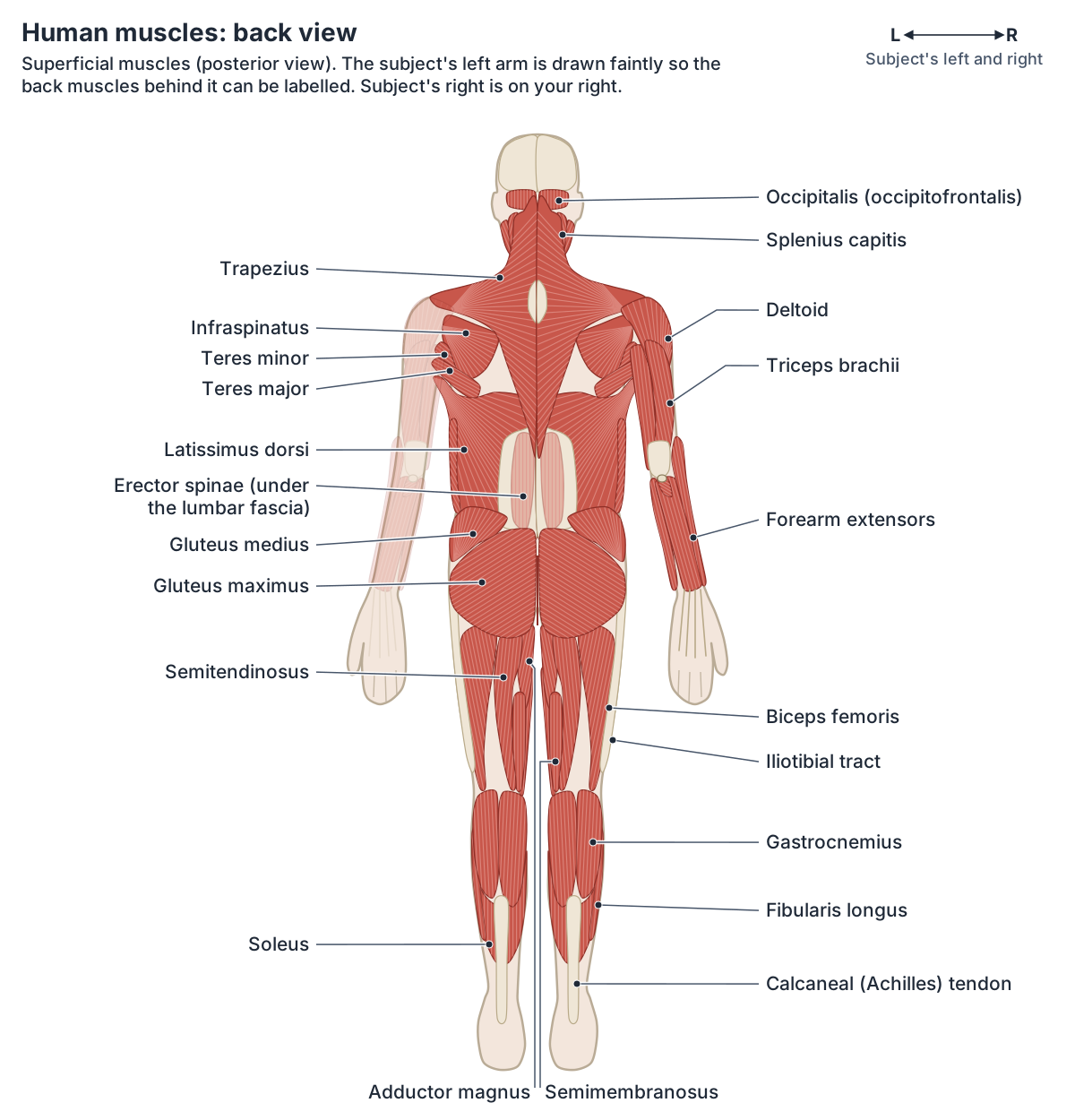
The trapezius covers the upper back and neck; it shrugs the shoulders, pulls the shoulder blades together and tilts the head back. The broad, triangular latissimus dorsi covers the lower back, and the triceps brachii on the back of the arm extends the forearm. (OpenStax Anatomy and Physiology 2e, 11.5: Muscles of the Pectoral Girdle and Upper Limbs.)
The erector spinae group forms most of the muscle mass of the back and is the main extensor of the vertebral column. In the diagram it lies under a sheet of fascia in the lower back. (OpenStax Anatomy and Physiology 2e, 11.3: Axial Muscles of the Head, Neck, and Back.)
The gluteus maximus is the largest of the gluteal muscles, with the gluteus medius deep to it. The hamstrings on the back of the thigh, biceps femoris, semitendinosus and semimembranosus, flex the knee, and the superficial calf muscles insert on the calcaneal (Achilles) tendon. (OpenStax Anatomy and Physiology 2e, 11.6: Appendicular Muscles of the Pelvic Girdle and Lower Limbs.)
The layers of back muscles, from trapezius to the deep spinal muscles, are covered in Back muscles.
Major surface muscles in the diagrams
| Muscle | Where it is | What it does |
|---|---|---|
| Pectoralis major | Front of the upper chest | Brings the elbows together and up, as in an uppercut |
| Deltoid | Cap of the shoulder | Main abductor of the arm |
| Biceps brachii | Front of the arm | Flexes the forearm, helps supinate it |
| Triceps brachii | Back of the arm | Extends the forearm |
| Trapezius | Upper back and neck | Shrugs and pulls the shoulder blades together |
| Latissimus dorsi | Lower back | Moves the elbow back and spreads the elbows apart |
| Rectus abdominis | Front of the abdomen | Flexes the vertebral column, as in sitting up |
| External oblique | Side of the abdomen | Outermost layer of the abdominal wall |
| Gluteus maximus | Buttock | Largest gluteal muscle |
| Quadriceps femoris | Front of the thigh | Extends the knee |
| Hamstrings | Back of the thigh | Flex the knee |
| Gastrocnemius and soleus | Calf | Insert on the calcaneal (Achilles) tendon |
Descriptions from OpenStax Anatomy and Physiology 2e, sections 11.4 to 11.6.
Organ systems diagram: the body's systems at a glance
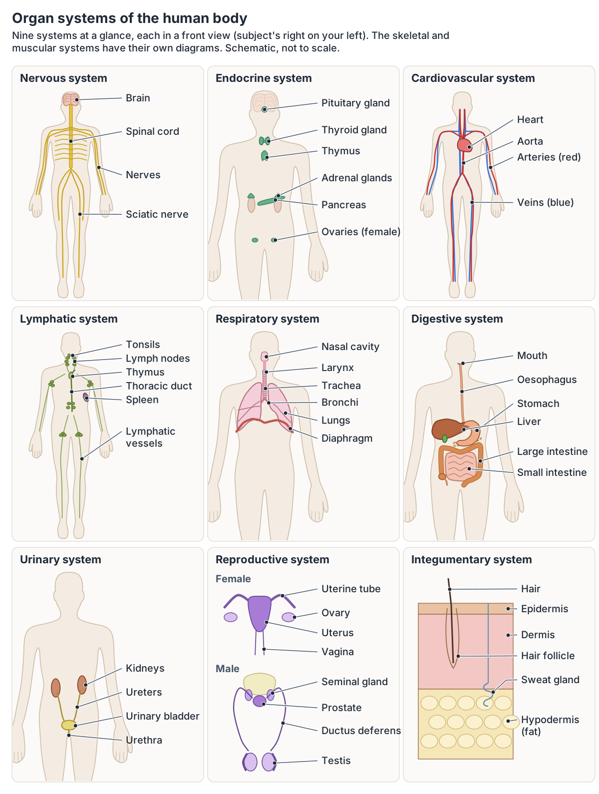
An organ system is a group of organs that work together to perform major functions or meet the body's needs. OpenStax describes eleven: integumentary, skeletal, muscular, nervous, endocrine, cardiovascular, lymphatic, respiratory, digestive, urinary and reproductive. (OpenStax Anatomy and Physiology 2e, 1.2: Structural Organization of the Human Body.)
Counts differ between sources. Cleveland Clinic says the number of body systems varies between 10 and 12 depending on who you ask; its own list of 12 adds an exocrine system, and it does not count the immune system as a separate organ system. (Cleveland Clinic: The Human Body.)
Kenhub describes 11 systems, or 10 when the skeletal and muscular systems are combined into one musculoskeletal system. (Kenhub: Human body systems.)
Five organs are often called vital, because the body cannot survive long without any one of them unless it is supported medically: the heart, brain, kidneys, liver and lungs. (Wakim S, Grewal M. Human Biology, 10.4: Human Organs and Organ Systems (LibreTexts).)
The heart, arteries, veins and the two circuits of blood flow are drawn in the guide to the Circulatory system.
Human body systems chart
| System | Main organs | Main job |
|---|---|---|
| Integumentary | Skin, hair, nails | Encloses the body; holds sensory receptors |
| Skeletal | Bones, joints, cartilage | Supports the body; with muscles, enables movement |
| Muscular | Skeletal muscles, tendons | Movement; helps maintain body temperature |
| Nervous | Brain, spinal cord, peripheral nerves | Detects and processes information, triggers responses |
| Endocrine | Pituitary, thyroid, adrenal glands, pancreas, ovaries, testes | Secretes hormones that regulate body processes |
| Cardiovascular | Heart, blood vessels | Delivers oxygen and nutrients; evens out temperature |
| Lymphatic | Thymus, spleen, lymph nodes and vessels | Returns fluid to the blood; defends against pathogens |
| Respiratory | Nasal passages, trachea, lungs | Delivers oxygen to the blood, removes carbon dioxide |
| Digestive | Stomach, liver, gallbladder, intestines | Processes food, removes undigested waste |
| Urinary | Kidneys, ureters, bladder, urethra | Controls water balance, removes wastes from the blood |
| Reproductive | Ovaries, uterus; testes, prostate | Produces sex hormones and gametes |
The eleven organ systems as described in OpenStax Anatomy and Physiology 2e, section 1.2, with endocrine glands from section 17.1, urinary organs from section 25.2 and reproductive organs from Kenhub.
Nervous and endocrine systems
The central nervous system is the brain and spinal cord; the brain sits in the cranial cavity and the spinal cord runs through the vertebral column. Everything else, the nerves and ganglia, is the peripheral nervous system. Together they sense changes, integrate the information and trigger responses. (OpenStax Anatomy and Physiology 2e, 12.1: Basic Structure and Function of the Nervous System.)
Endocrine glands are ductless and release hormones into the surrounding fluid, from where the blood carries them. The pituitary and pineal glands lie in the brain, the thyroid wraps around the front of the trachea, four parathyroid glands sit on the back of the thyroid, an adrenal gland sits on top of each kidney, the pancreas lies near the centre of the abdomen, and the ovaries and testes are endocrine organs too. (OpenStax Anatomy and Physiology 2e, 17.1: An Overview of the Endocrine System.)
The lobes, brainstem and cerebellum are shown in 3D in the guide to Brain anatomy.
Cardiovascular and lymphatic systems
The heart has four chambers, two atria and two ventricles. Its pulmonary circuit carries blood to and from the lungs, and its systemic circuit carries oxygenated blood to almost every tissue and returns it to the heart. In the diagrams, arteries are red and veins blue. (OpenStax Anatomy and Physiology 2e, 19.1: Heart Anatomy.)
The lymphatic system is made of lymphatic vessels, bean-shaped lymph nodes, the thymus between the sternum and the aorta, the spleen, red bone marrow, the tonsils in the pharynx and mucosa-associated lymphoid tissue. Lymph nodes cluster in the groin, armpits, neck, chest and abdomen. (OpenStax Anatomy and Physiology 2e, 21.1: Anatomy of the Lymphatic and Immune Systems.)
Lymph returns to the blood in the neck: the right lymphatic duct drains the upper right side of the body into the right subclavian vein, and the thoracic duct drains the rest into the left subclavian vein. (OpenStax Anatomy and Physiology 2e, 21.1: Anatomy of the Lymphatic and Immune Systems.)
Lymph node groups, lymphatic vessels and the lymphoid organs are mapped in the guide to the Lymphatic system.
Respiratory system
Air enters mainly through the nose and passes the pharynx, larynx, trachea and bronchi to the lungs. The conducting zone moves air and is not directly involved in gas exchange; the respiratory zone, in the lungs, is where oxygen and carbon dioxide are exchanged. (OpenStax Anatomy and Physiology 2e, 22.1: Organs and Structures of the Respiratory System.)
The dome-shaped diaphragm sits at the base of the lungs and the thoracic cavity, and each lung is wrapped in a two-layered serous membrane, the pleura. (OpenStax Anatomy and Physiology 2e, 22.2: The Lungs.)
The airway from the nose to the alveoli is drawn and explained in the guide to the Respiratory system.
Digestive system
The alimentary canal runs from the mouth to the anus through the pharynx, oesophagus, stomach and small and large intestines. It is about 7.62 m (25 ft) long in life and closer to 10.67 m (35 ft) when measured after death. The teeth, tongue, salivary glands, liver, gallbladder and pancreas are accessory organs. (OpenStax Anatomy and Physiology 2e, 23.1: Overview of the Digestive System.)
The small intestine is about 3 m (10 ft) long in a living person and has three parts, the duodenum, jejunum and ileum. The large intestine is about 7.6 cm (3 in) wide, more than twice the width of the small intestine. (OpenStax Anatomy and Physiology 2e, 23.5: The Small and Large Intestines.)
Each organ of the gastrointestinal tract is followed in order in the guide to the Digestive system.
Urinary system
Urine passes from the kidneys down the ureters, each about 30 cm long, to the bladder and out through the urethra, which averages about 20 cm in males and about 4 cm in females. (OpenStax Anatomy and Physiology 2e, 25.2: Gross Anatomy of Urine Transport.)
Each kidney is about 11 to 14 cm long, 6 cm wide and 4 cm thick. (OpenStax Anatomy and Physiology 2e, 25.3: Gross Anatomy of the Kidney.)
The kidneys, ureters, bladder and urethra are covered together in the guide to the Urinary system.
Reproductive system
The female reproductive organs include the ovaries, uterine (fallopian) tubes, uterus, vagina and vulva. The male organs include the testes, epididymis, ductus deferens, seminal vesicles, prostate, penis and urethra. (Kenhub: Human body systems.)
In both sexes the reproductive system produces sex hormones and gametes. The two testes lie within the scrotum, and the mammary glands within the breasts belong to the female system. (OpenStax Anatomy and Physiology 2e, 1.2: Structural Organization of the Human Body.)
The female body and its organs, with a female 3D model, are covered in the guide to Female anatomy.
Integumentary system: skin, hair and nails
The skin has two main layers, the epidermis of closely packed epithelial cells and the dermis below it, with the hypodermis of loose connective and fatty tissue beneath. Together with accessory structures such as hair follicles and sweat glands, the skin makes up the integumentary system. (OpenStax Anatomy and Physiology 2e, 5.1: Layers of the Skin.)
The integumentary system encloses the internal structures and holds many sensory receptors; it includes the hair, skin and nails. (OpenStax Anatomy and Physiology 2e, 1.2: Structural Organization of the Human Body.)
The epidermis, dermis and hypodermis are shown in 3D in Layers of the skin.
Human anatomy images: drawings, photographs and scans
Labelled drawings like the ones above simplify the body so the parts can be named. Real images of anatomy come from dissection photographs and medical imaging. The U.S. National Library of Medicine's Visible Human Project made complete, detailed 3D datasets of a male and a female body from MRI, CT and photographs of thin cross-sections: 1,871 CT and anatomical sections of the male at 1 mm intervals, and 5,189 anatomical images of the female at 0.33 mm. The male set was released in 1994, the female in 1995, and since 2019 no licence is needed to download them. (U.S. National Library of Medicine: The Visible Human Project.)
On an X-ray, tissues absorb radiation differently: bones absorb the most and look white, fat and other soft tissues look grey, and air-filled areas such as the lungs look black. (MedlinePlus: X-Rays.)
CT builds cross-sectional images, called slices, from an X-ray source that moves in a circle around the patient; stacked together, the slices form a 3D image. (National Institute of Biomedical Imaging and Bioengineering: Computed Tomography (CT).)
MRI is best for the soft, non-bony parts of the body, such as the brain, spinal cord, nerves, muscles, ligaments and tendons, and it does not use ionising radiation. (National Institute of Biomedical Imaging and Bioengineering: Magnetic Resonance Imaging (MRI).)
How cross-sections are cut (sagittal, coronal and transverse) is explained in the guide to Anatomical planes.
How to see each body system in the 3D model
The 3D model at the top of this page is built from BodyParts3D, a dictionary-type anatomy database of 3D structures based on a whole-body model of an adult human male, released under a CC BY licence. (BodyParts3D, Database Center for Life Science.)
It opens with the brain and the organs of the chest and abdomen highlighted inside a faint skeleton. Use the coloured system buttons to switch whole systems on or off, for example Muscular to cover the skeleton with muscles or Cardiovascular to add the heart and vessels, and the Skin and Muscles sliders to see through the outer layers. The Front, Back, Left, Right and Top buttons turn the body, Hover labels names each part under the pointer, and clicking a part opens its own page. Turn off Highlight topic to see every structure in full colour, and use the Female switch to see the female reproductive organs and breasts.
The model has gaps that the diagrams fill: it has no thyroid gland, rectus abdominis or latissimus dorsi, and no chewing muscles or muscles of facial expression apart from the platysma, so use the organ and muscle diagrams for those.
Using the rotatable model as a figure-drawing reference, with proportions and landmarks, is covered in Anatomy drawing reference.
Human body diagram in the 3D model
- Adrenal gland
- The adrenal (suprarenal) gland is a small endocrine gland on top of each kidney.
- Angular gyrus
- The angular gyrus is a cortical region of the inferior parietal lobule that arches over the end of the superior temporal sulcus.
- Anterior part of superior temporal gyrus
- This is the anterior portion of the superior temporal gyrus, the uppermost gyrus on the lateral surface of the temporal lobe.
- Appendix
- The vermiform appendix is a narrow blind-ended tube arising from the cecum.
- Ascending colon
- The ascending colon is the part of the large intestine rising from the cecum to the right colic (hepatic) flexure.
- Caudate lobe of liver
- The caudate lobe (Couinaud segment I) is a small liver lobe on the posterior surface between the inferior vena cava and the fissure for the ligamentum venosum.
- Cecum
- The caecum is the first part of the large intestine, a blind-ending pouch in the right lower abdomen below the point where the small intestine joins the colon.
- Cerebellum
- The cerebellum is the large structure at the back of the brain that coordinates movement, posture and balance and takes part in motor learning.
- Descending colon
- The descending colon passes down from the left colic (splenic) flexure to the sigmoid colon.
- Distal part of ileum
- The distal part of the ileum is a segment of the lower small intestine.
- Distal part of jejunum
- The distal part of the jejunum is a segment of the middle small intestine.
- Duodenum
- The duodenum is the first and shortest part of the small intestine, about 20 to 25 cm long and C-shaped.
- Esophagus
- The esophagus is a muscular tube about 22 to 25 cm long that carries swallowed food from the pharynx to the stomach.
- Gallbladder
- The gallbladder is a pear-shaped sac that stores and concentrates bile.
- Hepatovenous segment II
- Couinaud segment II is the superior part of the left lateral section of the liver.
- Hepatovenous segment III
- Couinaud segment III is the inferior part of the left lateral section of the liver.
- Hepatovenous segment IV
- Couinaud segment IV is the left medial section of the liver, also called the quadrate lobe in its inferior part.
- Hepatovenous segment V
- Couinaud segment V is the inferior part of the right anterior section of the liver.
- Hepatovenous segment VI
- Couinaud segment VI is the inferior part of the right posterior section of the liver.
- Hepatovenous segment VII
- Couinaud segment VII is the superior part of the right posterior section of the liver.
- Hepatovenous segment VIII
- Couinaud segment VIII is the superior part of the right anterior section of the liver.
- Inferior frontal gyrus
- The inferior frontal gyrus is the lowest of the three frontal gyri on the lateral frontal lobe, and contains Broca's area on the dominant side.
- Inferior temporal gyrus
- The inferior temporal gyrus is the lowest gyrus on the lateral surface of the temporal lobe.
- Kidney
- The kidney is a bean-shaped organ that filters blood to form urine and regulate fluid, electrolytes and blood pressure.
- Lobe of thymus
- The thymus lobes are the two lobes of the thymus gland, where T lymphocytes mature.
- Lower lobe of left lung
- The lower lobe of the left lung lies below and behind the oblique fissure. It forms the back and the base of the left lung, resting on the left dome of the diaphragm.
- Lower lobe of right lung
- The lower lobe of the right lung lies below and behind the oblique fissure. It forms the back and the base of the right lung, resting on the right dome of the diaphragm above the liver.
- Middle frontal gyrus
- The middle frontal gyrus is the central of the three frontal gyri on the lateral surface of the frontal lobe.
- Middle lobe of right lung
- The middle lobe is the wedge of the right lung that lies between the horizontal and oblique fissures, at the front and side of the lower chest. Only the right lung has one.
- Middle part of ileum
- The middle part of the ileum is a segment of the lower small intestine.
- Middle part of jejunum
- The middle part of the jejunum is a segment of the middle small intestine.
- Middle temporal gyrus
- The middle temporal gyrus is the central gyrus on the lateral surface of the temporal lobe.
- Occipital lobe
- The occipital lobe is the posterior lobe of the cerebral hemisphere, and the primary visual centre.
- Pancreas
- The pancreas is a soft, elongated gland with both exocrine and endocrine functions.
- Postcentral gyrus
- The postcentral gyrus is the primary somatosensory cortex of the parietal lobe.
- Posterior part of superior temporal gyrus
- This is the posterior portion of the superior temporal gyrus, important for hearing and language.
- Precentral gyrus
- The precentral gyrus is the primary motor cortex of the frontal lobe.
- Proximal part of ileum
- The proximal part of the ileum is a segment of the lower small intestine.
- Proximal part of jejunum
- The proximal part of the jejunum is a segment of the middle small intestine.
- Rectum
- The rectum is the final straight portion of the large intestine before the anal canal.
- Sigmoid colon
- The sigmoid colon is the S-shaped last part of the colon. It runs from the end of the descending colon at the pelvic brim to the rectum.
- Spleen
- The spleen is the largest lymphoid organ, filtering blood and supporting immune responses.
- Stomach
- The stomach is a J-shaped, expandable part of the digestive tract that mixes food with acid and enzymes.
- Superior frontal gyrus
- The superior frontal gyrus is the uppermost gyrus of the frontal lobe, extending onto its medial surface.
- Superior parietal lobule
- The superior parietal lobule is a parietal association area above the intraparietal sulcus.
- Supramarginal gyrus
- The supramarginal gyrus is part of the inferior parietal lobule that arches over the end of the lateral sulcus.
- Trachea
- The trachea (windpipe) is a flexible air tube extending from the larynx to the bronchi.
- Transverse colon
- The transverse colon is the part of the large intestine that crosses the abdomen between the right and left colic flexures.
- Upper lobe of left lung
- The upper lobe of the left lung is one of the left lung's two lobes. It lies above and in front of the oblique fissure, forms the apex of the lung and includes the lingula.
- Upper lobe of right lung
- The upper lobe of the right lung is the top of the right lung's three lobes. It lies above the horizontal fissure and above the upper part of the oblique fissure, and forms the apex of the lung.
- Ureter
- The ureter is a muscular tube carrying urine from the renal pelvis to the urinary bladder.
- Urinary bladder
- The urinary bladder is a hollow muscular organ that stores urine.
- Wall of atrium
- The muscular walls of the right and left atria, the thin-walled receiving chambers of the heart.
- Wall of ventricle
- The muscular wall of a ventricle of the heart.
Frequently asked questions
What are the main parts of the human body?
The body has an axial part, the head, neck and trunk, and four limbs. The trunk contains the chest (thorax), abdomen and pelvis. Each upper limb has the arm, forearm, wrist and hand; each lower limb has the thigh, knee, leg, ankle and foot. Inside, the organs are grouped into eleven organ systems, from the skin and skeleton to the reproductive system.
How many bones and muscles are in the human body?
An adult skeleton has 206 bones: 80 in the axial skeleton (skull, vertebral column and thoracic cage) and 126 in the limbs and their girdles. There are more than 600 muscles, which together make up about 40 percent of body weight. Each arm and each leg has 30 bones.
How many organ systems are there in the human body?
OpenStax and Kenhub describe eleven: integumentary, skeletal, muscular, nervous, endocrine, cardiovascular, lymphatic, respiratory, digestive, urinary and reproductive. Other sources count 10 to 12, depending on whether they combine the skeletal and muscular systems, add an exocrine system, or count the immune system separately.
What are the five vital organs?
The heart, brain, kidneys, liver and lungs are often called the five vital organs, because the body cannot survive long if any one of them stops working, unless it is supported medically. The heart pumps blood, the brain controls the body, the kidneys filter blood, the liver processes nutrients and makes bile, and the lungs exchange gases.
Why is the right side of the body on the left of an anatomy diagram?
Right and left in anatomy always mean the subject's right and left. In a front view you face the person, so their right side is on your left, as when you look at someone standing in front of you. In a back view you see the body from behind, so their right side is on your right. The whole-body diagrams here carry an R and L arrow.
Where can I find real images of human anatomy?
Real anatomy images come from dissection photographs and medical imaging. The U.S. National Library of Medicine's Visible Human Project offers free CT, MRI and cross-section photographs of a male and a female body. X-rays show bones in white, CT shows the body in cross-sectional slices and MRI shows soft tissues such as the brain and muscles.
References
- OpenStax. Anatomy and Physiology 2e, §1.2 Structural Organization of the Human Body. Rice University, 2022.
- OpenStax. Anatomy and Physiology 2e, §1.6 Anatomical Terminology. Rice University, 2022.
- OpenStax. Anatomy and Physiology 2e, §5.1 Layers of the Skin. Rice University, 2022.
- OpenStax. Anatomy and Physiology 2e, §7.1 Divisions of the Skeletal System. Rice University, 2022.
- OpenStax. Anatomy and Physiology 2e, §7.3 The Vertebral Column. Rice University, 2022.
- OpenStax. Anatomy and Physiology 2e, §7.4 The Thoracic Cage. Rice University, 2022.
- OpenStax. Anatomy and Physiology 2e, §8.1 The Pectoral Girdle. Rice University, 2022.
- OpenStax. Anatomy and Physiology 2e, §8.2 Bones of the Upper Limb. Rice University, 2022.
- OpenStax. Anatomy and Physiology 2e, §8.3 The Pelvic Girdle and Pelvis. Rice University, 2022.
- OpenStax. Anatomy and Physiology 2e, §8.4 Bones of the Lower Limb. Rice University, 2022.
- OpenStax. Anatomy and Physiology 2e, §11.2 Naming Skeletal Muscles. Rice University, 2022.
- OpenStax. Anatomy and Physiology 2e, §11.3 Axial Muscles of the Head, Neck, and Back. Rice University, 2022.
- OpenStax. Anatomy and Physiology 2e, §11.4 Axial Muscles of the Abdominal Wall and Thorax. Rice University, 2022.
- OpenStax. Anatomy and Physiology 2e, §11.5 Muscles of the Pectoral Girdle and Upper Limbs. Rice University, 2022.
- OpenStax. Anatomy and Physiology 2e, §11.6 Appendicular Muscles of the Pelvic Girdle and Lower Limbs. Rice University, 2022.
- OpenStax. Anatomy and Physiology 2e, §12.1 Basic Structure and Function of the Nervous System. Rice University, 2022.
- OpenStax. Anatomy and Physiology 2e, §17.1 An Overview of the Endocrine System. Rice University, 2022.
- OpenStax. Anatomy and Physiology 2e, §19.1 Heart Anatomy. Rice University, 2022.
- OpenStax. Anatomy and Physiology 2e, §21.1 Anatomy of the Lymphatic and Immune Systems. Rice University, 2022.
- OpenStax. Anatomy and Physiology 2e, §22.1 Organs and Structures of the Respiratory System. Rice University, 2022.
- OpenStax. Anatomy and Physiology 2e, §22.2 The Lungs. Rice University, 2022.
- OpenStax. Anatomy and Physiology 2e, §23.1 Overview of the Digestive System. Rice University, 2022.
- OpenStax. Anatomy and Physiology 2e, §23.5 The Small and Large Intestines. Rice University, 2022.
- OpenStax. Anatomy and Physiology 2e, §23.6 Accessory Organs in Digestion: The Liver, Pancreas, and Gallbladder. Rice University, 2022.
- OpenStax. Anatomy and Physiology 2e, §25.2 Gross Anatomy of Urine Transport. Rice University, 2022.
- OpenStax. Anatomy and Physiology 2e, §25.3 Gross Anatomy of the Kidney. Rice University, 2022.
- National Cancer Institute. SEER Training Modules: Muscle Groups.
- National Cancer Institute. SEER Training Modules: Skeletal System.
- Cleveland Clinic. Muscle.
- Cleveland Clinic. The Human Body: How Does It Work?
- Kenhub. Human body systems: overview, anatomy, functions.
- Wakim S, Grewal M. Human Biology, §10.4 Human Organs and Organ Systems. LibreTexts.
- Texas Tech University Health Sciences Center El Paso. Medical Gross Anatomy: Topographical anatomy of the thorax.
- Texas Tech University Health Sciences Center El Paso. Medical Gross Anatomy: Topographical anatomy of the abdomen.
- Texas Tech University Health Sciences Center El Paso. Medical Gross Anatomy: Topographical anatomy of the back.
- U.S. National Library of Medicine. The Visible Human Project.
- MedlinePlus, U.S. National Library of Medicine. X-Rays.
- National Institute of Biomedical Imaging and Bioengineering. Computed Tomography (CT).
- National Institute of Biomedical Imaging and Bioengineering. Magnetic Resonance Imaging (MRI).
- BodyParts3D. Database Center for Life Science, Japan.
- Mitsuhashi N. et al. (2009). BodyParts3D: 3D structure database for anatomical concepts. Nucleic Acids Research 37, D782–D785. Data © The Database Center for Life Science, CC BY 4.0.
- Radiopaedia. Oblique fissure (surface anatomy used to place the oblique fissures in the 3D lungs).
- Radiopaedia. Horizontal fissure (surface anatomy used to place the horizontal fissure in the 3D right lung).
Related anatomy
- Organs of the bodyHuman anatomy organs explained with a labelled internal organs diagram: organs by body system and location, left vs…
- Skeletal systemExplore a realistic 3D human skeleton: the 206 bones, axial vs appendicular skeleton, bone types, bony landmarks and…
- Muscular systemExplore an interactive 3D muscle model: how muscles move bones, how they are named, the upper body and leg muscles…
- Body regionsRegional terms in anatomy with front and back body-region diagrams: cephalic, cervical, brachial, femoral, crural and…
- Male anatomyMale anatomy diagram and 3D model: male body organs, urethra and bladder, pelvis, Adam's apple, muscle, pelvic floor…
- Female anatomyFemale anatomy explained with a labelled body diagram and 3D model: reproductive organs, vulva, breasts, pelvis…
Explore the whole body in the interactive 3D human anatomy model, or browse the A–Z list of human structures.