

What is the endocrine system?
OpenStax defines the endocrine system as the cells, tissues and organs that secrete hormones as a primary or secondary function. Endocrine glands are ductless: they secrete their hormones straight into the fluid around them, and the interstitial fluid and blood vessels then carry the hormones through the body. (OpenStax Anatomy and Physiology 2e, 17.1: An Overview of the Endocrine System.)
A hormone is a chemical messenger. The blood carries it to its target cells, which have receptors (docking molecules) for that hormone; when the hormone binds its receptor, it sets off changes inside the target cell. (Hiller-Sturmhöfel S, Bartke A. The Endocrine System: An Overview. Alcohol Health Res World. 1998.)
Sources do not agree on one list of glands. OpenStax names five endocrine glands (the pituitary, thyroid, parathyroid, adrenal and pineal glands), describes the pancreas as a gland with both digestive and hormone-secreting cells, and treats the hypothalamus, thymus, heart, kidneys, stomach, small intestine, liver, skin, ovaries and testes as organs with endocrine function; it adds that fat (adipose tissue) and even bone make hormones. (OpenStax Anatomy and Physiology 2e, 17.1: An Overview of the Endocrine System.)
MedlinePlus lists nine endocrine glands: the adrenal glands, hypothalamus, pancreatic islets, ovaries, parathyroids, pineal, pituitary, testes and thyroid. NIDDK speaks of eight major glands and a 1998 review of seven. That is why quizzes ask for '5', '7' or '8' glands: the number depends on what each source calls a gland, so learn the glands rather than a count. (MedlinePlus Medical Encyclopedia: Endocrine glands.)
Where every major organ sits, from brain to bladder, on a labelled front view. Organs of the body.
Endocrine system diagram: how to label the glands
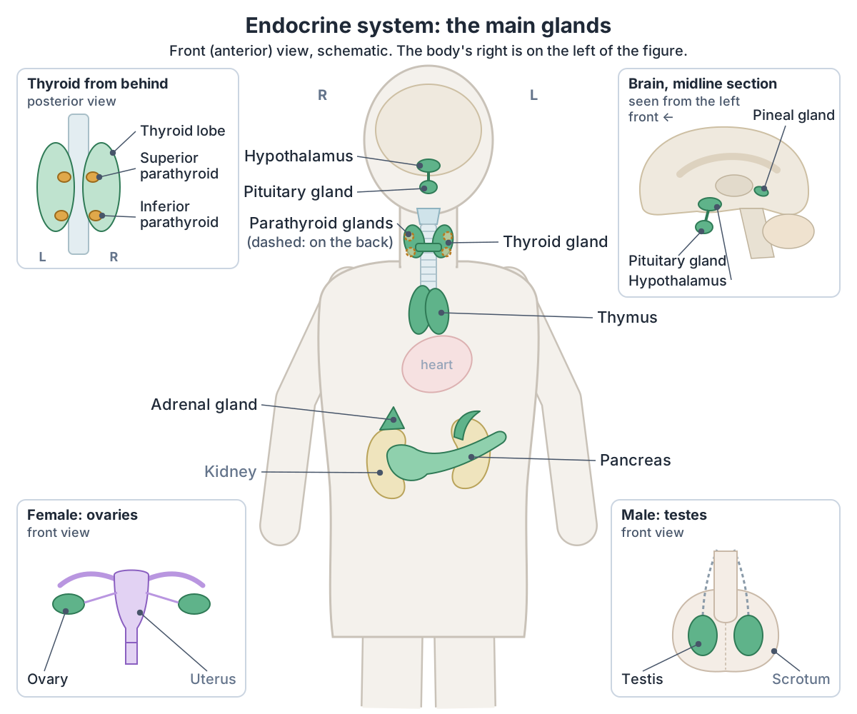
Endocrine system diagrams are usually drawn from the front, with the subject's right on the viewer's left, as in the labelled map at the top of this page. Work from the head down:
1. Hypothalamus: an almond-sized part deep in the brain, below the thalamus and directly above the pituitary gland. (Cleveland Clinic: Hypothalamus.)
2. Pituitary gland: a pea-sized gland that hangs below the hypothalamus, behind the bridge of the nose, in a hollow of the sphenoid bone called the sella turcica. (Cleveland Clinic: Pituitary Gland.)
3. Pineal gland: a small cone-shaped gland in the middle of the brain that projects backwards from the third ventricle. Other structures hide it in a front view, so our map shows it in a midline section of the brain. (NCI SEER Training Modules: Pituitary and Pineal Glands.)
4. Thyroid gland: a butterfly-shaped gland in the front of the neck, spanning about the C5 to T1 vertebrae. Its two lobes are joined by a central isthmus and wrap around the cricoid cartilage and upper rings of the trachea. (TeachMeAnatomy: The Thyroid Gland.)
5. Parathyroid glands: tiny round glands, usually four, embedded in the back (posterior surface) of the thyroid. They cannot be seen from the front, so diagrams show them dashed or in a view from behind. (OpenStax Anatomy and Physiology 2e, 17.5: The Parathyroid Glands.)
6. Thymus: in the upper chest behind the breastbone (sternum), between the lungs, just in front of and above the heart. (Cleveland Clinic: Thymus.)
7. Adrenal glands: one above the upper pole of each kidney. The right gland is shaped like a pyramid and the left is semilunar (half-moon-shaped). (Kenhub: Suprarenal (adrenal) glands.)
8. Pancreas: lies across the back wall of the upper abdomen, behind the stomach, and stretches from the region of the duodenum (on the body's right) to the spleen (on the left). (NCI SEER Training Modules: Pancreas (Islets of Langerhans).)
9. Ovaries (female): small oval glands located on either side of the uterus. (Cleveland Clinic: Endocrine System.)
10. Testes (male): paired oval glands, each suspended in the scrotum by its own spermatic cord. (Kenhub: Endocrine system.)
One body shows only one pair of gonads, so maps add the other pair as a small figure, as ours does.
Labelled diagrams of the organs, skeleton, muscles and organ systems. Human body diagram.
Endocrine vs exocrine glands
The difference is the duct. Exocrine glands have ducts that carry their secretion to a surface: sweat, sebaceous and mammary glands are exocrine, and so are the glands that make digestive enzymes. Endocrine glands have no ducts to carry their product to a surface; their hormones go into the blood instead. (NCI SEER Training Modules: Introduction to the Endocrine System.)
The pancreas is the classic gland that is both. It is mainly an exocrine gland: its acinar cells secrete digestive enzymes. Scattered through it are small clusters of endocrine cells, the pancreatic islets (formerly called the islets of Langerhans), whose alpha cells make glucagon and beta cells make insulin. (OpenStax Anatomy and Physiology 2e, 17.9: The Endocrine Pancreas.)
The MSD Manual puts it simply: endocrine glands release their hormones directly into the bloodstream, and the pancreas is both an endocrine and an exocrine gland. (MSD Manual Consumer Version: Endocrine Glands.)
Head, neck, body and tail, the ducts, the acini and islets, and the blood supply in 3D. Pancreas anatomy.
Endocrine system vs nervous system
Both systems carry messages around the body, and OpenStax says that together they are primarily responsible for homeostasis, the stable internal state the body needs. The nervous system uses electrical and chemical signals over short distances, and its target cells respond within milliseconds. The endocrine system uses only chemical signals, which can travel long distances in the blood, and its responses take longer to start. (OpenStax Anatomy and Physiology 2e, 17.1: An Overview of the Endocrine System.)
The effects also last different lengths of time. The NCI SEER training module says a nerve signal's effect is short, measured in seconds, and local, while the action of the endocrine system is measured in minutes, hours or weeks. Kenhub similarly says endocrine signals can last from a few hours to a few weeks. (NCI SEER Training Modules: Introduction to the Endocrine System.)
The two systems meet in the hypothalamus, which Cleveland Clinic calls the main link between the endocrine system and the nervous system. It is part of the brain, yet it makes hormones and controls the pituitary gland. (Cleveland Clinic: Hypothalamus.)
The lobes and deep parts of the brain, including the diencephalon that holds the hypothalamus. Brain anatomy.
Endocrine system vs nervous system
| Feature | Endocrine system | Nervous system |
|---|---|---|
| Signal | Chemical only: hormones | Electrical and chemical: nerve impulses and neurotransmitters |
| Distance travelled | Long or short (through the blood) | Always short |
| How fast a response starts | Fast or slow | Always fast, within milliseconds |
| How long the effect lasts | Minutes, hours or weeks | Seconds |
| What it targets | Internal environment | Internal and external environment |
From OpenStax 17.1 (Table 17.1) and the NCI SEER endocrine module.
Endocrine glands and their hormones
The table below lists each main endocrine gland, where it is, its main hormones and what they do. OpenStax's own table of endocrine glands leaves out the hypothalamus and thymus, which it describes in other sections; both are included here. (OpenStax Anatomy and Physiology 2e, 17.2: Hormones.)
Counts of pituitary hormones differ between sources. OpenStax says the anterior lobe produces seven hormones, because it adds beta endorphin, and describes melanocyte-stimulating hormone (MSH) from the zone between the lobes. Cleveland Clinic lists six anterior-lobe hormones and two that the posterior lobe stores and releases. The table gives the six anterior hormones that every source lists. (OpenStax Anatomy and Physiology 2e, 17.3: The Pituitary Gland and Hypothalamus.)
Some hormones have two names in different books: the adrenal medulla's adrenaline is also called epinephrine, and noradrenaline is also called norepinephrine. (OpenStax Anatomy and Physiology 2e, 17.6: The Adrenal Glands.)
Endocrine glands, where they are and the hormones they make
| Gland | Where it is | Main hormones | Main effects | Sources |
|---|---|---|---|---|
| Hypothalamus | Deep in the brain, below the thalamus and directly above the pituitary gland | Releasing and inhibiting hormones (TRH, CRH, GnRH, GHRH, somatostatin, dopamine); also makes ADH and oxytocin | Switch the anterior pituitary's hormones on or off; its ADH and oxytocin are stored in and released from the posterior pituitary | OpenStax 17.3; Cleveland Clinic (hypothalamus) |
| Pituitary gland: anterior lobe | Hangs below the hypothalamus on a stalk (infundibulum), in the sella turcica of the sphenoid bone | Growth hormone (GH), prolactin, TSH, ACTH, FSH, LH | GH: growth of body tissues; prolactin: milk production; TSH: thyroid hormone release; ACTH: adrenal cortex hormones; FSH and LH: gamete and sex hormone production in the gonads | OpenStax 17.3; SEER; Cleveland Clinic (pituitary) |
| Pituitary gland: posterior lobe | Back of the pituitary, joined to the hypothalamus by the stalk | Stores and releases antidiuretic hormone (ADH, vasopressin) and oxytocin made by the hypothalamus | ADH: water reabsorption by the kidneys; oxytocin: uterine contractions in childbirth and milk release | OpenStax 17.3; SEER; Cleveland Clinic (pituitary) |
| Pineal gland | Small midline gland deep in the brain, at the back of the third ventricle, between the superior colliculi | Melatonin | Helps set the sleep-wake (circadian) cycle | OpenStax 17.2, 17.7; SEER; Kenhub |
| Thyroid gland | Front of the neck, below the larynx and in front of the trachea (about C5 to T1); two lobes joined by an isthmus | Thyroxine (T4) and triiodothyronine (T3); calcitonin | T3 and T4 raise the basal metabolic rate; calcitonin lowers blood calcium | OpenStax 17.2, 17.4; SEER; TeachMeAnatomy; Kenhub |
| Parathyroid glands | Usually four, two superior and two inferior, on the back of the thyroid lobes | Parathyroid hormone (PTH) | Raises blood calcium | OpenStax 17.5; SEER; Kenhub |
| Thymus | Upper chest behind the sternum, in front of and above the heart | Thymosins | Help T lymphocytes (T cells) develop | OpenStax 17.10; SEER; TeachMeAnatomy; Cleveland Clinic (thymus) |
| Adrenal glands: cortex | Outer layer of each adrenal gland, on top of each kidney | Aldosterone; cortisol; small amounts of androgens | Aldosterone: keeps sodium and water; cortisol: raises blood glucose and drives the long-term stress response | OpenStax 17.6; SEER; TeachMeAnatomy |
| Adrenal glands: medulla | Centre of each adrenal gland | Adrenaline (epinephrine) and noradrenaline (norepinephrine) | Fight-or-flight response to short-term stress | OpenStax 17.6; SEER; TeachMeAnatomy |
| Pancreas (pancreatic islets) | Across the back of the upper abdomen, behind the stomach, from the duodenum to the spleen | Insulin (beta cells) and glucagon (alpha cells) | Insulin lowers blood glucose; glucagon raises it | OpenStax 17.9; SEER |
| Ovaries | In the pelvis, one on each side of the uterus | Oestrogens (estrogens) and progesterone | Female sexual development, the menstrual cycle and preparing the uterus for pregnancy | OpenStax 17.8; SEER; TeachMeAnatomy; Cleveland Clinic |
| Testes | In the scrotum, outside the abdomen | Testosterone | Male sexual development and sperm production | OpenStax 17.8; SEER; Cleveland Clinic; Kenhub |
Each row was checked against the sources named in its last column (full titles in the references). Different sources count the glands differently; see 'What is the endocrine system?'.
What the 3D endocrine model shows, and what it lacks
The 3D model at the top of the page opens on the adult male body (BodyParts3D) and highlights the hypothalamus, pituitary gland, pineal body, both lobes of the thymus, both adrenal glands, the pancreas and the testes, with the skeleton shown faintly for orientation. Key labels name the glands. The pineal body is tiny and sits deep in the middle of the brain, so it is hidden in the front view: turn the model to the side to see it behind the hypothalamus.
The model has no thyroid or parathyroid glands, so those appear only in the 2D gland map. It shows whole organs, not their inner parts: the pancreatic islets, the adrenal cortex and medulla, and the hormone-making cells of the heart, kidneys and gut are not modelled separately. The ovaries exist only in the female body; the uterus guide (linked below) opens that body with the uterus, uterine tubes and ovaries highlighted.
The kidneys are not highlighted, but each adrenal gland sits on the superior aspect of a kidney, and the right kidney lies a little lower than the left because the liver displaces it. Switch on the Urinary system in the viewer to see the kidneys under the adrenal glands. (OpenStax Anatomy and Physiology 2e, 25.3: Gross Anatomy of the Kidney.)
The female body in 3D, with the uterus, uterine tubes and ovaries highlighted. Uterus and ovaries.
How do hormones work?
Hormones come in two chemical families. Those made from amino acids are the amines (made from tyrosine or tryptophan: thyroid hormones, adrenaline and noradrenaline, and melatonin), the peptides (short chains such as ADH) and the proteins (longer chains such as growth hormone and insulin). Those made from lipids are the steroids, built from cholesterol: cortisol, aldosterone, testosterone and the oestrogens. (OpenStax Anatomy and Physiology 2e, 17.2: Hormones.)
Steroid hormones do not dissolve in water. Because blood is mostly water, they travel to their target cells bound to transport proteins. Being lipid-soluble, they cross the cell membrane and bind receptors inside the cell. Thyroid hormones are the one amino-acid-derived exception: they are lipid-soluble too and also bind receptors inside the cell. (OpenStax Anatomy and Physiology 2e, 17.2: Hormones.)
Inside the cell, the hormone-receptor complex acts as a switch for particular genes, increasing or decreasing the making of their messenger RNA and so changing which proteins the cell produces. (OpenStax Biology 2e, 37.2: How Hormones Work.)
Water-soluble hormones cannot cross the cell membrane, so they bind a receptor on the cell surface. In the cAMP pathway the receptor activates a G protein, the G protein activates the enzyme adenylyl cyclase, and that enzyme turns ATP into cyclic AMP (cAMP). cAMP is the second messenger: it activates protein kinases inside the cell. The enzyme phosphodiesterase then breaks cAMP down, ending the signal. (OpenStax Anatomy and Physiology 2e, 17.2: Hormones.)
Each step of such a pathway activates many molecules, so a small amount of hormone can produce a large response. Cells also adjust how sensitive they are: with prolonged low hormone levels they make more receptors (up-regulation), and with rising levels they make fewer (down-regulation). (OpenStax Biology 2e, 37.2: How Hormones Work.)
What makes a gland release its hormone? OpenStax lists three kinds of stimulus: humoral (a change in the blood level of a non-hormone chemical, such as a nutrient or an ion), hormonal (another hormone, as when TSH makes the thyroid release its hormones) and neural (nerve signals, as when the sympathetic nervous system triggers the fight-or-flight response). Hormones also affect one another: one may allow another to act (permissive), two may add together (synergistic) or two may oppose each other (antagonistic). (OpenStax Anatomy and Physiology 2e, 17.2: Hormones.)
Not every chemical signal travels in the blood. A paracrine signal acts on neighbouring cells, such as histamine released by immune cells in the airways, and an autocrine signal acts on the same cell that released it. (OpenStax Anatomy and Physiology 2e, 17.1: An Overview of the Endocrine System.)
Some anterior pituitary hormones exist mainly to control other endocrine glands: TSH, ACTH, FSH and LH are called tropic hormones because they turn the function of other endocrine glands on or off. (OpenStax Anatomy and Physiology 2e, 17.3: The Pituitary Gland and Hypothalamus.)
Lipid-soluble vs water-soluble hormones
| Lipid-soluble hormones | Water-soluble hormones | |
|---|---|---|
| Examples | Steroids (cortisol, aldosterone, testosterone, oestrogens) and thyroid hormones | Peptides and proteins (ADH, insulin, growth hormone) and most amines (adrenaline) |
| In the blood | Carried bound to transport proteins | Dissolve in the water of the blood |
| Can it enter the target cell? | Yes | No |
| Where the receptor is | Inside the cell | On the cell surface |
| What happens next | The hormone-receptor complex switches target genes on or off | A second messenger such as cAMP activates protein kinases inside the cell |
From OpenStax Anatomy and Physiology 2e 17.2 and OpenStax Biology 2e 37.2.
The hypothalamus and pituitary gland: the control centre
OpenStax calls the hypothalamus–pituitary complex the command centre of the endocrine system. The hypothalamus is part of the diencephalon, anterior and inferior to the thalamus. The pituitary hangs from it on a stalk called the infundibulum and is cradled in the sella turcica of the sphenoid bone. A network of small blood vessels, the hypophyseal portal system, carries hypothalamic hormones straight to the anterior pituitary without their first entering the general circulation. (OpenStax Anatomy and Physiology 2e, 17.3: The Pituitary Gland and Hypothalamus.)
The pituitary has two parts. The anterior lobe (adenohypophysis) is glandular tissue that makes its own hormones. The posterior lobe (neurohypophysis) is neural tissue: it makes no hormones itself but stores and releases two that are made by nerve cells in the hypothalamus, ADH and oxytocin, which travel down the nerve fibres of the stalk. (OpenStax Anatomy and Physiology 2e, 17.3: The Pituitary Gland and Hypothalamus.)
The hypothalamus communicates with the anterior lobe through a network of blood vessels and with the posterior lobe through the pituitary stalk. Its releasing hormones switch anterior pituitary hormones on: GHRH for growth hormone, GnRH for FSH and LH, CRH for ACTH and TRH for TSH. Dopamine signals the pituitary to stop releasing prolactin, and somatostatin prevents the secretion of growth hormone. (Cleveland Clinic: Hypothalamus.)
Because its hormones control the functions of many other endocrine glands, the pituitary is sometimes called the master gland. The name hides the fact that the hypothalamus, in turn, secretes hormones that control the pituitary. (MSD Manual Consumer Version: Endocrine Glands.)
The hypophysis in the sella turcica: lobes, stalk, relations and hormones in 3D. Pituitary gland anatomy.
Hypothalamic hormones and the pituitary hormones they control
| Hypothalamic hormone | Effect on the anterior pituitary | Then acts on |
|---|---|---|
| Thyrotropin-releasing hormone (TRH) | Releases thyroid-stimulating hormone (TSH) | Thyroid gland |
| Corticotropin-releasing hormone (CRH) | Releases adrenocorticotropic hormone (ACTH) | Adrenal cortex |
| Gonadotropin-releasing hormone (GnRH) | Releases FSH and LH | Ovaries or testes |
| Growth hormone-releasing hormone (GHRH) | Releases growth hormone (GH) | Body tissues |
| Somatostatin (growth hormone-inhibiting hormone) | Inhibits growth hormone release | Body tissues |
| Dopamine (prolactin-inhibiting hormone) | Inhibits prolactin release | Mammary glands |
From OpenStax 17.3 and Cleveland Clinic (hypothalamus).
Feedback loops: how hormone levels stay balanced
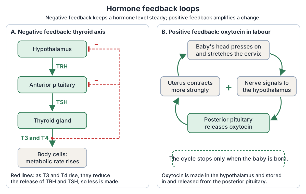
Many values in the body, hormone levels among them, are held near a set point by negative feedback, a mechanism that reverses any change away from the set point. A feedback loop has a sensor that monitors a value, a control centre that compares it with the normal range, and an effector that makes the change. (OpenStax Anatomy and Physiology 2e, 1.5: Homeostasis.)
In the endocrine system, negative feedback means that a rising hormone level inhibits further secretion of that hormone. OpenStax's example is the glucocorticoids such as cortisol: the hypothalamus and pituitary direct the adrenal glands to release them, and as their concentration in the blood rises, the hypothalamus and pituitary reduce their signalling. (OpenStax Anatomy and Physiology 2e, 17.2: Hormones.)
The thyroid axis is a classic negative feedback loop. 1. Low blood levels of T3 and T4 stimulate the hypothalamus to release TRH. 2. TRH makes the anterior pituitary secrete TSH. 3. TSH makes the thyroid secrete T3 and T4. 4. As T3 and T4 rise, they reduce the production and secretion of TSH, so less T3 and T4 is made. (OpenStax Anatomy and Physiology 2e, 17.4: The Thyroid Gland.)
Some loops need no pituitary. When blood glucose is high, beta cells in the pancreatic islets release insulin; as glucose falls, further insulin release is inhibited. Low glucose triggers glucagon, and rising glucose inhibits further glucagon release. (OpenStax Anatomy and Physiology 2e, 17.9: The Endocrine Pancreas.)
Blood calcium works the same way: low blood calcium triggers parathyroid hormone (PTH) from the parathyroid glands, and rising calcium inhibits further PTH release. (OpenStax Anatomy and Physiology 2e, 17.5: The Parathyroid Glands.)
Positive feedback does the opposite: it intensifies a change instead of reversing it. Blood clotting is a non-hormonal example: each step of clotting triggers the release of more clotting substances. (OpenStax Anatomy and Physiology 2e, 1.5: Homeostasis.)
Oxytocin in childbirth is one of the few hormonal examples. The baby presses against the cervix, which stimulates the release of oxytocin from the posterior pituitary (the hypothalamus makes it). Oxytocin travels to the uterus and makes it contract; the contractions cause more oxytocin release, and the cycle continues until the baby is born. (Cleveland Clinic: Oxytocin.)
Lobes, isthmus, blood supply, nerves and the parathyroid glands, with labelled diagrams. Thyroid gland anatomy.
Thyroid and parathyroid glands
The thyroid is a butterfly-shaped gland in front of the trachea, just below the larynx: a central isthmus joins its left and right lobes. It is made mostly of follicles that produce thyroxine (T4) and triiodothyronine (T3). Both depend on iodine, and many cells convert T4 into the more potent T3 by removing an iodine atom. Thyroid hormones influence the basal metabolic rate and raise body heat. (OpenStax Anatomy and Physiology 2e, 17.4: The Thyroid Gland.)
Parafollicular (C) cells of the thyroid secrete calcitonin, which lowers blood calcium and so opposes the parathyroid glands. (NCI SEER Training Modules: Thyroid and Parathyroid Glands.)
OpenStax notes, though, that calcitonin usually plays only a small part in normal calcium balance. (OpenStax Anatomy and Physiology 2e, 17.4: The Thyroid Gland.)
Most people have four parathyroid glands, but some have more in the neck or chest. Their chief cells release parathyroid hormone (PTH) when blood calcium is low. PTH raises calcium by acting on bone, by making the kidney tubules reabsorb more calcium and by starting the production of calcitriol, which increases calcium absorption from the gut. (OpenStax Anatomy and Physiology 2e, 17.5: The Parathyroid Glands.)
TeachMeAnatomy places the superior pair at the middle of the back of each thyroid lobe and the inferior pair near the lower poles. The number of glands varies, and the inferior glands can lie as low as the superior mediastinum in the chest. (TeachMeAnatomy: The Parathyroid Glands.)
The thyroid, larynx, trachea and the muscles, vessels and nerves around them. Neck anatomy.
Adrenal glands: cortex and medulla
Each adrenal gland sits on the upper pole of a kidney, enclosed with it in the renal fascia but separated from it by a small amount of fibrous tissue. (TeachMeAnatomy: The Adrenal Glands.)
Each gland has an outer cortex of glandular tissue and an inner medulla of nervous tissue. The cortex has three zones: the zona glomerulosa makes mineralocorticoids, chiefly aldosterone; the zona fasciculata makes glucocorticoids, chiefly cortisol; and the zona reticularis makes small amounts of androgens. (OpenStax Anatomy and Physiology 2e, 17.6: The Adrenal Glands.)
The glands lie in the posterior abdomen, behind the peritoneum, between the upper inner (superomedial) part of each kidney and the diaphragm. The right gland is pyramid-shaped and the left semilunar. (TeachMeAnatomy: The Adrenal Glands.)
Aldosterone conserves sodium and water, and cortisol raises blood glucose. The adrenal medulla develops from neural tissue and secretes adrenaline (epinephrine) and noradrenaline (norepinephrine) when sympathetic nerves stimulate it, particularly during stress. (NCI SEER Training Modules: Adrenal (Suprarenal) Gland.)
The two parts handle different kinds of stress. The medulla answers acute, short-term stress, the fight-or-flight response. For long-term stress, the hypothalamus releases CRH, the anterior pituitary releases ACTH and the adrenal cortex releases cortisol, which promotes the breakdown of glycogen to glucose. (OpenStax Anatomy and Physiology 2e, 17.6: The Adrenal Glands.)
Right vs left gland, the cortex zones, the medulla and the blood supply in 3D. Adrenal glands anatomy.
Pineal gland and thymus
The pineal gland's cells (pinealocytes) make melatonin, an amine hormone made from serotonin. Light reaching the retinas inhibits melatonin production; in darkness production rises and causes drowsiness. Melatonin may influence the body's circadian rhythms, and after travel across several time zones it can take up to several days for melatonin production to adjust to the new light-dark pattern (jet lag). (OpenStax Anatomy and Physiology 2e, 17.7: The Pineal Gland.)
The NCI SEER module describes the pineal gland as a small cone-shaped structure that extends backwards from the third ventricle of the brain. Its melatonin passes into the cerebrospinal fluid and then the blood, and affects reproductive development and daily cycles. (NCI SEER Training Modules: Pituitary and Pineal Glands.)
The thymus's endocrine role is to make a group of hormones called thymosins, which help T lymphocytes develop. It is more active in infancy and early childhood than later in life, and it shrinks with age. OpenStax adds a caution: thymosins are also found in other tissues, so they cannot be called purely thymic hormones. (OpenStax Anatomy and Physiology 2e, 17.10: Organs with Secondary Endocrine Functions.)
The thymus lies mainly in the superior mediastinum, behind the manubrium of the sternum, and in some people reaches up into the neck. After puberty it decreases in size and is slowly replaced by fat. (TeachMeAnatomy: The Thymus.)
Where the pineal gland sits in the brain and how light controls its melatonin. Pineal gland (pineal body).
Ovaries and testes (the gonads)
The gonads belong to both the reproductive and the endocrine systems. The main hormone of the testes is testosterone, a steroid that drives the development of male sex characteristics and sperm production. The ovaries mainly make oestrogens (estradiol, estriol and estrone) and progesterone, which helps regulate the menstrual cycle. Both gonads also make inhibin, which inhibits FSH release. (OpenStax Anatomy and Physiology 2e, 17.8: Gonadal and Placental Hormones.)
The ovaries make oestrogen and progesterone in response to the pituitary gonadotrophins LH and FSH. They lie in the pelvis, attached to the back of the broad ligament of the uterus by a fold called the mesovarium. (TeachMeAnatomy: The Ovaries.)
Progesterone causes the lining of the uterus to thicken in preparation for pregnancy. Most testosterone comes from the testes, but a small amount is also made by the adrenal cortex, and testosterone secretion is regulated by negative feedback. (NCI SEER Training Modules: Gonads.)
In the 3D model the testes are highlighted in the default male body. The ovaries are in the female body only and are not highlighted on this page; the uterus guide shows them highlighted in 3D.
The testis and epididymis in section, with the seminiferous tubules, Leydig cells and blood supply. Testis anatomy.
Organs with endocrine tissue
Many organs whose main job is something else also contain hormone-making cells. Cells in the walls of the heart's atria secrete atrial natriuretic peptide (ANP), which signals the kidneys to reabsorb less sodium and so lowers blood volume and blood pressure. (OpenStax Anatomy and Physiology 2e, 17.10: Organs with Secondary Endocrine Functions.)
The lining of the stomach releases gastrin when food arrives, which stimulates the production of hydrochloric acid and the enzyme pepsin. The lining of the small intestine releases secretin and cholecystokinin; cholecystokinin makes the gallbladder contract and release bile, and stimulates the pancreas to secrete digestive enzymes. (NCI SEER Training Modules: Other Endocrine Glands.)
The kidneys release the enzyme renin when blood flow to them falls, which leads to aldosterone release; they make erythropoietin (EPO) when oxygen is low, triggering red blood cell production in the bone marrow; and they make calcitriol from vitamin D3, which helps the gut absorb calcium. (OpenStax Anatomy and Physiology 2e, 17.10: Organs with Secondary Endocrine Functions.)
Body fat releases leptin, which helps keep body weight stable over the long term by regulating hunger and giving a feeling of fullness; it acts mainly on the hypothalamus. (Cleveland Clinic: Leptin.)
During pregnancy the placenta makes hormones too. Human chorionic gonadotropin (hCG) promotes progesterone production and reduces the mother's immune response to the fetus; human placental lactogen helps prepare the breasts for milk production; and relaxin is thought to soften and widen the pubic symphysis before childbirth. (OpenStax Anatomy and Physiology 2e, 17.8: Gonadal and Placental Hormones.)
The liver makes insulin-like growth factor 1, the immediate stimulus for growth, and angiotensinogen, the precursor of angiotensin, which raises blood pressure. The skin turns cholesterol into cholecalciferol (a vitamin D precursor) under ultraviolet light, and bone cells release osteocalcin, which increases insulin production. (OpenStax Anatomy and Physiology 2e, 17.10: Organs with Secondary Endocrine Functions.)
Location, coverings, internal parts and hilum of the kidneys in 3D. Kidney anatomy.
Organs with endocrine tissue and their hormones
| Organ | Hormone | Main effect |
|---|---|---|
| Heart (atria) | Atrial natriuretic peptide (ANP) | Makes the kidneys excrete more sodium, lowering blood volume and pressure |
| Stomach | Gastrin | Stimulates stomach acid |
| Small intestine | Secretin and cholecystokinin (CCK) | Bicarbonate and enzymes from the pancreas; bile from the gallbladder |
| Kidneys | Erythropoietin (EPO), calcitriol; the enzyme renin | EPO: red blood cell production; calcitriol: calcium absorption; renin: leads to aldosterone release |
| Liver | Insulin-like growth factor 1 (IGF-1), angiotensinogen | IGF-1: body growth; angiotensinogen: precursor of angiotensin, which raises blood pressure |
| Adipose tissue (body fat) | Leptin, adiponectin | Leptin: satiety signals to the brain; adiponectin: reduces insulin resistance |
| Placenta (pregnancy only) | Human chorionic gonadotropin (hCG); also hPL and relaxin | hCG promotes progesterone production during pregnancy |
From OpenStax 17.8 and 17.10, checked against the NCI SEER module and Cleveland Clinic.
What does the endocrine system do? 8 functions
Cleveland Clinic sums up the system's main job as releasing hormones into the blood while continuously monitoring their levels. Sources group the results in slightly different ways; each of the eight functions below is named by at least two of the sources on this page. (Cleveland Clinic: Endocrine System.)
1. Controls metabolism and body heat. Thyroid hormones influence the basal metabolic rate, the energy the body uses at rest, and their calorigenic effect raises body temperature. (OpenStax Anatomy and Physiology 2e, 17.4: The Thyroid Gland.)
2. Controls growth and development. Growth hormone from the anterior pituitary stimulates the growth of bones, muscles and other organs by promoting protein synthesis. (NCI SEER Training Modules: Pituitary and Pineal Glands.)
3. Drives sexual development and reproduction. At puberty oestrogens promote breast development and the maturing of the uterus and vagina, progesterone prepares the uterine lining for pregnancy, and testosterone drives male development. (NCI SEER Training Modules: Gonads.)
4. Balances water and salt. ADH from the hypothalamus helps the kidneys keep the right amount of water, avoiding dehydration or over-hydration, and aldosterone keeps the levels of electrolytes such as sodium in the body fluids steady. (OpenStax Biology 2e, 37.3: Regulation of Body Processes.)
5. Keeps blood glucose steady. Insulin helps body cells take up glucose, lowering blood glucose, while glucagon makes the liver turn stored glycogen back into glucose, raising it. (OpenStax Anatomy and Physiology 2e, 17.9: The Endocrine Pancreas.)
6. Keeps blood calcium steady. Parathyroid hormone is the main hormone that raises blood calcium. Calcitonin from the thyroid can lower it, but OpenStax notes that calcitonin usually plays only a small part in normal calcium balance. The right calcium level is needed for muscle contraction and nerve impulses. (OpenStax Anatomy and Physiology 2e, 17.4: The Thyroid Gland.)
7. Responds to stress. Adrenaline and noradrenaline from the adrenal medulla drive the short-term fight-or-flight response, and cortisol from the adrenal cortex handles long-term stress. (OpenStax Anatomy and Physiology 2e, 17.6: The Adrenal Glands.)
8. Sets the sleep-wake cycle. The pineal gland releases melatonin as light fades, causing drowsiness, and less in daylight, promoting wakefulness. (OpenStax Anatomy and Physiology 2e, 17.7: The Pineal Gland.)
Clinical sources add mood: NIDDK says the endocrine system affects growth and development, metabolism, sexual function and mood. (NIDDK: Endocrine Diseases.)
The thymus, spleen, tonsils and lymph nodes that make and house the immune cells. Lymphatic system.
Fun facts about the endocrine system
1. The posterior pituitary makes no hormones of its own. It stores and releases ADH and oxytocin, which the hypothalamus makes. (Cleveland Clinic: Pituitary Gland.)
2. The heart is an endocrine organ. When blood pressure rises, it releases two hormones, A-type and B-type natriuretic peptide. (Cleveland Clinic: Endocrine System.)
3. Body fat is an endocrine tissue. Adipose cells release leptin in amounts proportional to the body's fat stores, and leptin promotes a feeling of fullness (satiety). (OpenStax Anatomy and Physiology 2e, 17.10: Organs with Secondary Endocrine Functions.)
4. The placenta is a temporary organ that forms in the uterus during pregnancy. The first hormone it releases, human chorionic gonadotropin (hCG), is the hormone that pregnancy tests measure. (Cleveland Clinic: Placenta.)
5. The thymus is at its biggest around puberty. After that it slowly shrinks and is replaced by fat. (Cleveland Clinic: Thymus.)
6. Thyroid hormones cannot be made without iodine, and a goitre (an enlarged thyroid) can be the visible sign of iodine deficiency. (OpenStax Anatomy and Physiology 2e, 17.4: The Thyroid Gland.)
7. The adrenal medulla develops from neural tissue, and its hormones are released when sympathetic nerves stimulate it, particularly in stressful situations. (NCI SEER Training Modules: Adrenal (Suprarenal) Gland.)
8. Melatonin is made from serotonin, and light switches its production off. That is why jet lag after crossing several time zones can take up to several days to settle. (OpenStax Anatomy and Physiology 2e, 17.7: The Pineal Gland.)
9. Cleveland Clinic calls the digestive tract the largest endocrine-related organ system; gastrin from the stomach is one example of its hormones. (Cleveland Clinic: Endocrine System.)
Common endocrine disorders
If hormone levels are too high or too low, a person may have an endocrine disease or disorder. The notes below are educational only; anyone worried about a hormone problem should see a clinician. (NIDDK: Endocrine Diseases.)
Diabetes mellitus is a disease in which blood glucose is too high. In type 1 diabetes the body makes little or no insulin, because the immune system destroys the insulin-making cells of the pancreas. In type 2 diabetes the cells do not use insulin properly. (NIDDK: What Is Diabetes?.)
The CDC says about 90% to 95% of Americans with diabetes have type 2 diabetes, in which cells do not respond normally to insulin (insulin resistance). (CDC: Type 2 Diabetes.)
In hypothyroidism (underactive thyroid) the thyroid does not make enough thyroid hormone; common symptoms include tiredness, weight gain, trouble tolerating cold and a slowed heart rate. NIDDK names Hashimoto's disease, an autoimmune disorder, as the most common cause. (NIDDK: Hypothyroidism (Underactive Thyroid).)
In hyperthyroidism (overactive thyroid) the gland makes more thyroid hormone than the body needs; symptoms can include weight loss despite an increased appetite, a rapid or irregular heartbeat, sweating and trouble tolerating heat. Graves' disease, also autoimmune, is the most common cause. (NIDDK: Hyperthyroidism (Overactive Thyroid).)
Adrenal disorders show the two directions as well: Cushing's syndrome (too much cortisol over a long time) is marked by high blood glucose, while Addison's disease (too few corticosteroids such as cortisol) is a rare disorder that causes low blood glucose and low blood sodium. (OpenStax Anatomy and Physiology 2e, 17.6: The Adrenal Glands.)
The most common cause of Cushing's syndrome is long-term, high-dose glucocorticoid medicine. Of the other cases, about 8 in 10 come from a pituitary tumour that makes too much ACTH, and this type is called Cushing's disease. (NIDDK: Cushing's Syndrome.)
Pituitary disorders often involve growth hormone or ADH. Too much growth hormone in childhood causes gigantism (excessive growth); in adults, who have stopped growing, it causes acromegaly, with growth of the bones of the face, hands and feet. Too little growth hormone in childhood causes growth impairment, called growth hormone deficiency (formerly pituitary dwarfism). Diabetes insipidus, a different disease from diabetes mellitus, is caused by chronic underproduction of ADH, which leads to chronic dehydration. (OpenStax Anatomy and Physiology 2e, 17.3: The Pituitary Gland and Hypothalamus.)
Endocrine societies have proposed renaming this form of diabetes insipidus arginine vasopressin deficiency. (Working Group for Renaming Diabetes Insipidus. Changing the name of diabetes insipidus: a position statement. Endocr J. 2022.)
Too much parathyroid hormone (hyperparathyroidism) can greatly decrease bone density, leading to fractures. (OpenStax Anatomy and Physiology 2e, 17.5: The Parathyroid Glands.)
Underactive vs overactive thyroid: typical symptoms
| Hypothyroidism (underactive) | Hyperthyroidism (overactive) | |
|---|---|---|
| Thyroid hormone | Too little | Too much |
| Metabolic rate | Low | High |
| Weight | Weight gain | Weight loss despite an increased appetite |
| Temperature | Trouble tolerating cold | Sweating, trouble tolerating heat |
| Heart | Slowed heart rate | Rapid or irregular heartbeat |
| Commonest cause (NIDDK) | Hashimoto's disease (autoimmune) | Graves' disease (autoimmune) |
Educational summary from NIDDK and OpenStax 17.4; symptoms vary between people and have other causes. Not a diagnostic tool.
Endocrine system in the 3D model
- Adrenal gland
- The adrenal (suprarenal) gland is a small endocrine gland on top of each kidney.
- Hypothalamus
- The hypothalamus is a small diencephalic region that regulates homeostasis, autonomic function and endocrine control.
- Lobe of thymus
- The thymus lobes are the two lobes of the thymus gland, where T lymphocytes mature.
- Pancreas
- The pancreas is a soft, elongated gland with both exocrine and endocrine functions.
- Pineal body
- The pineal body (gland) is a small pinecone-shaped endocrine gland in the brain.
- Pituitary gland
- The pituitary gland (hypophysis) is a pea-sized endocrine gland that regulates many other glands.
- Testis
- The testis is the male gonad that produces sperm and testosterone.
Frequently asked questions
What are the 7 major glands of the endocrine system?
There is no single official count. A 1998 review lists seven: the hypothalamus, pituitary gland, thyroid gland, parathyroid glands, adrenal glands, pancreas and gonads (ovaries or testes). OpenStax names five glands (pituitary, thyroid, parathyroid, adrenal and pineal) and also describes the pancreas as a gland with hormone-secreting cells, while MedlinePlus lists nine, adding the pineal gland and counting the ovaries and testes separately. Learn the glands and their hormones rather than a number.
What are the main functions of the endocrine system?
Its hormones control metabolism and body heat, growth and development, sexual development and reproduction, the balance of water, salt and blood pressure, blood glucose, blood calcium, the response to stress and the sleep-wake cycle. Clinical sources also link hormones to mood. The system works by releasing hormones into the blood and adjusting their levels through feedback.
Which gland is called the master gland?
The pituitary gland is often called the master gland because its hormones control many other endocrine glands, including the thyroid, the adrenal cortex and the gonads. It is not fully in charge, though: the hypothalamus, just above it, controls the pituitary with releasing and inhibiting hormones, and makes the two hormones the posterior pituitary releases.
What is the difference between endocrine and exocrine glands?
Exocrine glands have ducts that carry their secretions to a surface, such as sweat glands to the skin or salivary glands into the mouth. Endocrine glands are ductless: they release hormones into the surrounding fluid and blood, which carry them around the body. The pancreas is both, with exocrine enzyme-making cells and endocrine islets.
Is the pancreas part of the endocrine system?
Yes, but only partly. Most of the pancreas is exocrine: its acinar cells make digestive enzymes that drain through a duct into the duodenum. Its pancreatic islets (islets of Langerhans) are endocrine: their beta cells release insulin, which lowers blood glucose, and their alpha cells release glucagon, which raises it. So the pancreas belongs to both the digestive and endocrine systems.
How is the endocrine system different from the nervous system?
The nervous system uses electrical impulses and neurotransmitters over short distances, and its targets respond within milliseconds; the effects last seconds. The endocrine system uses hormones carried in the blood, which can reach distant organs, and its responses start more slowly and last minutes, hours or weeks. The hypothalamus links the two systems.
What are signs of an endocrine problem?
It depends on the gland and on whether there is too much or too little hormone. An underactive thyroid can cause tiredness, weight gain and feeling cold, and an overactive thyroid weight loss, a fast heartbeat and heat intolerance. Diabetes means blood glucose is too high. These signs have many causes, so anyone worried should see a doctor.
References
- OpenStax. Anatomy and Physiology 2e, §17.1 An Overview of the Endocrine System. Rice University, 2022.
- OpenStax. Anatomy and Physiology 2e, §17.2 Hormones. Rice University, 2022.
- OpenStax. Anatomy and Physiology 2e, §17.3 The Pituitary Gland and Hypothalamus. Rice University, 2022.
- OpenStax. Anatomy and Physiology 2e, §17.4 The Thyroid Gland. Rice University, 2022.
- OpenStax. Anatomy and Physiology 2e, §17.5 The Parathyroid Glands. Rice University, 2022.
- OpenStax. Anatomy and Physiology 2e, §17.6 The Adrenal Glands. Rice University, 2022.
- OpenStax. Anatomy and Physiology 2e, §17.7 The Pineal Gland. Rice University, 2022.
- OpenStax. Anatomy and Physiology 2e, §17.8 Gonadal and Placental Hormones. Rice University, 2022.
- OpenStax. Anatomy and Physiology 2e, §17.9 The Endocrine Pancreas. Rice University, 2022.
- OpenStax. Anatomy and Physiology 2e, §17.10 Organs with Secondary Endocrine Functions. Rice University, 2022.
- OpenStax. Anatomy and Physiology 2e, §1.5 Homeostasis. Rice University, 2022.
- OpenStax. Anatomy and Physiology 2e, §25.3 Gross Anatomy of the Kidney. Rice University, 2022.
- OpenStax. Biology 2e, §37.2 How Hormones Work. Rice University, 2018.
- OpenStax. Biology 2e, §37.3 Regulation of Body Processes. Rice University, 2018.
- National Cancer Institute SEER Training Modules. Introduction to the Endocrine System.
- National Cancer Institute SEER Training Modules. Pituitary and Pineal Glands.
- National Cancer Institute SEER Training Modules. Thyroid and Parathyroid Glands.
- National Cancer Institute SEER Training Modules. Adrenal (Suprarenal) Gland.
- National Cancer Institute SEER Training Modules. Pancreas (Islets of Langerhans).
- National Cancer Institute SEER Training Modules. Gonads.
- National Cancer Institute SEER Training Modules. Other Endocrine Glands.
- Hiller-Sturmhöfel S, Bartke A. The Endocrine System: An Overview. Alcohol Health and Research World. 1998;22(3):153–164.
- Baxter R. Endocrine system. Kenhub, reviewed 7 September 2023.
- Kenhub. Suprarenal (adrenal) glands.
- TeachMeAnatomy. The Parathyroid Glands.
- TeachMeAnatomy. The Thymus.
- TeachMeAnatomy. The Thyroid Gland.
- TeachMeAnatomy. The Adrenal Glands.
- TeachMeAnatomy. The Ovaries.
- Cleveland Clinic. Endocrine System. Updated 22 November 2023.
- Cleveland Clinic. Hypothalamus. Updated 16 March 2022.
- Cleveland Clinic. Pituitary Gland. Updated 4 April 2022.
- Cleveland Clinic. Oxytocin. Updated 27 March 2022.
- Cleveland Clinic. Thymus. Updated 15 May 2022.
- Cleveland Clinic. Kidney. Updated 5 November 2025.
- Cleveland Clinic. Leptin. Updated 29 January 2025.
- Cleveland Clinic. Placenta. Updated 15 December 2025.
- Young WF Jr. Endocrine Glands. MSD Manual Consumer Version, reviewed February 2025.
- MedlinePlus Medical Encyclopedia. Endocrine glands. Reviewed 24 April 2025.
- National Institute of Diabetes and Digestive and Kidney Diseases (NIDDK). Endocrine Diseases.
- NIDDK. Hypothyroidism (Underactive Thyroid). Last reviewed March 2021.
- NIDDK. Hyperthyroidism (Overactive Thyroid). Last reviewed August 2021.
- NIDDK. What Is Diabetes? Last reviewed April 2023.
- Centers for Disease Control and Prevention. Type 2 Diabetes. 15 May 2024.
- NIDDK. Cushing's Syndrome.
- Working Group for Renaming Diabetes Insipidus; Arima H, Cheetham T, Christ-Crain M, et al. Changing the name of diabetes insipidus: a position statement of The Working Group for Renaming Diabetes Insipidus. Endocr J. 2022;69(11):1281-1284.
- Mitsuhashi N. et al. (2009). BodyParts3D: 3D structure database for anatomical concepts. Nucleic Acids Research 37, D782–D785. Data © The Database Center for Life Science, CC BY 4.0.
Related anatomy
- Pituitary glandPituitary gland (hypophyseal gland) anatomy: the hypophyseal fossa and sella turcica, relations, lobes, portal blood…
- Thyroid glandThyroid gland anatomy with labelled diagrams: lobes, isthmus, pyramidal lobe, arteries and veins, recurrent laryngeal…
- Adrenal glandAnatomy of the adrenal glands with diagrams and a 3D model: right vs left shape, cortex zones and medulla, hormones…
- PancreasPancreas anatomy: head, uncinate process, neck, body and tail, ducts, acini and islets, blood supply and variants…
- TestisTesticle anatomy with a labeled testis diagram and 3D model: the tunica vaginalis and albuginea, lobules, seminiferous…
- UterusUterus anatomy with a labeled uterus (womb) diagram and 3D model: fundus, body, isthmus and cervix, wall layers…
Explore the whole body in the interactive 3D human anatomy model, or browse the A–Z list of human structures.