

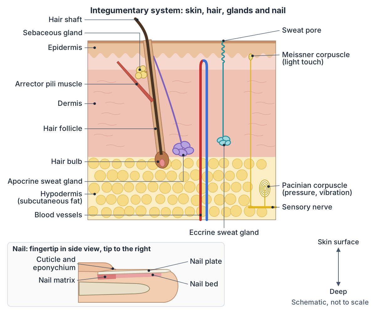
What is the integumentary system?
The integumentary system is the skin together with its accessory structures, and its overall job is to protect the body. The skin is a true organ, even if it does not look like one: it is made of several tissues that work together as a single structure. (OpenStax Anatomy and Physiology 2e, 5.1: Layers of the Skin.)
The accessory structures, also called skin appendages, are the hair, nails, sweat glands and sebaceous glands. They are derived from the epidermis and dermis, the two layers of the skin. (Yousef H, Miao JH, Alhajj M, Badri T. Histology, Skin Appendages. StatPearls, 2023.)
The name comes from integument, a word for a natural covering such as a skin, shell or rind. It is from the Latin integumentum, meaning a covering. (Dictionary.com: integument.)
All the organ systems of the body, and where each main organ sits. Organs of the body.
Integumentary system organs and parts
The skin is the main organ of the integumentary system. With its accessory structures it forms the largest organ system in the human body, covering about 1.5 to 2 square metres in an adult. (OpenStax Anatomy and Physiology 2e, Chapter 5: The Integumentary System (Introduction).)
Cleveland Clinic describes the system as the skin, nails, hair and glands, along with the nerves and blood vessels that support them. It lists four groups of glands: sweat (sudoriferous) glands, sebaceous glands that make sebum (oil), ceruminous glands in the ear that secrete earwax, and mammary glands, which in females produce milk after giving birth. (Cleveland Clinic: Integumentary System.)
Some sources draw the boundary more widely. Kenhub describes the integumentary system as the skin and its appendages, the subcutaneous tissue, the deep fascia, the mucocutaneous junctions (places where a mucous membrane changes into skin) and the breasts. (Kenhub: Integumentary system.)
The dermis carries the skin's blood vessels and nerves: its deep reticular layer is well supplied with blood vessels and has a rich sensory and sympathetic nerve supply, while the epidermis above it has no blood vessels of its own. (OpenStax Anatomy and Physiology 2e, 5.1: Layers of the Skin.)
Parts of the integumentary system
| Part | Where it is | Main job |
|---|---|---|
| Epidermis | Outer layer of the skin; keratinised epithelium with no blood vessels | Waterproof barrier; makes keratin and melanin; makes vitamin D in UV light |
| Dermis | Under the epidermis; papillary and reticular layers of connective tissue | Strength (collagen) and stretch (elastin); holds blood vessels, nerves, hair follicles and glands |
| Hypodermis (subcutaneous layer) | Under the dermis; loose connective tissue and fat | Stores fat, insulates and cushions; connects the skin to the fascia over bones and muscles |
| Hair | Grows from follicles over most of the body, but not on the palms, soles or lips | Protection, touch and temperature control |
| Nails | On the backs of the fingertips and toes | Protect the ends of the fingers and toes; help with grip and fine movements |
| Sweat glands (eccrine and apocrine) | In the dermis; eccrine almost everywhere, apocrine mainly in the armpits and genital region | Sweat for cooling (eccrine); thicker sweat that opens into hair follicles (apocrine) |
| Sebaceous glands | Most open into hair follicles | Sebum (oil) that lubricates and waterproofs skin and hair |
| Ceruminous glands | Skin of the outer, cartilaginous ear canal | A secretion that forms earwax with sebum |
| Mammary glands | Breasts; well developed in females | Milk |
| Sensory receptors | Epidermis, dermis and hypodermis | Detect touch, vibration, temperature and pain |
The skin's layers: epidermis, dermis and hypodermis
The skin has two main layers: the epidermis, made of closely packed epithelial cells, and the dermis beneath it. Under the dermis lies the hypodermis, which is mostly loose connective tissue and fat. (OpenStax Anatomy and Physiology 2e, 5.1: Layers of the Skin.)
Not every source draws the line in the same place. StatPearls says the skin has 3 layers, the epidermis, dermis and hypodermis, and the MSD Manual and Cleveland Clinic also count the hypodermis as the skin's third layer. This is a difference in naming, not in the anatomy. (Yousef H, Alhajj M, Fakoya AO, Sharma S. Anatomy, Skin (Integument), Epidermis. StatPearls, 2024.)
The epidermis is a keratinised stratified squamous epithelium with no blood vessels. Most of the body is covered by thin skin, whose epidermis has four layers (strata); from deep to superficial these are the stratum basale, stratum spinosum, stratum granulosum and stratum corneum. The thick skin of the palms and soles has a fifth layer, the stratum lucidum, between the stratum granulosum and the stratum corneum. (OpenStax Anatomy and Physiology 2e, 5.1: Layers of the Skin.)
The epidermis contains several kinds of cell. Keratinocytes make and store the tough protein keratin. Melanocytes in the stratum basale make the pigment melanin, which colours the skin and hair and protects the DNA of epidermal cells from ultraviolet (UV) damage. Merkel cells act as touch receptors. (OpenStax Anatomy and Physiology 2e, 5.1: Layers of the Skin.)
Langerhans cells are dendritic (antigen-presenting) immune cells: they take up microbes and foreign material in the epidermis and carry antigens to nearby lymph nodes to start an immune response. (Neagu M et al. Langerhans Cells: Revising Their Role in Skin Pathologies. J Pers Med 2022.)
Skin colour depends mainly on melanin. Melanocytes in the stratum basale make it and pass it to the surrounding keratinocytes in vesicles called melanosomes. Eumelanin is black or brown and pheomelanin is reddish, and people with darker skin have melanocytes that make more melanin. Two other pigments, carotene and the haemoglobin in the blood, also affect skin colour. Sun exposure stimulates the melanocytes to make more melanin, which builds up in the keratinocytes as a tan; melanin production peaks about 10 days after exposure, and the extra melanin protects the DNA of epidermal cells from UV damage. (OpenStax Anatomy and Physiology 2e, 5.1: Layers of the Skin.)
The dermis is made of two layers of connective tissue. The thin papillary layer, of loose areolar tissue, pushes up into the epidermis as finger-like dermal papillae and holds many small blood vessels, nerve fibres and touch receptors called Meissner corpuscles. The much thicker reticular layer is dense irregular connective tissue: its collagen fibres give the skin strength and its elastin fibres let it stretch. (OpenStax Anatomy and Physiology 2e, 5.1: Layers of the Skin.)
The hypodermis (also called the subcutaneous layer or superficial fascia) connects the skin to the fascia over the bones and muscles. Its fat stores energy and insulates and cushions the body. (OpenStax Anatomy and Physiology 2e, 5.1: Layers of the Skin.)
On average the skin is about 2 millimetres thick. It is thinner on sensitive areas such as the eyelids and thicker where it takes more stress, such as the soles of the feet. (Cleveland Clinic: Integumentary System.)
A labelled 3D block of the epidermis, dermis and hypodermis, with the strata of the epidermis. Skin layers in 3D.
Hair: shaft, follicle and growth cycle
A hair is a filament of keratin that grows out of the epidermis. The hair shaft is the part not anchored in the follicle. The root sits in a hair follicle and ends deep in the dermis at the hair bulb, where a layer of dividing cells, the hair matrix, surrounds the hair papilla. New cells added at the bulb push the hair up the follicle, so the hair you can see is completely dead and made entirely of keratin. (OpenStax Anatomy and Physiology 2e, 5.2: Accessory Structures of the Skin.)
A hair follicle, its sebaceous gland and its arrector pili muscle together form the pilosebaceous unit. The arrector pili is a bundle of smooth muscle that runs from the follicle to the papillary dermis just below the epidermis. Sympathetic nerves make it contract in the cold, which makes the hair stand up and raises goosebumps. (Martel JL, Miao JH, Badri T, Fakoya AO. Anatomy, Hair Follicle. StatPearls, 2024.)
Hair grows in a cycle. In the growth phase (anagen) cells divide rapidly at the root for years; OpenStax gives 2 to 7 years. A short transition phase (catagen) of 2 to 3 weeks follows, then a resting phase (telogen) of about 2 to 4 months, after which a new anagen phase begins. (OpenStax Anatomy and Physiology 2e, 5.2: Accessory Structures of the Skin.)
A 2023 review gives similar times for scalp hair: anagen about 2 to 8 years, catagen about 2 weeks and telogen about 2 to 3 months. It adds a fourth phase, exogen, in which the old hair is shed. (Natarelli N, Gahoonia N, Sivamani RK. Integrative and Mechanistic Approach to the Hair Growth Cycle and Hair Loss. J Clin Med 2023.)
Hair grows about 0.3 to 0.35 mm a day: OpenStax gives 0.3 mm a day during anagen and StatPearls 0.35 mm. Estimates of normal daily hair loss vary: OpenStax gives about 50 hairs a day and StatPearls around 100. (Murphrey MB, Agarwal S, Zito PM. Anatomy, Hair. StatPearls, 2023.)
Hair grows on nearly every part of the body; the palms, the soles, the lips and the urogenital openings are the usual exceptions. Hair helps with temperature control, touch and protection from UV radiation. (Brown TM, Krishnamurthy K. Histology, Hair and Follicle. StatPearls, 2023.)
The layers of the hair shaft and follicle, the hair bulb and the growth cycle, with labelled diagrams. Hair anatomy.
Nails: plate, matrix and nail bed
A nail consists of the nail plate, the nail folds, the nail matrix, the nail bed and the hyponychium. (Kenhub: Integumentary system.)
The nail plate (OpenStax calls it the nail body) is made of densely packed dead keratinocytes. It forms at the nail root, where a matrix of dividing cells lets the nail grow continuously. The lateral nail folds overlap and anchor the sides, and a crescent-shaped area near the base is the lunula. The nail bed under the plate is rich in blood vessels, which makes the nail look pink, and the hyponychium is the area under the free edge. (OpenStax Anatomy and Physiology 2e, 5.2: Accessory Structures of the Skin.)
The fold of skin at the base of the nail, the proximal nail fold, ends in the eponychium, which leaves a thin layer of dead cells, the cuticle, on the nail plate (OpenStax treats cuticle and eponychium as one term). (Shelverton TA. The Nail Unit: Essential Anatomy and Physiology for the Beauty Industry. NailKnowledge, 2024.)
Nails protect the backs of the tips of the fingers and toes and help with grip, fine finger movements and scratching. Like hair, nails are ectodermal appendages. (Brahs AB, Bolla SR. Histology, Nail. StatPearls, 2023.)
Nails grow slowly. In a study of 22 healthy young American adults, fingernails grew an average of 3.47 mm a month and toenails 1.62 mm a month. A 2026 review gives round figures of about 3 mm a month for fingernails and 1 mm a month for toenails. (Yaemsiri S, Hou N, Slining MM, He K. Growth rate of human fingernails and toenails in healthy American young adults. J Eur Acad Dermatol Venereol 2010.)
The parts of a fingernail and toenail, from the matrix and lunula to the hyponychium, with labelled diagrams. Nail anatomy.
Glands of the integumentary system
Sweat (sudoriferous) glands are of two types, eccrine and apocrine. Both lie in the dermis and end in a coiled secretory part. Eccrine glands open directly onto the skin surface, while apocrine glands open into hair follicles. (Patel BC, Treister AD, McCausland C, Lio PA, Jozsa F. Anatomy, Skin, Sudoriferous Gland. StatPearls, 2023.)
Eccrine glands make a watery, hypotonic sweat that cools the body. They are found all over the skin but are especially dense on the palms, the soles and the forehead. Their sweat is mostly water, with some salt, antibodies, traces of metabolic waste and dermcidin, a peptide that kills microbes. (OpenStax Anatomy and Physiology 2e, 5.2: Accessory Structures of the Skin.)
Apocrine sweat glands are larger than eccrine glands and lie deeper in the dermis. They are found mainly where hair is dense, such as the armpits and the genital region. Their sweat also contains organic compounds, which make it thicker; skin bacteria break these down, which causes body odour. (OpenStax Anatomy and Physiology 2e, 5.2: Accessory Structures of the Skin.)
StatPearls lists the apocrine gland sites as the armpit (axilla), the anogenital region, the external ear canal and the areola around the nipple. (Patel BC, Treister AD, McCausland C, Lio PA, Jozsa F. Anatomy, Skin, Sudoriferous Gland. StatPearls, 2023.)
Sebaceous glands are oil glands found over almost all of the skin except the palms and soles, and most of them open into hair follicles. (Liu Y et al. Comparative Characterization of Human Meibomian Glands, Free Sebaceous Glands, and Hair-Associated Sebaceous Glands. Int J Mol Sci 2024.)
Sebaceous glands release sebum, a mixture of lipids that lubricates and waterproofs the skin and hair and contains fatty acids that are antibacterial. Hormones stimulate sebum secretion, and many of these hormones do not become active until puberty. (OpenStax Anatomy and Physiology 2e, 5.2: Accessory Structures of the Skin.)
Sebaceous glands secrete in an unusual, holocrine way: the gland cells, called sebocytes, are deliberately destroyed as their product is released. Sebum helps keep the skin lubricated and defended against its surroundings and against infection. (Hoover E, Aslam S, Krishnamurthy K. Physiology, Sebaceous Glands. StatPearls, 2022.)
Ceruminous glands are modified apocrine sweat glands in the skin of the outer, cartilaginous part of the ear canal. They secrete a watery fluid that combines with the output of the sebaceous glands there to form cerumen (earwax). (Saunders TFC, Monksfield P. A Case of Conservatively Managed Invasive Ceruminoma and a Review of the Literature. Case Rep Otolaryngol 2014.)
Earwax moisturises the skin of the ear canal, helps protect it from infection and forms a barrier against water, foreign bodies and even insects. (Sevy JO, Hohman MH, Singh A. Cerumen Impaction Removal. StatPearls, 2023.)
Mammary glands are modified sweat glands that produce breast milk. (OpenStax Anatomy and Physiology 2e, 27.2: Anatomy and Physiology of the Ovarian Reproductive System.)
StatPearls classes the mammary gland as an apocrine gland, because the tip of each secretory cell and part of its cytoplasm become part of the secretion. The gland is present in both sexes, but it is well developed in females and rudimentary in males. (Khan YS, Fakoya AO, Sajjad H. Anatomy, Thorax: Mammary Gland. StatPearls, 2025.)
The mammary gland's lobes, ducts, nipple and areola, and how milk is made and released. Breast anatomy.
Glands of the skin compared
| Gland | Where | Opens onto | Secretion |
|---|---|---|---|
| Eccrine sweat gland | Almost all skin; most dense on the palms, soles and forehead | The skin surface | Watery sweat (water, some salt, traces of waste) that cools the body |
| Apocrine sweat gland | Armpits and anogenital region; StatPearls adds the ear canal and areola | Hair follicles | Thicker sweat with organic compounds |
| Sebaceous gland | Almost all skin except the palms and soles; mostly beside hair follicles | Mostly hair follicles | Sebum, a mixture of lipids |
| Ceruminous gland | Outer, cartilaginous part of the ear canal | The ear canal | Watery fluid that mixes with sebum to form earwax (cerumen) |
| Mammary gland | Breasts | The nipple, through lactiferous ducts | Milk |
What does the integumentary system do? 7 functions
OpenStax groups the functions of the integumentary system under four headings: protection, sensory function, thermoregulation and vitamin D synthesis. Other sources also list excretion, immune defence and water balance, which gives the seven functions below. (OpenStax Anatomy and Physiology 2e, 5.3: Functions of the Integumentary System.)
1. Protection: the skin shields the body from wind, water and UV sunlight. Layers of keratin and glycolipids in the stratum corneum make it a barrier against water loss, and it is the first line of defence against grit, microbes and harmful chemicals. (OpenStax Anatomy and Physiology 2e, 5.3: Functions of the Integumentary System.)
2. Sensation: nerve structures in the epidermis, dermis and hypodermis detect touch, surface temperature and pain. Meissner corpuscles respond to light touch, Pacinian corpuscles to vibration and Merkel cells to touch, and the hair root plexus around each follicle senses when a hair is disturbed. (OpenStax Anatomy and Physiology 2e, 5.3: Functions of the Integumentary System.)
3. Temperature control: when the body is warm, sweat glands release sweat, which cools the body as it evaporates, and arterioles in the dermis widen so that heat carried by the blood is lost through the skin. In the cold the arterioles narrow to save heat, especially in the fingers and the tip of the nose. (OpenStax Anatomy and Physiology 2e, 5.3: Functions of the Integumentary System.)
4. Vitamin D synthesis: when the epidermis is exposed to UV light it makes cholecalciferol (vitamin D3) from a derivative of cholesterol. The liver converts it to calcidiol and the kidneys convert that to calcitriol, the active form. Vitamin D is needed for normal absorption of calcium and phosphorus, which healthy bones require. (OpenStax Anatomy and Physiology 2e, 5.3: Functions of the Integumentary System.)
5. Excretion: the skin excretes sebum, sweat and other waste from the body. (Cleveland Clinic: Integumentary System.)
6. Immune defence: Langerhans cells in the epidermis are dendritic (antigen-presenting) immune cells that take up microbes and foreign material and carry antigens to nearby lymph nodes to start an immune response. (Neagu M et al. Langerhans Cells: Revising Their Role in Skin Pathologies. J Pers Med 2022.)
The papillary dermis also contains phagocytes, defensive cells that help fight bacteria and other infections. (OpenStax Anatomy and Physiology 2e, 5.1: Layers of the Skin.)
7. Water and electrolyte balance: the MSD Manual lists maintaining the body's water balance and electrolyte balance among the skin's main functions, and StatPearls includes the control of insensible fluid loss. (MSD Manual Consumer Version: Structure and Function of the Skin.)
How does the integumentary system work with other body systems?
Nervous system: receptors in the skin send touch, temperature and pain signals to the central nervous system (the brain and spinal cord), and motor nerves supply the arrector pili muscles and the glands. (OpenStax Anatomy and Physiology 2e, 5.3: Functions of the Integumentary System.)
The hypothalamus in the brain acts as the body's thermostat: it monitors body temperature and, through sympathetic nerves, starts the right responses in the skin, such as sweating and changes in blood flow. (OpenStax Anatomy and Physiology 2e, 24.6: Energy and Heat Balance.)
Circulatory (cardiovascular) system: StatPearls describes temperature control as teamwork, in which thermoreceptors lead to changes in blood flow to the skin, in sweating and in body hair. (Kim JY, Dao H. Physiology, Integument. StatPearls, 2023.)
Skeletal system: vitamin D made in the skin is needed to absorb calcium and phosphorus for healthy bones. Without enough of it, children can develop rickets, a painful condition in which the bones are misshapen and the legs bow. (OpenStax Anatomy and Physiology 2e, 5.3: Functions of the Integumentary System.)
Immune system: Cleveland Clinic describes the skin as the first line of defence against bacteria and infection, and notes that it sends white blood cells to injuries to begin healing. (Cleveland Clinic: Integumentary System.)
Respiratory system: the tiny hairs in the nose filter out dust and other particles before they are breathed into the lungs. (Cleveland Clinic: Integumentary System.)
Endocrine system: sebum secretion is stimulated by hormones, many of which do not become active until puberty, so sebaceous glands are relatively inactive in childhood. (OpenStax Anatomy and Physiology 2e, 5.2: Accessory Structures of the Skin.)
Reproductive system: the mammary glands make milk. Prolactin from the anterior pituitary stimulates milk production in the glandular tissue, and oxytocin causes the milk to be ejected. (NCI SEER Training Modules: Mammary Glands.)
The strip of skin that each spinal nerve supplies, with a labelled dermatome map. Dermatomes.
Integumentary system facts and figures
The skin is the body's largest organ. Kenhub gives its area as about 2 square metres, in line with the 1.5 to 2 square metres for an adult in OpenStax. (Kenhub: Integumentary system.)
Its weight is quoted very differently. Cleveland Clinic gives about 6 pounds (about 2.7 kg) or more, and the National Cancer Institute's SEER training site also gives about six pounds, while OpenStax says the skin makes up about 16 percent of adult body weight. (NCI SEER Training Modules: Anatomy of the Skin.)
Eccrine sweat glands are found almost everywhere on the body, with the highest concentration on the palms and soles. (Patel BC, Treister AD, McCausland C, Lio PA, Jozsa F. Anatomy, Skin, Sudoriferous Gland. StatPearls, 2023.)
The five layers of soft tissue over the skull, from the hair-bearing skin down to the pericranium. Scalp anatomy.
Integumentary system facts
| Fact | Figure |
|---|---|
| Largest organ system | The skin and its accessory structures |
| Skin surface area (adult) | About 1.5 to 2 square metres |
| Skin weight | About 6 pounds (2.7 kg) in some sources; about 16% of body weight in OpenStax |
| Average skin thickness | About 2 mm; thinner on the eyelids, thicker on the soles |
| Layers of the epidermis | 4 in thin skin; 5 in the thick skin of the palms and soles |
| Hair growth | About 0.3 to 0.35 mm a day |
| Hairs shed | About 50 to 100 a day |
| Scalp hair growth phase (anagen) | About 2 to 8 years |
| Fingernail growth | About 3 to 3.5 mm a month |
| Toenail growth | About 1 to 1.6 mm a month |
| Densest eccrine sweat glands | Palms and soles |
| Skin with no hair | Palms, soles and lips |
Each figure is cited in the sections of this page; where sources differ, the range covers both.
Common integumentary system conditions
Cleveland Clinic groups the conditions of the integumentary system by the part they affect: the skin (for example acne, eczema, psoriasis, burns and skin cancer), the hair (such as alopecia areata), the nails (such as onychomycosis) and the glands (such as hyperhidrosis). (Cleveland Clinic: Integumentary System.)
Skin cancers are named after the cells they start in. Basal cell carcinoma starts in the stratum basale, squamous cell carcinoma affects the keratinocytes of the stratum spinosum, and melanoma is an uncontrolled growth of melanocytes, the pigment cells. (OpenStax Anatomy and Physiology 2e, 5.4: Diseases, Disorders, and Injuries of the Integumentary System.)
Burns are classed by how deep they go. Superficial (first-degree) burns are limited to the epidermis, partial-thickness (second-degree) burns involve part of the dermis, and full-thickness (third-degree) burns extend through the whole dermis into the fat beneath. (Merck Manual Professional Version: Burns.)
Acne usually occurs on skin rich in sebaceous glands, such as the face and back, when too much sebum and keratin block hair follicles. Eczema shows as dry, itchy patches of skin that look like a rash. (OpenStax Anatomy and Physiology 2e, 5.4: Diseases, Disorders, and Injuries of the Integumentary System.)
Alopecia areata is a non-scarring autoimmune condition that causes hair loss on the scalp or the body. Onychomycoses are fungal infections of the nails. (Kenhub: Integumentary system.)
In hyperhidrosis a person sweats when the body does not need to cool down. It can affect the whole body or just certain areas. (NHS: Excessive sweating (hyperhidrosis).)
This section is for learning only. Anyone with a new, changing or worrying problem with their skin, hair or nails should see a doctor or another clinician.
What the 3D model shows, and what it lacks
The 3D view uses BodyParts3D, a database that represents anatomical structures as 3D data on a whole-body model of an adult man. On this page it highlights the skin and the hair of the head; the model also has a separate pubic hair part. (Mitsuhashi N et al. BodyParts3D: 3D structure database for anatomical concepts. Nucleic Acids Res 2009.)
The model's skin is a single outer surface. It has no epidermis, dermis or hypodermis, and no hair follicles, glands or nails, so those parts are shown in the labelled diagram at the top of this page. The Female button switches to the female body, which has its own skin surface from the Human Reference Atlas.
Integumentary system in the 3D model
- Hair of head
- The hair of the head is made of keratinised hair shafts growing from follicles in the scalp.
- Skin
- The skin is the body's largest organ, forming a protective covering.
Frequently asked questions
What is the integumentary system?
The integumentary system is the body's outer covering: the skin plus its accessory structures, which are the hair, nails, sweat glands and sebaceous (oil) glands, together with the nerves and blood vessels that supply them. The skin is its main organ. The name comes from integument, a natural covering such as a skin, shell or rind. The system protects the body, senses touch, temperature and pain, helps control body temperature and makes vitamin D.
What are the organs of the integumentary system?
The skin is the main organ. Its accessory structures are the hair, the nails and four kinds of gland: sweat glands (eccrine and apocrine), sebaceous glands, ceruminous glands in the ear canal and mammary glands in the breasts. The skin itself has an epidermis and a dermis, with the fatty hypodermis beneath; some sources count the hypodermis as a third layer of the skin.
What are the 7 functions of the integumentary system?
Sources group them differently, but seven functions cover what they list: protection against water loss, microbes, chemicals and UV light; sensing touch, temperature and pain; temperature control by sweating and changing skin blood flow; vitamin D synthesis; excretion of sweat and sebum; immune defence by Langerhans cells and phagocytes; and water and electrolyte balance. OpenStax uses four headings, and Cleveland Clinic lists eight points.
How many layers of skin are there?
The skin has two main layers, the epidermis on top and the dermis beneath, with the fatty hypodermis below them; StatPearls, the MSD Manual and Cleveland Clinic count the hypodermis as a third layer. The epidermis itself has four layers (strata) in thin skin and five in the thick skin of the palms and soles, which adds the stratum lucidum. Counting those five strata with the dermis and hypodermis gives seven.
How does the integumentary system regulate body temperature?
When the body is warm, eccrine sweat glands release sweat, which cools the body as it evaporates, and arterioles in the dermis widen so that heat carried by the blood is lost through the skin. In the cold the arterioles narrow to keep heat in, especially in the fingers and the tip of the nose. The hypothalamus monitors body temperature and starts these responses through the sympathetic nervous system.
What glands are in the integumentary system?
Eccrine sweat glands make watery sweat for cooling and are most dense on the palms and soles. Apocrine sweat glands, mainly in the armpits and genital region, open into hair follicles and make thicker sweat. Sebaceous glands make oily sebum. Ceruminous glands in the ear canal help form earwax, and mammary glands, which are modified sweat glands, make milk.
Is the skin the largest organ in the body?
Yes. The skin is the body's largest organ, and with its accessory structures it is the largest organ system. In an adult it covers about 1.5 to 2 square metres and is about 2 mm thick on average, thinner on the eyelids and thicker on the soles. Its weight is quoted differently: about 6 pounds (2.7 kg) in some sources, about 16 percent of body weight in OpenStax.
What are some fun facts about the integumentary system?
The hair you can see is dead keratin, pushed up by living cells in the hair bulb. Hair grows about 0.3 to 0.35 mm a day, and we lose roughly 50 to 100 hairs a day. Fingernails grow about 3 to 3.5 mm a month, two to three times faster than toenails. The palms and soles have no hair but have the densest eccrine sweat glands. Goosebumps appear when tiny arrector pili muscles contract.
References
- Betts JG, et al. Anatomy and Physiology 2e, Chapter 5 Introduction: The Integumentary System. OpenStax, Rice University, 2022.
- Betts JG, et al. Anatomy and Physiology 2e, §5.1 Layers of the Skin. OpenStax, Rice University, 2022.
- Betts JG, et al. Anatomy and Physiology 2e, §5.2 Accessory Structures of the Skin. OpenStax, Rice University, 2022.
- Betts JG, et al. Anatomy and Physiology 2e, §5.3 Functions of the Integumentary System. OpenStax, Rice University, 2022.
- Betts JG, et al. Anatomy and Physiology 2e, §5.4 Diseases, Disorders, and Injuries of the Integumentary System. OpenStax, Rice University, 2022.
- Betts JG, et al. Anatomy and Physiology 2e, §14.1 Sensory Perception. OpenStax, Rice University, 2022.
- Betts JG, et al. Anatomy and Physiology 2e, §24.6 Energy and Heat Balance. OpenStax, Rice University, 2022.
- Betts JG, et al. Anatomy and Physiology 2e, §27.2 Anatomy and Physiology of the Ovarian Reproductive System. OpenStax, Rice University, 2022.
- Kim JY, Dao H. Physiology, Integument. In: StatPearls. StatPearls Publishing, 2023.
- Lopez-Ojeda W, Pandey A, Alhajj M, Oakley AM. Anatomy, Skin (Integument). In: StatPearls. StatPearls Publishing, 2022.
- Yousef H, Alhajj M, Fakoya AO, Sharma S. Anatomy, Skin (Integument), Epidermis. In: StatPearls. StatPearls Publishing, 2024.
- Yousef H, Miao JH, Alhajj M, Badri T. Histology, Skin Appendages. In: StatPearls. StatPearls Publishing, 2023.
- Patel BC, Treister AD, McCausland C, Lio PA, Jozsa F. Anatomy, Skin, Sudoriferous Gland. In: StatPearls. StatPearls Publishing, 2023.
- Hoover E, Aslam S, Krishnamurthy K. Physiology, Sebaceous Glands. In: StatPearls. StatPearls Publishing, 2022.
- Martel JL, Miao JH, Badri T, Fakoya AO. Anatomy, Hair Follicle. In: StatPearls. StatPearls Publishing, 2024.
- Murphrey MB, Agarwal S, Zito PM. Anatomy, Hair. In: StatPearls. StatPearls Publishing, 2023.
- Brown TM, Krishnamurthy K. Histology, Hair and Follicle. In: StatPearls. StatPearls Publishing, 2023.
- Brahs AB, Bolla SR. Histology, Nail. In: StatPearls. StatPearls Publishing, 2023.
- Khan YS, Fakoya AO, Sajjad H. Anatomy, Thorax: Mammary Gland. In: StatPearls. StatPearls Publishing, 2025.
- Sevy JO, Hohman MH, Singh A. Cerumen Impaction Removal. In: StatPearls. StatPearls Publishing, 2023.
- Rad A. Integumentary system. Kenhub, last reviewed 19 September 2023.
- Cleveland Clinic. Integumentary System. Medically reviewed, last updated 25 April 2022.
- National Cancer Institute SEER Training Modules. Anatomy of the Skin (Melanoma).
- National Cancer Institute SEER Training Modules. Mammary Glands.
- Benedetti J. Structure and Function of the Skin. MSD Manual Consumer Version, reviewed August 2026.
- Natarelli N, Gahoonia N, Sivamani RK. Integrative and Mechanistic Approach to the Hair Growth Cycle and Hair Loss. J Clin Med. 2023;12(3):893.
- Yaemsiri S, Hou N, Slining MM, He K. Growth rate of human fingernails and toenails in healthy American young adults. J Eur Acad Dermatol Venereol. 2010;24(4):420–423.
- Pinos-León V, Vasconez-Gonzalez J, Izquierdo-Condoy JS, et al. Minoxidil Beyond Hair: A Comprehensive Review of Its Effects on Nail Growth. J Clin Med. 2026;15(5):1792.
- Liu Y, Butovich IA, Garreis F, et al. Comparative Characterization of Human Meibomian Glands, Free Sebaceous Glands, and Hair-Associated Sebaceous Glands Based on Biomarkers, Analysis of Secretion Composition, and Gland Morphology. Int J Mol Sci. 2024;25(6):3109.
- Neagu M, Constantin C, Jugulete G, et al. Langerhans Cells: Revising Their Role in Skin Pathologies. J Pers Med. 2022;12(12):2072.
- Shelverton TA. The Nail Unit: Essential Anatomy and Physiology for the Beauty Industry. NailKnowledge, 2024.
- Jeong H, Noh H, Kim CH. Middle Ear Ceruminous Gland Adenoma Obstructing the Eustachian Tube Orifice. Case Rep Otolaryngol. 2021;2021:5987353.
- Saunders TFC, Monksfield P. A Case of Conservatively Managed Invasive Ceruminoma and a Review of the Literature. Case Rep Otolaryngol. 2014;2014:897540.
- Merck Manual Professional Version. Burns.
- NHS. Excessive sweating (hyperhidrosis).
- Dictionary.com. Integument.
- Mitsuhashi N, Fujieda K, Tamura T, Kawamoto S, Takagi T, Okubo K. BodyParts3D: 3D structure database for anatomical concepts. Nucleic Acids Res. 2009;37(Database issue):D782–D785.
- Mitsuhashi N. et al. (2009). BodyParts3D: 3D structure database for anatomical concepts. Nucleic Acids Research 37, D782–D785. Data © The Database Center for Life Science, CC BY 4.0.
Related anatomy
- Hair anatomyHair anatomy with labelled diagrams: shaft layers, follicle root sheaths, bulb and dermal papilla, arrector pili, hair…
- Nail anatomyNail anatomy with labelled diagrams: nail plate, matrix, lunula, cuticle vs eponychium, nail bed, hyponychium and…
- Breast anatomyBreast anatomy with labelled diagrams and a 3D model: lobes, milk ducts, Cooper's ligaments, nipple, areola, blood…
- ScalpScalp anatomy with labelled diagrams: the five SCALP layers, arteries, veins and danger area, nerves, lymph, why scalp…
- DermatomesDermatome anatomy with front and back dermatome maps: every level from C2 to S5, key landmarks, dermatomes vs…
- Nervous systemThe nervous system explained with a labeled diagram: CNS vs PNS, somatic and autonomic divisions, organs, neurons…
Explore the whole body in the interactive 3D human anatomy model, or browse the A–Z list of human structures.