

What is the nervous system?
The nervous system is the body's major controlling, regulating and communicating system. Through its receptors it keeps the body in touch with the world outside and with conditions inside, it is the centre of mental activity such as thought, learning and memory, and together with the endocrine system it keeps the internal environment stable (homeostasis). (SEER Training Modules (NIH National Cancer Institute): Introduction to the Nervous System.)
Like other body systems it is built from organs, mainly the brain, spinal cord, nerves and ganglia. These organs are made of nervous tissue together with blood and connective tissue. Nervous tissue has two main kinds of cell: neurons, which carry signals, and glial cells, which support them. (SEER Training Modules (NIH National Cancer Institute): Introduction to the Nervous System.)
Although its parts are often called separate 'nervous systems', there is really only one, highly integrated nervous system in the body. It is divided in two ways: by where the parts are, into the central and peripheral nervous systems, and by what they do, into divisions such as the sensory and motor divisions and the somatic and autonomic nervous systems. (SEER Training Modules (NIH National Cancer Institute): Organization of the Nervous System.)
A small region at the base of the brain links the nervous system with the hormone system. The hypothalamus is largely involved in regulating homeostasis and is in charge of both the autonomic nervous system and the endocrine system, which it controls through the anterior pituitary gland. (OpenStax Anatomy and Physiology 2e, 13.2: The Central Nervous System.)
Nervous system facts at a glance
| Fact | Detail |
|---|---|
| Main parts | Central nervous system (brain and spinal cord) and peripheral nervous system (nerves and ganglia) |
| Cell types | Neurons, which carry signals, and glial cells, which support them: 4 types of glial cell in the CNS and 2 in the PNS. A 2009 count found about 86 billion neurons and a similar number of non-neuronal cells in the adult human brain alone; the often-quoted 100 billion was never based on a direct count |
| Regions of the brain | 4: cerebrum, diencephalon, brainstem and cerebellum |
| Cranial nerves | 12 pairs, numbered I to XII |
| Spinal nerves | 31 pairs: 8 cervical, 12 thoracic, 5 lumbar, 5 sacral and 1 coccygeal |
| End of the spinal cord | About L1 to L2 in the average adult (normal range T12 to L3) |
| Main nerve plexuses | 4: cervical, brachial, lumbar and sacral |
| Largest nerve | The sciatic nerve, from the sacral plexus |
| Cranial nerve with the longest course | The vagus nerve (X), which reaches the abdomen |
| Fastest signals | Up to about 120 metres per second in the largest myelinated fibres; about 0.5 to 2.5 metres per second in unmyelinated C fibres |
Sources: SEER Training Modules; OpenStax A&P 2e 12.2, 13.2 and 13.4; Kenhub (nervous system, conus medullaris, sciatic nerve, vagus nerve); TeachMeAnatomy (sciatic and vagus nerves); Kiernan (Western University) and Yap et al. 2020 for conduction speeds; Azevedo et al. 2009 for the neuron count.
What does the nervous system do? 5 functions
The nervous system has three general, overlapping functions: sensory input, integration and motor output (OpenStax calls them sensation, integration and response). It detects a change, decides what it means and then makes muscles or glands act. Two broader roles follow from these: keeping the body's internal environment stable, and producing thought, learning and memory. (SEER Training Modules (NIH National Cancer Institute): Introduction to the Nervous System.)
1. Sensory input (sensation). Millions of sensory receptors detect changes, called stimuli, inside and outside the body: temperature, light and sound from the outside world, and pressure, pH, carbon dioxide and electrolyte levels inside it. This information is turned into electrical signals called nerve impulses and sent to the brain. (SEER Training Modules (NIH National Cancer Institute): Introduction to the Nervous System.)
2. Integration. The brain and spinal cord combine incoming signals with other sensations, memories, learning and emotion, and decide on a response. OpenStax uses a batter facing a pitch as its example: seeing the ball does not automatically make the batter swing, because its speed and path, and the state of the game, are weighed first. (OpenStax Anatomy and Physiology 2e, 12.1: Basic Structure and Function of the Nervous System.)
3. Motor output (response). The nervous system sends signals to effectors, the muscles and glands that carry out the response. It can make all three types of muscle work: skeletal muscle moves the skeleton, cardiac muscle speeds the heart during exercise, and smooth muscle moves food along the digestive tract. Producing sweat to lower body temperature is a response of glands. (OpenStax Anatomy and Physiology 2e, 12.1: Basic Structure and Function of the Nervous System.)
4. Homeostasis. The autonomic part of the nervous system works automatically and continuously, without conscious effort, on heart rate, breathing rate, blood pressure, body temperature and other activities of the internal organs that keep the internal environment stable. (SEER Training Modules (NIH National Cancer Institute): The Peripheral Nervous System.)
5. Thought, memory and emotion. The nervous system is the centre of all mental activity, including thought, learning and memory. These higher functions also feed into integration, so what you remember and how you feel shape how you respond. (SEER Training Modules (NIH National Cancer Institute): Introduction to the Nervous System.)
Cleveland Clinic describes the same work in everyday terms: the nervous system manages thoughts, memory, learning and feelings; movement, balance and coordination; the senses; sleep; heartbeat and breathing patterns; the response to stress, including sweating; and digestion. (Cleveland Clinic: Nervous System.)
Central vs peripheral nervous system (CNS vs PNS)
The central nervous system (CNS) is made of two organs, the brain and the spinal cord. The brain lies in the cranial vault of the skull and the spinal cord in the vertebral canal, and the two are continuous at the foramen magnum, the large opening in the base of the skull. The organs of the peripheral nervous system (PNS) are the nerves and ganglia. (SEER Training Modules (NIH National Cancer Institute): Organization of the Nervous System.)
Kenhub describes the CNS as the integration and command centre of the body and the PNS as the conduit between the CNS and the body. The PNS consists of 12 pairs of cranial nerves, 31 pairs of spinal nerves and many small clusters of neuron cell bodies called ganglia. (Kenhub: The nervous system.)
The boundary is not perfectly sharp. Some parts of the PNS lie inside the skull or the vertebral canal, and the same axons can change name as they cross the line: OpenStax calls the axons that leave the eye the optic nerve and, inside the cranium, the optic tract, with the name changing at the optic chiasm. OpenStax treats the chiasm as the border. (OpenStax Anatomy and Physiology 2e, 12.1: Basic Structure and Function of the Nervous System.)
The optic nerve itself, though, is technically CNS tissue, an outgrowth of the brain myelinated by oligodendrocytes. (Kenhub: Optic nerve.)
The same kinds of structure have different names on each side. A cluster of neuron cell bodies is a nucleus in the CNS but a ganglion in the PNS, and a bundle of axons is a tract in the CNS but a nerve in the PNS. Regions rich in cell bodies and dendrites form grey matter; regions rich in axons form white matter, which looks white because the axons are wrapped in fatty (lipid-rich) myelin. (OpenStax Anatomy and Physiology 2e, 12.1: Basic Structure and Function of the Nervous System.)
Different glial cells make the myelin on each side. In the CNS one oligodendrocyte provides myelin for segments of several axons; in the PNS each Schwann cell wraps a single segment of just one axon. (OpenStax Anatomy and Physiology 2e, 12.2: Nervous Tissue.)
Central vs peripheral nervous system (CNS vs PNS)
| Feature | Central nervous system (CNS) | Peripheral nervous system (PNS) |
|---|---|---|
| Organs | Brain and spinal cord | 12 pairs of cranial nerves, 31 pairs of spinal nerves, and ganglia |
| Where it lies | In the skull and the vertebral canal, wrapped in meninges and cerebrospinal fluid | Throughout the body outside the brain and spinal cord (a few parts lie inside the skull or vertebral canal) |
| Cluster of neuron cell bodies | Nucleus | Ganglion |
| Bundle of axons | Tract | Nerve |
| Cells that make myelin | Oligodendrocytes (each myelinates segments of several axons) | Schwann cells (each myelinates one segment of one axon) |
| Other glial cells | Astrocytes, microglia and ependymal cells | Satellite cells |
| Connective tissue coverings | Meninges: dura mater, arachnoid and pia mater | Epineurium, perineurium and endoneurium in each nerve |
| Main job | Integration and command: processes information and issues commands | Carries signals between the CNS and the rest of the body |
Sources: OpenStax A&P 2e 12.1, 12.2 and 13.4; SEER Training Modules (CNS and PNS); Kenhub.
Divisions of the nervous system: somatic, autonomic and enteric
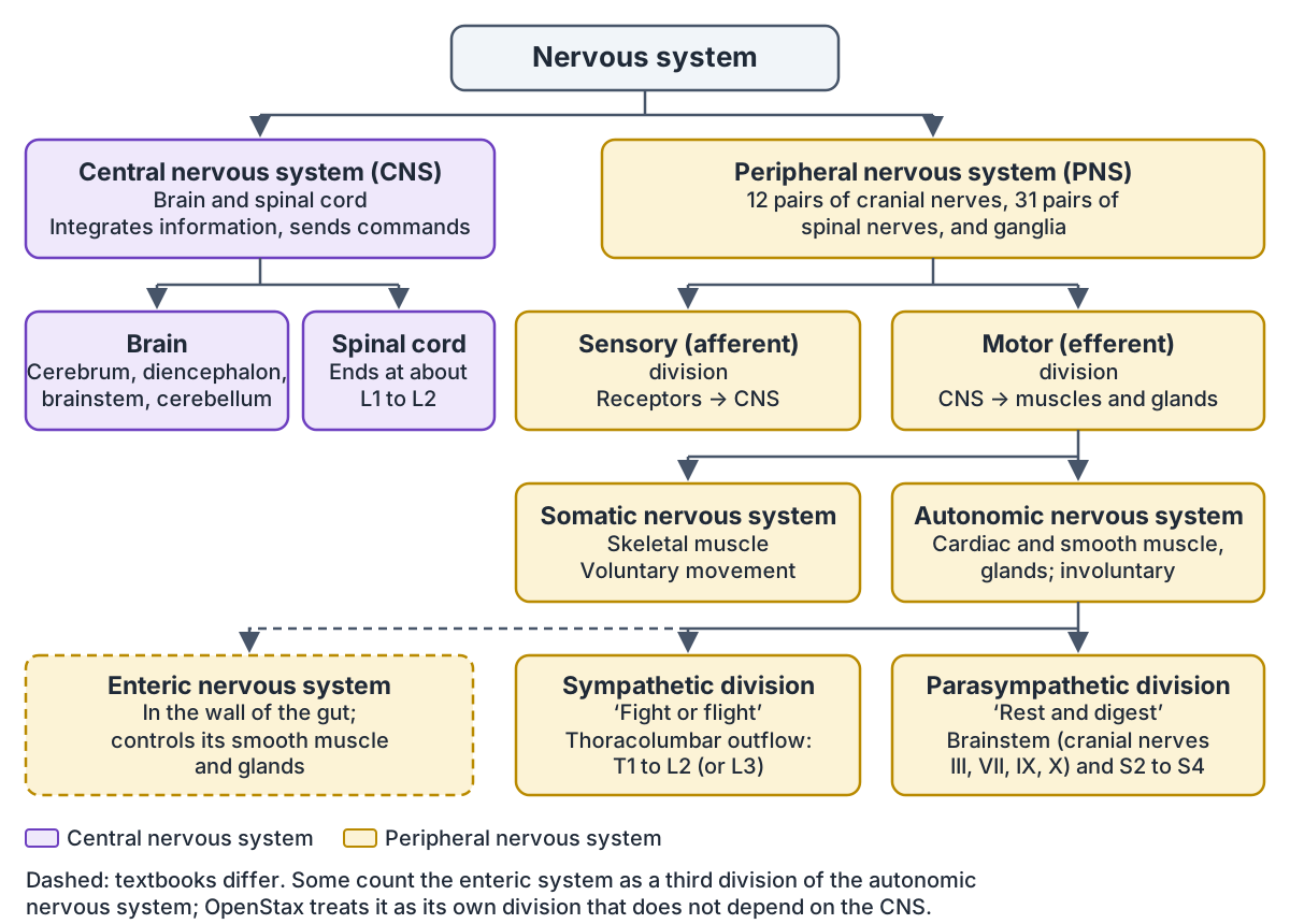
The PNS has a sensory (afferent) division, which carries impulses from receptors to the CNS, and a motor (efferent) division, which carries impulses from the CNS out to muscles and glands. The motor division is split again into the somatic nervous system and the autonomic nervous system. (SEER Training Modules (NIH National Cancer Institute): Organization of the Nervous System.)
The somatic nervous system is responsible for conscious perception and voluntary movement through skeletal muscle, which is why it is sometimes called the voluntary nervous system. Not every somatic response is a decision, though: jumping when a friend shouts 'Boo!' is a reflex that uses skeletal muscle. (OpenStax Anatomy and Physiology 2e, 12.1: Basic Structure and Function of the Nervous System.)
The autonomic nervous system is the involuntary part: its nerves supply cardiac muscle, smooth muscle and gland cells, and its two divisions work together to maintain homeostasis. The sympathetic division prepares the body for increased activity, the 'fight-or-flight' response; its fibres have a thoracolumbar origin, from spinal cord segments T1 to L2 or L3. The parasympathetic division conserves energy for 'rest and digest' activities; its fibres have a craniosacral outflow, from the brainstem and spinal cord segments S2 to S4. (Kenhub: The nervous system.)
Parasympathetic fibres leave the brainstem in four cranial nerves: the oculomotor (III), facial (VII), glossopharyngeal (IX) and vagus (X) nerves. (Kenhub: Autonomic nervous system.)
The enteric nervous system (ENS) controls the smooth muscle and glands of the digestive system from within the gut wall. OpenStax describes it as a large part of the PNS that does not depend on the CNS, and adds that it is sometimes valid to count it as part of the autonomic system because it forms part of the autonomic output that regulates digestion. (OpenStax Anatomy and Physiology 2e, 12.1: Basic Structure and Function of the Nervous System.)
Kenhub's summary instead lists the enteric system as a third division of the autonomic nervous system, beside the sympathetic and parasympathetic divisions. Both arrangements appear in textbooks, so use the one your course follows. (Kenhub: The nervous system.)
The sympathetic trunk, the parasympathetic cranial and sacral outflow and the effect of each division on every organ are covered in the guide to the Autonomic nervous system.
Organs of the nervous system
The main organs of the nervous system are the brain, the spinal cord, the nerves and the ganglia. The brain and spinal cord are the organs of the CNS; the nerves and ganglia are the organs of the PNS. Nerves are bundles of nerve fibres, and ganglia are small knots of nerve cell bodies outside the CNS. (SEER Training Modules (NIH National Cancer Institute): Organization of the Nervous System.)
The input comes from sensory receptors. Kenhub groups the senses into somatic senses (touch, pressure, vibration, pain, temperature, stretch and position sense from the skin, muscles and joints), visceral senses from the internal organs, and the special senses, such as vision. (Kenhub: The nervous system.)
The brain has four major regions: the cerebrum, the diencephalon (which includes the thalamus and hypothalamus), the brainstem (midbrain, pons and medulla oblongata) and the cerebellum. The cerebrum is covered by a folded layer of grey matter, the cerebral cortex, and is split into left and right hemispheres joined by the white matter of the corpus callosum. (OpenStax Anatomy and Physiology 2e, 13.2: The Central Nervous System.)
Each region has its own jobs. The thalamus relays information between the cerebral cortex and the rest of the nervous system, the hypothalamus regulates homeostasis, the pons and medulla regulate the cardiovascular and respiratory systems, and the cerebellum compares the commands sent by the cerebrum with sensory feedback from the body so that movement can be corrected. (OpenStax Anatomy and Physiology 2e, 13.2: The Central Nervous System.)
The CNS is protected by bone, membranes and fluid. Inside the skull and vertebral canal it is wrapped in three meninges: the tough outer dura mater, the web-like arachnoid mater and the thin pia mater, which follows every fold of the brain's surface. Cerebrospinal fluid (CSF), filtered from the blood by the choroid plexuses, circulates through the ventricles and fills the subarachnoid space between the arachnoid and the pia, where it cushions the brain and spinal cord before draining back into the blood. (OpenStax Anatomy and Physiology 2e, 13.3: Circulation and the Central Nervous System.)
The blood-brain barrier, to which astrocytes contribute, lets nutrients such as glucose and amino acids into nervous tissue but keeps out many other substances, including many drugs. (OpenStax Anatomy and Physiology 2e, 12.2: Nervous Tissue.)
The lobes, gyri, deep nuclei, brainstem and cerebellum are labelled in 3D and in diagrams in the guide to Brain anatomy.
The spinal cord and the 31 pairs of spinal nerves
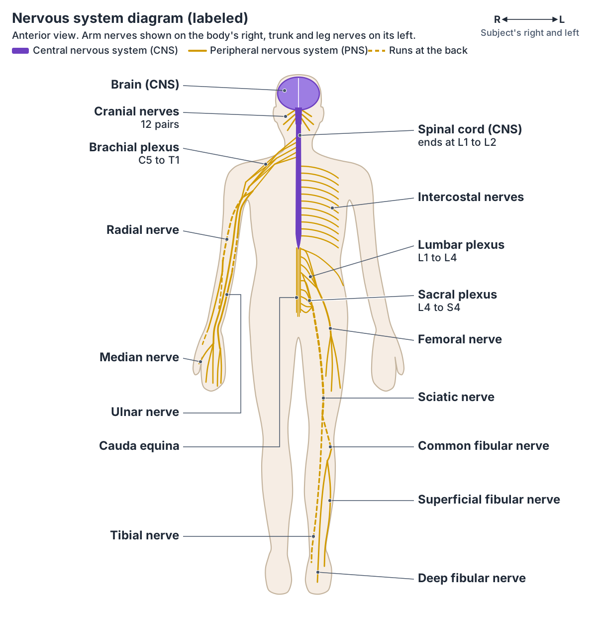
The spinal cord does not run the whole length of the spine. In the average adult it ends between the first and second lumbar vertebrae (L1 to L2), where it tapers into the conus medullaris, although the tip can lie as high as T12 or as low as L3. Below it, the cauda equina is made of the spinal nerve roots L2 to S5 and the coccygeal nerve. (Kenhub: Conus medullaris and cauda equina.)
The cord is shorter than the vertebral column because it hardly grows longer after the first or second year of life while the skeleton keeps growing. The nerve roots for the lowest segments therefore run down the canal below the cord's end as a long bundle that resembles a horse's tail, the cauda equina. (OpenStax Anatomy and Physiology 2e, 13.2: The Central Nervous System.)
The spinal cord has two main jobs. It is a conduction pathway: sensory impulses travel up to the brain in ascending tracts and motor impulses travel down in descending tracts. It is also a reflex centre, where many reflexes are completed without involving the higher brain centres. (SEER Training Modules (NIH National Cancer Institute): The Central Nervous System.)
There are 31 pairs of spinal nerves: 8 cervical (C1 to C8), 12 thoracic (T1 to T12), 5 lumbar (L1 to L5), 5 sacral (S1 to S5) and 1 coccygeal. There are 8 cervical nerves but only 7 cervical vertebrae because C1 leaves between the skull (occipital bone) and the first vertebra, while C8 leaves between the seventh cervical and the first thoracic vertebra. (OpenStax Anatomy and Physiology 2e, 13.4: The Peripheral Nervous System.)
Each spinal nerve joins the cord by a dorsal root and a ventral root. The cell bodies of its sensory neurons sit in the dorsal root ganglion, and those of its motor neurons sit in the grey matter of the cord. The two roots join just before the nerve leaves the vertebral column, so every spinal nerve is a mixed nerve, carrying both sensory and motor fibres. (SEER Training Modules (NIH National Cancer Institute): The Peripheral Nervous System.)
The cord's cross-section, tracts, meninges, conus medullaris and cauda equina are covered in the guide to the Spinal cord.
The 12 pairs of cranial nerves
There are 12 pairs of cranial nerves, numbered with Roman numerals: olfactory (I), optic (II), oculomotor (III), trochlear (IV), trigeminal (V), abducens (VI), facial (VII), vestibulocochlear (VIII), glossopharyngeal (IX), vagus (X), accessory (XI) and hypoglossal (XII). (Kenhub: The nervous system.)
Three are sensory (I, II and VIII), five are motor (III, IV, VI, XI and XII) and four are mixed (V, VII, IX and X). They supply mainly the head and neck. (Kenhub: The nervous system.)
Each nerve's exit from the skull, its fibres and what it supplies, with a diagram of the underside of the brain, are in the guide to the Cranial nerves.
The vagus nerve: a cranial nerve that reaches the abdomen
The vagus nerve (cranial nerve X) has the longest course of all the cranial nerves. It leaves the skull through the jugular foramen, runs down the neck in the carotid sheath, passes through the thorax and enters the abdomen through the oesophageal hiatus of the diaphragm. It is the main parasympathetic outflow to the heart and the gastrointestinal organs. (TeachMeAnatomy: The Vagus Nerve (CN X).)
Kenhub calls it the longest and most complex of the cranial nerves and links it mainly with the parasympathetic division of the autonomic nervous system, supplying the organs of the chest and abdomen. (Kenhub: Vagus nerve.)
Its branches in the neck, thorax and abdomen are traced with a diagram in the guide to the Vagus nerve.
Nerve plexuses and the nerves of the arm
Spinal nerves supply the whole body below the head, and also the back of the scalp (C2 and C3), while cranial nerves, mainly the trigeminal nerve, supply the face and the front of the scalp. (TeachMeAnatomy: The Scalp.)
Some spinal nerves run straight to their targets; others first interlace with each other to form networks called plexuses. There are four major plexuses: cervical, brachial, lumbar and sacral. (Kenhub: The nervous system.)
The thoracic spinal nerves T2 to T11 are not part of any plexus. Instead they give rise to the intercostal nerves, which run between the ribs. (OpenStax Anatomy and Physiology 2e, 13.4: The Peripheral Nervous System.)
Kenhub counts the intercostal nerves slightly differently: eleven of them arise segmentally from the anterior rami of T1 to T11, and the twelfth thoracic nerve is called the subcostal nerve because it runs below the last rib. The anterior ramus of T1 splits: its larger branch joins the brachial plexus and its smaller branch becomes the first intercostal nerve. (Kenhub: Intercostal nerves.)
The brachial plexus is formed by the anterior rami of spinal nerves C5, C6, C7, C8 and T1. It is described in five parts, roots, trunks, divisions, cords and branches, and ends in five major nerves to the upper limb: the musculocutaneous, axillary, median, radial and ulnar nerves. (TeachMeAnatomy: The Brachial Plexus.)
The median nerve forms from the medial and lateral cords. It runs down the arm beside the brachial artery, enters the front of the forearm through the cubital fossa, descends between flexor digitorum superficialis and flexor digitorum profundus and passes into the hand through the carpal tunnel. (TeachMeAnatomy: The Median Nerve.)
The ulnar nerve continues from the medial cord (roots C8 and T1). It runs down the medial (inner) side of the arm, passes behind the elbow between the medial epicondyle and the olecranon, follows the ulna down the forearm and enters the hand through the ulnar (Guyon's) canal. (TeachMeAnatomy: The Ulnar Nerve.)
The radial nerve continues from the posterior cord. It runs in the radial groove on the back of the humerus, wraps around the bone laterally and passes in front of the lateral epicondyle at the elbow, where it divides into a deep (motor) branch and a superficial (sensory) branch. It supplies triceps brachii and the extensor muscles of the forearm. (TeachMeAnatomy: The Radial Nerve.)
Every branch of the brachial plexus, with roots, muscles and skin supplied and a labelled plexus diagram, is in the guide to the Nerves of the upper limb.
Nerve plexuses and their main nerves
| Plexus | Spinal nerves (anterior rami) | Main nerves | Main area supplied |
|---|---|---|---|
| Cervical plexus | C1 to C4 | Phrenic nerve to the diaphragm; skin and muscle branches of the neck | Neck and diaphragm |
| Brachial plexus | C5 to T1 | Musculocutaneous, axillary, radial, median and ulnar nerves | Upper limb |
| Lumbar plexus | L1 to L4, with a contribution from T12 | Femoral and obturator nerves; iliohypogastric, ilioinguinal, genitofemoral and lateral cutaneous nerve of the thigh | Lower abdominal wall, front and inner thigh |
| Sacral plexus | L4 to S4 | Sciatic nerve (dividing into the tibial and common fibular nerves), superior and inferior gluteal nerves, posterior cutaneous nerve of the thigh, pudendal nerve | Pelvis, buttock, back of the thigh, most of the leg and foot, perineum |
Root values follow TeachMeAnatomy and Kenhub. OpenStax gives wider ranges for the cervical (C1 to C5) and brachial (C4 to T1) plexuses and says the lumbar plexus arises from all the lumbar spinal nerves. Thoracic spinal nerves T2 to T11 are not part of any plexus (T1 joins the brachial plexus and T12 contributes to the lumbar plexus).
Lumbar and sacral plexuses: the nerves of the leg
The lumbar plexus forms within the psoas major muscle from the anterior rami of L1, L2, L3 and L4, with a contribution from T12. Its branches include the obturator nerve (L2 to L4), which supplies the muscles of the medial thigh, and the femoral nerve. (TeachMeAnatomy: The Lumbar Plexus.)
The femoral nerve (L2 to L4) is the largest branch of the lumbar plexus. It passes under the inguinal ligament into the femoral triangle, where it lies lateral to the femoral vessels, and supplies the muscles at the front of the thigh, including the quadriceps. Its saphenous branch supplies the skin of the medial side of the leg and foot. (TeachMeAnatomy: The Femoral Nerve.)
The sacral plexus lies on the posterior wall of the pelvis, in front of the piriformis muscle. It is formed by the anterior rami of S1 to S4, joined by fibres from L4 and L5 that arrive as the lumbosacral trunk. Its main branches are the superior and inferior gluteal nerves, the sciatic nerve (L4 to S3), the posterior cutaneous nerve of the thigh and the pudendal nerve. (TeachMeAnatomy: The Sacral Plexus.)
The sciatic nerve is the largest nerve in the body. It leaves the pelvis through the greater sciatic foramen, enters the back of the thigh deep to the long head of biceps femoris and, at the apex of the popliteal fossa behind the knee, divides into the tibial and common fibular nerves. (TeachMeAnatomy: The Sciatic Nerve.)
The tibial nerve descends into the posterior compartment of the leg and passes behind the medial malleolus through the tarsal tunnel to reach the sole of the foot. (Kenhub: Tibial nerve.)
The common fibular (common peroneal) nerve runs down and outwards across the popliteal fossa towards the head of the fibula, wraps around the neck of the fibula and divides into the superficial fibular nerve, for the muscles of the lateral compartment of the leg, and the deep fibular nerve, mainly for the anterior compartment and the dorsum of the foot. (Kenhub: Common fibular (peroneal) nerve.)
The lumbar and sacral plexuses, each leg nerve's roots and the skin areas they supply are mapped in the guide to the Nerves of the lower limb.
Dermatomes: the skin map of the spinal nerves
A dermatome is the area of skin whose sensation is carried by one spinal nerve to one segment of the spinal cord. Myotomes are similar but carry motor signals to muscles. When a spinal nerve is pressed on, it can cause weakness in the muscles it supplies and pain and numbness in its dermatome. (Kenhub: Spinal nerves.)
A front and back dermatome map from C2 to S5, with its key landmarks, is in the guide to Dermatomes.
Neurons and glial cells: the cells of the nervous system
Nerve tissue has two main types of cell. Neurons are the conducting cells that transmit impulses. Glial cells (neuroglia) do not conduct nerve impulses; instead they support, nourish and protect the neurons. (SEER Training Modules (NIH National Cancer Institute): Nerve Tissue.)
A neuron has a cell body (soma), which contains the nucleus; dendrites, usually highly branched processes where other neurons communicate with it; and a single axon that carries signals to its target cells. The axon leaves the cell body at the axon hillock and ends in axon terminals with synaptic end bulbs. Many axons are wrapped in myelin, with gaps called nodes of Ranvier. (OpenStax Anatomy and Physiology 2e, 12.2: Nervous Tissue.)
Neurons are also grouped by the direction they carry signals. Sensory (afferent) neurons carry impulses from receptors to the CNS, motor (efferent) neurons carry impulses from the CNS to muscles and glands, and interneurons, which lie entirely within the CNS, link the two. (SEER Training Modules (NIH National Cancer Institute): Nerve Tissue.)
By shape, multipolar neurons have one axon and two or more dendrites and include every neuron that is not unipolar or bipolar. Bipolar neurons have two processes and are not very common. Unipolar neurons have a single process that splits into a peripheral and a central branch, and they are exclusively sensory, with their cell bodies almost always in sensory ganglia such as the dorsal root ganglia. In humans they are more precisely called pseudounipolar neurons; OpenStax notes that true unipolar cells are found only in invertebrates. (OpenStax Anatomy and Physiology 2e, 12.2: Nervous Tissue.)
The CNS has four types of glial cell: astrocytes, which support neurons and contribute to the blood-brain barrier; oligodendrocytes, which make myelin; microglia, the resident immune cells (macrophages) of the CNS; and ependymal cells, which in the choroid plexus help produce cerebrospinal fluid. The PNS has two: satellite cells, which surround neuron cell bodies in ganglia, and Schwann cells, which make myelin. (OpenStax Anatomy and Physiology 2e, 12.2: Nervous Tissue.)
A labelled 3D motor neuron with its dendrites, myelinated axon and neuromuscular junction is on the page Neuron anatomy.
Nerve anatomy: what is a nerve made of?
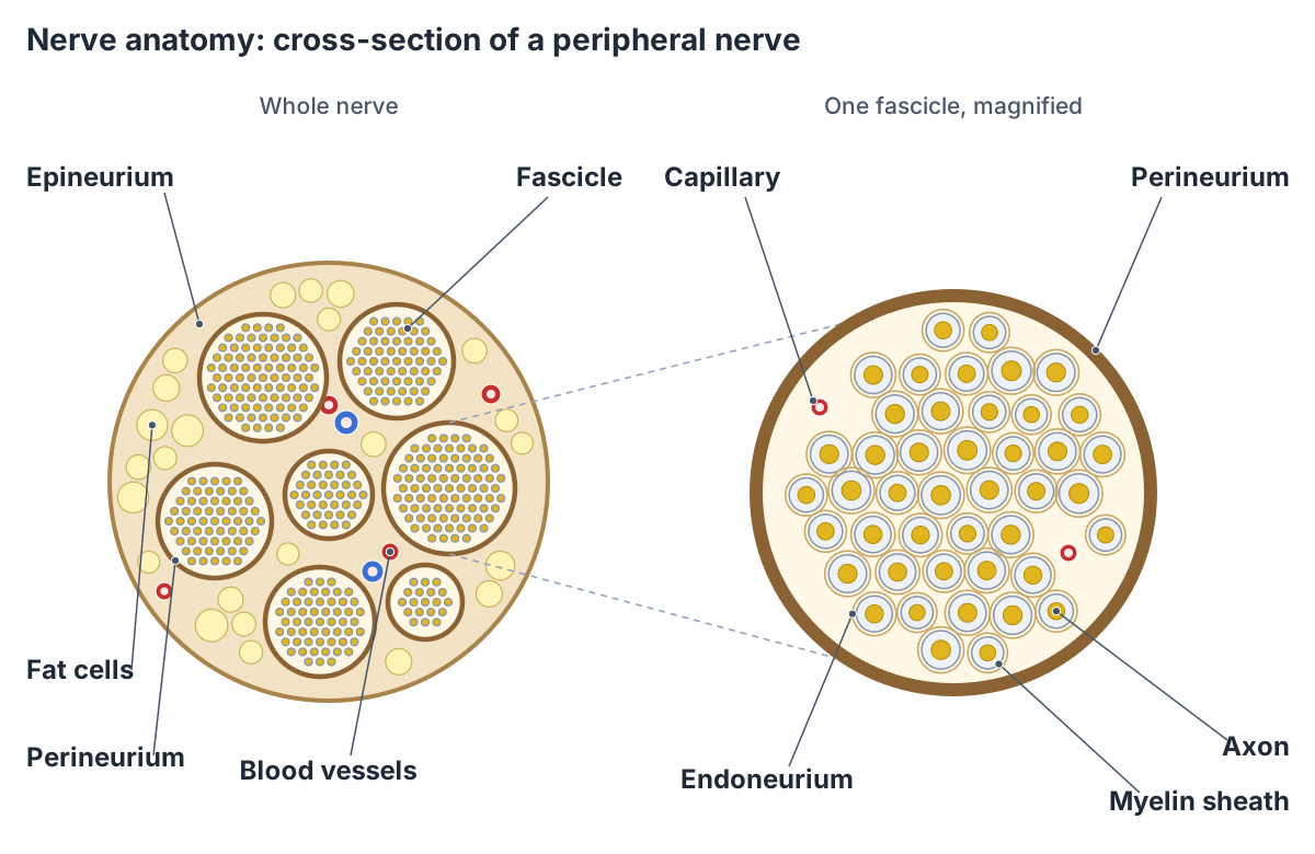
A nerve is a bundle of nerve fibres in the PNS surrounded by connective tissue, which can also enclose blood vessels. Sensory nerves contain only afferent fibres, motor nerves only efferent fibres, and mixed nerves contain both. (SEER Training Modules (NIH National Cancer Institute): The Peripheral Nervous System.)
Three layers of connective tissue wrap a nerve. The epineurium surrounds the whole nerve. Inside it the axons are grouped into bundles called fascicles, each wrapped in its own perineurium. Each individual axon is surrounded by loose connective tissue called endoneurium. OpenStax points out that these three layers resemble the connective tissue sheaths of a muscle. (OpenStax Anatomy and Physiology 2e, 13.4: The Peripheral Nervous System.)
The epineurium also contains fat cells, fibroblasts and small blood and lymph vessels, and some nerves, such as the sciatic nerve, carry a lot of fat in it. Arteries form a network of vessels in the epineurium, and branches pass through the perineurium to reach the endoneurium as arterioles and capillaries. The perineurium helps maintain the pressure inside each fascicle and contributes to a barrier around the fibres. (NYSORA: Connective Tissues of Peripheral Nerves.)
How does the nervous system work? Action potentials and synapses
Neurons send signals as action potentials, brief electrical events that are propagated along the axon. At rest the inside of a neuron is negative compared with the outside. OpenStax uses −70 mV as the typical resting membrane potential and notes that the exact value varies between cells. (OpenStax Anatomy and Physiology 2e, 12.4: The Action Potential.)
When a stimulus depolarises the membrane to a threshold of about −55 mV, voltage-gated sodium channels open, sodium ions rush in and the inside briefly becomes positive. Potassium channels then return the membrane towards rest, overshooting slightly (hyperpolarisation). The whole event lasts about 1 to 2 milliseconds. (OpenStax Anatomy and Physiology 2e, 12.4: The Action Potential.)
Action potentials are 'all or none'. A stimulus that does not reach threshold produces no action potential, and a stronger stimulus produces more action potentials, more quickly, not bigger ones. (OpenStax Anatomy and Physiology 2e, 12.4: The Action Potential.)
Myelin speeds the signal up. Along an unmyelinated axon the action potential spreads continuously, which is slow. Along a myelinated axon only the nodes of Ranvier depolarise, so the signal jumps from node to node (saltatory conduction), which is much faster. (OpenStax Anatomy and Physiology 2e, 12.4: The Action Potential.)
The thickest myelinated fibres, which supply skeletal muscle and carry signals from muscle spindles, conduct at up to about 120 metres per second. Thin unmyelinated C fibres, which carry pain and temperature signals, conduct at only about 0.5 to 2.5 metres per second. (Kiernan J. Nerve fibre types: size and conduction velocity of nerve fibers. Western University.)
Neurons pass signals on at synapses. At a chemical synapse, the arriving action potential opens voltage-gated calcium channels in the synaptic end bulb; calcium makes vesicles fuse with the membrane and release a neurotransmitter into the synaptic cleft. The transmitter fits its receptor on the next cell like a key in a lock, and is then cleared by enzymes or taken back up by neurons or glial cells. At an electrical synapse, ions pass directly from one cell to the next. (OpenStax Anatomy and Physiology 2e, 12.5: Communication Between Neurons.)
Each phase of the voltage change is shown on a labelled graph on the page Action potential diagram.
Reflexes: how the reflex arc works
A reflex is an immediate, automatic response to a stimulus. The pathway it uses, the reflex arc, has five parts in order: (1) a receptor that detects the stimulus and turns it into an electrical signal; (2) a sensory (afferent) neuron that carries the signal to the spinal cord or brainstem; (3) an integration centre that processes it; (4) a motor (efferent) neuron that carries the command out; and (5) an effector, the muscle or gland that responds. (Kenhub: Reflex arc.)
In the knee-jerk (patellar) reflex, the sensory neuron synapses directly with the motor neuron, so the reflex is monosynaptic: tapping the patellar tendon makes the quadriceps contract and the leg extend. Withdrawal reflexes are polysynaptic, with one or more interneurons between the sensory and motor neurons. When you pull your hand off something hot, the withdrawal is driven by the spinal cord and the brain registers the heat only afterwards. (Kenhub: Reflex arc.)
In the hot-stove withdrawal reflex, the connection in the spinal cord makes biceps brachii contract. At the same time an interneuron in the dorsal horn of the cord inhibits the motor neuron of the opposing triceps brachii, so the triceps does not resist and the arm pulls away faster. (OpenStax Anatomy and Physiology 2e, 14.3: Motor Responses.)
Common nervous system disorders
When part of the nervous system goes wrong, people can have trouble moving, speaking, swallowing, breathing or learning, or problems with memory, the senses or mood. MedlinePlus groups neurological diseases into inherited conditions such as Huntington's disease, problems of development such as spina bifida, degenerative diseases such as Parkinson's and Alzheimer's disease, diseases of the brain's blood vessels such as stroke, injuries to the brain and spinal cord, seizure disorders such as epilepsy, cancers such as brain tumours, and infections such as meningitis. (MedlinePlus (US National Library of Medicine): Neurologic Diseases.)
Single peripheral nerves can be affected too. Kenhub gives two classic examples: Bell's palsy affects the facial nerve, and carpal tunnel syndrome affects the median nerve where it is compressed in the carpal tunnel at the wrist. (Kenhub: The nervous system.)
Sciatica happens when the sciatic nerve, which runs from the lower back to the feet, is irritated or compressed. Its symptoms usually affect the buttock and the back of one leg, often including the foot and toes. (NHS: Sciatica.)
This section is for learning only and is not medical advice. Sudden drooping of one side of the face, weakness in an arm or difficulty speaking can be signs of a stroke, which is a medical emergency, so get emergency help at once; any other new or changing symptoms should be checked by a clinician. (NHS: Bell's palsy.)
Examples of nervous system disorders by the part affected
| Part affected | Type of problem | Examples |
|---|---|---|
| Brain | Disease of the blood vessels that supply it | Stroke |
| Brain | Nerve cells damaged or dying (degenerative disease) | Alzheimer's disease, Parkinson's disease |
| Brain | Seizure disorder | Epilepsy |
| Brain | Cancer | Brain tumours |
| Brain and spinal cord | Injury | Traumatic brain injury, spinal cord injury |
| Brain | Faulty genes | Huntington's disease |
| Spinal cord and its coverings | Problem with development | Spina bifida |
| Brain and spinal cord coverings | Infection | Meningitis |
| Cranial nerve | Inflammation and swelling of the facial nerve (VII) | Bell's palsy |
| Peripheral nerve | Pressure on the median nerve at the wrist | Carpal tunnel syndrome |
| Sciatic nerve or its spinal nerve roots | Irritation or compression | Sciatica |
Educational overview, not a diagnosis. Sources: MedlinePlus (neurologic diseases, Huntington's disease); Cleveland Clinic (nervous system, Bell's palsy); Kenhub; NHS (carpal tunnel syndrome, sciatica); OpenStax A&P 2e 13.4.
What the 3D nervous system model shows
The 3D view highlights the brain of the BodyParts3D adult male model and opens on its left side, where the gyri of the cerebral cortex are coloured by lobe and the midbrain, pons, medulla oblongata and cerebellum show below. Deeper parts are highlighted too but lie inside the brain: the insula, corpus callosum and fornix, the thalamus, hypothalamus and basal nuclei (caudate nucleus, putamen and globus pallidus), the hippocampus and amygdala, and the ventricles; turn the model or hide outer parts to reach them. The cranial nerves are represented by the optic nerves, optic chiasm and optic tracts and a few nerves of the orbit: the ophthalmic and trochlear nerves and the branches of the oculomotor nerve. Click any highlighted part to open its own page.
The model has no spinal cord, spinal nerves or limb nerves. It includes only a short piece of the central canal of the spinal cord, about 3.6 cm long, at the level of the medulla and the top of the neck, without the cord tissue around it. Use the labelled diagram at the top of the page for the spinal cord, plexuses and peripheral nerves.
The optic nerves show that the line between the CNS and PNS is partly a matter of naming. OpenStax calls the same axons the optic nerve where they leave the eye and the optic tract inside the cranium, with the name changing at the optic chiasm. (OpenStax Anatomy and Physiology 2e, 12.1: Basic Structure and Function of the Nervous System.)
Strictly, though, the optic nerve is an outgrowth of the brain: it is wrapped in meninges and CSF and has no Schwann cells (its myelin comes from oligodendrocytes), so most anatomy texts count it as part of the CNS. (Kenhub: Optic nerve.)
Nervous system in the 3D model
- Amygdala
- The amygdala is an almond-shaped nuclear complex in the medial temporal lobe that is central to processing emotionally significant stimuli.
- Angular gyrus
- The angular gyrus is a cortical region of the inferior parietal lobule that arches over the end of the superior temporal sulcus.
- Anterior part of superior temporal gyrus
- This is the anterior portion of the superior temporal gyrus, the uppermost gyrus on the lateral surface of the temporal lobe.
- Caudate nucleus
- The caudate nucleus is a C-shaped nucleus of the basal ganglia that, with the putamen, forms the striatum.
- Central canal of spinal cord
- The central canal is a narrow, CSF-filled channel running through the length of the spinal cord.
- Cerebellum
- The cerebellum is the large structure at the back of the brain that coordinates movement, posture and balance and takes part in motor learning.
- Choroid plexus of cerebral hemisphere
- The choroid plexus is a vascular, fringe-like tissue in the cerebral ventricles that produces cerebrospinal fluid.
- Cingulate gyrus
- The cingulate gyrus is a long arching gyrus on the medial surface of the cerebral hemisphere above the corpus callosum and a major part of the limbic lobe.
- Corpus callosum
- The corpus callosum is the largest white matter commissure, connecting the left and right cerebral hemispheres.
- Fornix of forebrain
- The fornix is a C-shaped white matter tract that carries the main output of the hippocampus.
- Fourth ventricle
- The fourth ventricle is a diamond-shaped, CSF-filled cavity of the hindbrain.
- Fusiform gyrus
- The fusiform gyrus is a long gyrus on the underside of the temporal and occipital lobes.
- Globus pallidus
- The globus pallidus is a basal ganglia nucleus that provides major inhibitory output to the thalamus.
- Hippocampus
- The hippocampus is a curved cortical structure in the medial temporal lobe that is essential for forming new long-term memories.
- Hypothalamus
- The hypothalamus is a small diencephalic region that regulates homeostasis, autonomic function and endocrine control.
- Inferior branch of oculomotor nerve
- The inferior division of the oculomotor nerve supplies three extraocular muscles and carries parasympathetic fibres to the ciliary ganglion.
- Inferior frontal gyrus
- The inferior frontal gyrus is the lowest of the three frontal gyri on the lateral frontal lobe, and contains Broca's area on the dominant side.
- Inferior temporal gyrus
- The inferior temporal gyrus is the lowest gyrus on the lateral surface of the temporal lobe.
- Insula
- The insula is a cortical region buried deep within the lateral sulcus, covered by the frontal, parietal and temporal opercula.
- Internal capsule
- The internal capsule is a V-shaped band of white matter carrying fibres between the cerebral cortex and the brainstem and spinal cord.
- Lateral ventricle
- The lateral ventricle is a C-shaped CSF-filled cavity within each cerebral hemisphere.
- Mammillary body
- The mammillary bodies are paired round bumps on the underside of the hypothalamus that are part of the memory circuit.
- Medulla oblongata
- The medulla oblongata is the lowest part of the brainstem, continuous with the spinal cord.
- Midbrain
- The midbrain is the smallest part of the brainstem and links the pons with the diencephalon.
- Middle frontal gyrus
- The middle frontal gyrus is the central of the three frontal gyri on the lateral surface of the frontal lobe.
- Middle temporal gyrus
- The middle temporal gyrus is the central gyrus on the lateral surface of the temporal lobe.
- Occipital lobe
- The occipital lobe is the posterior lobe of the cerebral hemisphere, and the primary visual centre.
- Ophthalmic nerve
- The ophthalmic nerve is the first and smallest division of the trigeminal nerve and is purely sensory.
- Optic chiasm
- The optic chiasm is the X-shaped junction where fibres from the two optic nerves partly cross.
- Optic nerve
- The optic nerve (CN II) carries visual information from the retina to the brain.
- Optic tract
- The optic tract is the pathway of visual fibres from the optic chiasm to the lateral geniculate body.
- Orbital gyrus
- The orbital gyri are the convolutions on the orbital (inferior) surface of the frontal lobe, resting on the roof of the orbit.
- Parahippocampal gyrus
- The parahippocampal gyrus is a gyrus on the medial temporal lobe, adjoining the hippocampus.
- Pineal body
- The pineal body (gland) is a small pinecone-shaped endocrine gland in the brain.
- Pituitary gland
- The pituitary gland (hypophysis) is a pea-sized endocrine gland that regulates many other glands.
- Pons
- The pons is the part of the brainstem between the midbrain and medulla that relays signals between the cerebrum and cerebellum.
- Postcentral gyrus
- The postcentral gyrus is the primary somatosensory cortex of the parietal lobe.
- Posterior part of superior temporal gyrus
- This is the posterior portion of the superior temporal gyrus, important for hearing and language.
- Precentral gyrus
- The precentral gyrus is the primary motor cortex of the frontal lobe.
- Putamen
- The putamen is the large lateral nucleus of the basal ganglia and part of the striatum.
- Superior branch of oculomotor nerve
- The superior division of the oculomotor nerve supplies the superior rectus and the levator palpebrae superioris.
- Superior frontal gyrus
- The superior frontal gyrus is the uppermost gyrus of the frontal lobe, extending onto its medial surface.
- Superior parietal lobule
- The superior parietal lobule is a parietal association area above the intraparietal sulcus.
- Supramarginal gyrus
- The supramarginal gyrus is part of the inferior parietal lobule that arches over the end of the lateral sulcus.
- Thalamus
- The thalamus is a paired diencephalic mass that relays sensory and motor information to the cerebral cortex.
- Third ventricle
- The third ventricle is a narrow midline CSF-filled cavity of the diencephalon.
- Trochlear nerve
- The trochlear nerve (CN IV) is a motor nerve supplying the superior oblique muscle of the eye.
Frequently asked questions
What are the two main parts of the nervous system?
The two main parts are the central nervous system (CNS) and the peripheral nervous system (PNS). The CNS is the brain and spinal cord, protected by the skull and vertebrae, the meninges and cerebrospinal fluid. The PNS is everything outside it: 12 pairs of cranial nerves, 31 pairs of spinal nerves and the ganglia, which link the CNS with the muscles, glands, skin and organs.
What are the 3 main functions of the nervous system?
The three main functions are sensation (sensory input), integration and response (motor output). Receptors detect changes inside and outside the body; the brain and spinal cord combine that information with memories, learning and emotion and decide what to do; then nerves carry signals to muscles and glands, which respond. The nervous system also keeps the internal environment stable and is the centre of thought and memory.
What are the main organs of the nervous system?
The main organs are the brain, the spinal cord, the nerves and the ganglia. The brain and spinal cord form the central nervous system; the nerves and ganglia, small clusters of nerve cell bodies outside the CNS, form the peripheral nervous system. The brain itself has four main regions: the cerebrum, diencephalon, brainstem and cerebellum.
What is the difference between the somatic and autonomic nervous system?
The somatic nervous system handles conscious sensation and voluntary movement, and its motor nerves supply skeletal muscle. The autonomic nervous system works automatically, without conscious effort, on cardiac muscle, smooth muscle and glands to control heart rate, breathing, blood pressure, digestion and body temperature. It has a sympathetic ('fight or flight') and a parasympathetic ('rest and digest') division.
How does the nervous system work?
Neurons carry information as action potentials, brief electrical impulses that start when the cell membrane reaches a threshold of about −55 mV and travel along the axon, jumping between nodes in myelinated fibres. At synapses the impulse releases a neurotransmitter that acts on the next cell. Chains of neurons link receptors, the brain and spinal cord, and the muscles and glands that respond.
How many nerves are in the human body?
Forty-three pairs of nerves connect directly to the brain and spinal cord: 12 pairs of cranial nerves and 31 pairs of spinal nerves (8 cervical, 12 thoracic, 5 lumbar, 5 sacral and 1 coccygeal). In the neck and limbs the spinal nerves regroup in four plexuses into named nerves, such as the median, ulnar, radial, femoral and sciatic nerves, which divide into many smaller branches.
What is a nerve made of?
A nerve is a bundle of nerve fibres (axons) held together by connective tissue. The epineurium wraps the whole nerve and contains fat and blood vessels. The fibres are grouped into fascicles, each wrapped in perineurium, and each individual fibre is surrounded by endoneurium. Sensory nerves carry only afferent fibres, motor nerves only efferent fibres and mixed nerves both.
Is the brain part of the nervous system?
Yes. The brain and the spinal cord together make up the central nervous system, and the two are continuous at the foramen magnum at the base of the skull. The brain has four major regions: the cerebrum, the diencephalon, the brainstem (midbrain, pons and medulla oblongata) and the cerebellum. The brain and nervous system are therefore one system, not two.
References
- OpenStax. Anatomy and Physiology 2e, §12.1 Basic Structure and Function of the Nervous System. Rice University, 2022.
- OpenStax. Anatomy and Physiology 2e, §12.2 Nervous Tissue. Rice University, 2022.
- OpenStax. Anatomy and Physiology 2e, §12.4 The Action Potential. Rice University, 2022.
- OpenStax. Anatomy and Physiology 2e, §12.5 Communication Between Neurons. Rice University, 2022.
- OpenStax. Anatomy and Physiology 2e, §13.2 The Central Nervous System. Rice University, 2022.
- OpenStax. Anatomy and Physiology 2e, §13.3 Circulation and the Central Nervous System. Rice University, 2022.
- OpenStax. Anatomy and Physiology 2e, §13.4 The Peripheral Nervous System. Rice University, 2022.
- OpenStax. Anatomy and Physiology 2e, §14.3 Motor Responses. Rice University, 2022.
- OpenStax. Anatomy and Physiology 2e, §15.1 Divisions of the Autonomic Nervous System. Rice University, 2022.
- SEER Training Modules: Introduction to the Nervous System. US National Institutes of Health, National Cancer Institute.
- SEER Training Modules: Organization of the Nervous System. US National Institutes of Health, National Cancer Institute.
- SEER Training Modules: The Central Nervous System. US National Institutes of Health, National Cancer Institute.
- SEER Training Modules: The Peripheral Nervous System. US National Institutes of Health, National Cancer Institute.
- SEER Training Modules: Nerve Tissue. US National Institutes of Health, National Cancer Institute.
- Vasković J. The nervous system. Kenhub.
- Kenhub. Histology of glial cells.
- Vasković J. Action potential. Kenhub.
- Karamitrou A. Reflex arc. Kenhub.
- Kenhub. Spinal nerves.
- Kenhub. Conus medullaris and cauda equina.
- Kenhub. Autonomic nervous system.
- Kenhub. Intercostal nerves.
- Kenhub. Vagus nerve.
- Kenhub. Sciatic nerve.
- Kenhub. Tibial nerve.
- Kenhub. Common fibular (peroneal) nerve.
- Kenhub. Optic nerve.
- TeachMeAnatomy. The Brachial Plexus.
- TeachMeAnatomy. The Median Nerve.
- TeachMeAnatomy. The Ulnar Nerve.
- TeachMeAnatomy. The Radial Nerve.
- TeachMeAnatomy. The Lumbar Plexus.
- TeachMeAnatomy. The Sacral Plexus.
- TeachMeAnatomy. The Femoral Nerve.
- TeachMeAnatomy. The Sciatic Nerve.
- TeachMeAnatomy. The Vagus Nerve (CN X).
- TeachMeAnatomy. The Scalp.
- NYSORA. Connective Tissues of Peripheral Nerves.
- Kiernan J. Nerve fibre types, Table 2: Size and conduction velocity of nerve fibers. Western University.
- Yap JYY, Keatch C, Lambert E, Woods W, Stoddart PR, Kameneva T. Critical Review of Transcutaneous Vagus Nerve Stimulation: Challenges for Translation to Clinical Practice. Front Neurosci. 2020;14:284 (Table 1, nerve fibre types).
- Azevedo FAC, Carvalho LRB, Grinberg LT, et al. Equal numbers of neuronal and nonneuronal cells make the human brain an isometrically scaled-up primate brain. J Comp Neurol. 2009;513(5):532-541.
- Byrne JH. Resting Potentials and Action Potentials. Neuroscience Online, McGovern Medical School at UTHealth.
- Cleveland Clinic. Nervous System. Last updated 16 November 2023.
- Cleveland Clinic. Bell's Palsy.
- MedlinePlus. Neurologic Diseases. US National Library of Medicine.
- MedlinePlus. Huntington's Disease. US National Library of Medicine.
- NHS. Bell's palsy.
- NHS. Carpal tunnel syndrome.
- NHS. Sciatica.
- Mitsuhashi N. et al. (2009). BodyParts3D: 3D structure database for anatomical concepts. Nucleic Acids Research 37, D782–D785. Data © The Database Center for Life Science, CC BY 4.0.
Related anatomy
- Brain anatomyBrain anatomy with a labeled brain diagram and 3D model: cerebrum and its four lobes, gyri, corpus callosum, thalamus…
- Spinal cordSpinal cord anatomy with labelled diagrams: cross-section, grey matter horns, spinothalamic and dorsal column tracts…
- Cranial nervesThe 12 cranial nerves in order with labelled diagrams, plus nerve anatomy of the face: facial nerve branches…
- Autonomic nervous systemThe autonomic nervous system explained: sympathetic vs parasympathetic outflow diagram, sympathetic trunk, effects on…
- Upper limb nervesBrachial plexus diagram with roots, trunks, cords and branches; axillary, musculocutaneous, median, ulnar and radial…
- Lower limb nervesLower limb nerves explained: lumbar and sacral plexus diagram, femoral, saphenous, sciatic, tibial, common peroneal…
Explore the whole body in the interactive 3D human anatomy model, or browse the A–Z list of human structures.